Co-reporter:Qi Sun, Minghui Li, Xiao Yang, Xi Xu, Junsong Wang, Jianfa Zhang
The Journal of Nutritional Biochemistry 2017 Volume 47(Volume 47) pp:
Publication Date(Web):1 September 2017
DOI:10.1016/j.jnutbio.2017.04.015
Previous studies suggest that dietary salecan (a water-soluble β-glucan) effectively reduces high-fat-diet-induced adiposity through disturbing bile-acid-promoted emulsification in mice. However, the effects of salecan on metabolic genes and metabolites involved in lipid accumulation are mostly unknown. Here, we confirmed that dietary 3% and 6% salecan for 4 weeks markedly decreased fat accumulation in liver and adipose tissue in high-fat-diet rats, displaying a decrease in mRNA levels of SREBP1-C, FAS, SCD1 and ACC1 involved in de novo lipogenesis and a reduction of levels of GPAT1, DGAT1 and DGAT2 related to triglyceride synthesis. Dietary salecan also increased the mRNA levels of PPARα and CYP7A1, which are related to fatty acid oxidation and cholesterol decomposition, respectively. In the 1H nuclear magnetic resonance metabolomic analysis, both the serum and liver metabolite profiles differed among the control groups, and the metabolic profiles of the salecan groups were shifted toward that of the low-fat-diet group. Metabolites analysis showed that salecan significantly increased hepatic glutathione and betaine levels which are related to regulation of cellular reactive oxygen species. These data demonstrate that dietary salecan not only disturbed fat digestion and absorption but also influenced lipid accumulation and metabolism in diet-induced obesity.Download high-res image (153KB)Download full-size image
Co-reporter:Linxiang Xu;Rui Cheng;Jing Li;Yang Wang
Applied Microbiology and Biotechnology 2017 Volume 101( Issue 2) pp:585-598
Publication Date(Web):2017 January
DOI:10.1007/s00253-016-7814-z
Salecan, a soluble β-1,3-d-glucan produced by a salt-tolerant strain Agrobacterium sp. ZX09, has been the subject of considerable interest in recent years because of its multiple bioactivities and unusual rheological properties in solution. In this study, both succinyl and pyruvyl substituent groups on salecan were identified by an enzymatic hydrolysis following nuclear magnetic resonance (NMR), HPLC, and MS analysis. The putative succinyltransferase gene (sleA) and pyruvyltransferase gene (sleV) were determined and cloned. Disruption of the sleA gene resulted in the absence of succinyl substituent groups on salecan. This defect could be complemented by expressing the sleA cloned in a plasmid. Thus, the sleA and sleV genes located in a 19.6-kb gene cluster may be involved in salecan biosynthesis. Despite the lack of succinyl substituents, the molecular mass of salecan generated by the sleA mutant did not substantially differ from that generated by the wild-type strain. Loss of succinyl substituents on salecan changed its rheological characteristics, especially a decrease in intrinsic viscosity.
Co-reporter:T Wang, Z Wang, P Yang, L Xia, M Zhou, S Wang, Jie Du and J Zhang
Cell Death & Disease 2016 7(4) pp:e2176
Publication Date(Web):2016-04-01
DOI:10.1038/cddis.2016.9
The severity of acute liver failure (ALF) induced by bacterial lipopolysaccharide (LPS) is associated with the hepatic innate immune response. The core circadian molecular clock modulates the innate immune response by controlling rhythmic pathogen recognition by the innate immune system and daily variations in cytokine gene expression. However, the molecular link between circadian genes and the innate immune system has remained unclear. Here, we showed that mice lacking the clock gene Per1 (Period1) are more susceptible to LPS/d-galactosamine (LPS/GalN)-induced macrophage-dependent ALF compared with wild-type (WT) mice. Per1 deletion caused a remarkable increase in the number of Kupffer cells (KCs) in the liver, resulting in an elevation of the levels of pro-inflammatory cytokines after LPS treatment. Loss of Per1 had no effect on the proliferation or apoptosis of macrophages; however, it enhanced the recruitment of macrophages, which was associated with an increase in CC chemokine receptor 2 (Ccr2) expression levels in monocytes/macrophages. Deletion of Ccr2 rescued d-GalN/LPS-induced liver injury in Per1−/− mice. We demonstrated that the upregulation of Ccr2 expression by Per1 deletion could be reversed by the synthetic peroxisome proliferator-activated receptor gamma (PPAR-γ) antagonist GW9662. Further analysis indicated that PER1 binds to PPAR-γ on the Ccr2 promoter and enhanced the inhibitory effect of PPAR-γ on Ccr2 expression. These results reveal that Per1 reduces hepatic macrophage recruitment through interaction with PPAR-γ and prevents an excessive innate immune response in endotoxin-induced liver injury.
Co-reporter:Linxiang Xu
Applied Microbiology and Biotechnology 2016 Volume 100( Issue 21) pp:9023-9036
Publication Date(Web):2016 November
DOI:10.1007/s00253-016-7836-6
Bacterial glucans have aroused increasing interest in commercial applications in the food and pharmaceutical sectors. A number of bacterial glucans have been reported over recent decades, and their structure, production, and functional properties have been extensively studied. In this paper, we review recent researches on bacterial glucans, with emphasis on the production, physical and chemical properties, and the new developments in food, biomedical, pharmaceutical, and other industrial applications.
Co-reporter:Y Zhan, Z Wang, P Yang, T Wang, L Xia, M Zhou, Y Wang, S Wang, Z Hua and J Zhang
Cell Death & Disease 2014 5(1) pp:e985
Publication Date(Web):2014-01-01
DOI:10.1038/cddis.2013.516
D-galactosamine (GalN)/lipopolysaccharide (LPS)-induced lethality and acute liver failure is dependent on endogenously produced inflammatory cytokines. Adenosine has been proven to be a central role in the regulation of inflammatory response. It is not entirely clear that which adenosine action is actually crucial to limiting inflammatory tissue destruction. Here we showed that GalN/LPS challenge elevated hepatic adenosine and induced lethality in adenosine receptor-deficient mice with equal efficiency as wild-type mice. In GalN/LPS-treated mice, pretreatment with adenosine 5′-monophosphate (5′-AMP) significantly elevated hepatic adenosine level and reduced mortality through decreasing cytokine and chemokine production. In RAW264.7 cells, 5′-AMP treatment inhibited the production of inflammatory cytokines, which is not mediated through adenosine receptors. 5′-AMP failed to attenuate LPS-induced nuclear factor-κB (NF-κB) p65 nuclear translocation, but reduced LPS-induced recruitment of NF-κB p65 to inflammatory gene promoters and decreased LPS-induced enrichment of H3K4 dimethylation at the tumor necrosis factor-α (TNF-α) promoter, which was involved in 5′-AMP-induced elevation of cellular adenosine and a decline of methylation potential. In vitro biochemical analysis revealed that adenosine directly attenuated recruitment of NF-κB to the TNF-α and interleukin-6 promoters. Our findings demonstrate that 5′-AMP-inhibiting inflammatory response is not mediated by adenosine receptors and it may represent a potential protective agent for amelioration of LPS-induced liver injury.
Co-reporter:Mengyi Zhou, Chuanliang Pu, Lin Xia, Xiaohong Yu, Bin Zhu, Rui Cheng, Linxiang Xu, Jianfa Zhang
Carbohydrate Polymers 2014 Volume 102() pp:772-779
Publication Date(Web):15 February 2014
DOI:10.1016/j.carbpol.2013.10.091
•The levels of lactate and SCFA in contents of cecum and colon as well as in feces were generally elevated as a result of Salecan consumption.•The amount of Lactobacillus and Bifidobacterium increased in cecal contents of mice fed Salecan.•Dietary incorporation of Salecan may be beneficial in improving gastrointestinal health.Salecan, a linear extracellular polysaccharide consisting of β-(1,3)-d-glucan, has potential applications in the food industry due to its excellent toxicological profile and rheological properties. The aim of the present study was to evaluate the effects of dietary supplementation with 8% Salecan on the gastrointestinal microbiota in mice. In the Salecan group, the following significant differences (p < 0.05) from the cellulose group were found: increased body weight gain, greater mass of cecum and cecal contents, and higher butyrate concentrations in the cecal and colonic contents at wk 4. Moreover, populations of Lactobacillus and Bifidobacterium increased 3- and 6-fold, respectively, in the cecal contents of mice consuming Salecan. These results suggest that the dietary incorporation of Salecan, by providing SCFAs and increasing beneficial microbiota, may be beneficial in improving gastrointestinal health, and have relevance to the use of Salecan as a dietary supplement for human consumption.
Co-reporter:Rui Cheng;Linxiang Xu;Shiming Wang;Yang Wang
Biotechnology Letters 2014 Volume 36( Issue 4) pp:797-803
Publication Date(Web):2014 April
DOI:10.1007/s10529-013-1413-1
A new β-1,3-1,4-glucanase gene (PlicA) was cloned from Paenibacillus sp. S09. The ORF contained 717 bp coding for a 238 amino acid protein. PlicA, expressed in Escherichia coli and purified by Ni2+-affinity chromatography, had optimum activity at 55 °C and pH 6.2. The specific activity toward barley β-glucan reached 7,055 U/mg. Km and Vmax values with barley β-glucan were 3.7 mg/ml and 3.3 × 10³ μmol/min mg, respectively. The enzyme exhibited acid- and alkali-tolerance with more than 80 % activity remaining after incubation for 4 h at pH 3.5–12. PlicA was salt-tolerant (>90 % activity retained in 4 M NaCl at 25 °C for 24 h) and salt-activated: activity rising 1.5-fold in 0.5 M NaCl. The thermostability was improved by NaCl and CaCl2. This is the first report of an acid-, alkali- and salt-tolerant bacterial β-1,3-1,4-glucanase with high catalytic efficiency.
Co-reporter:Peng Chen;Zhongqiu Wang;Liyan Zeng;Shiming Wang;Wei Dong;Aiqun Jia;Chun Cai
Journal of Applied Toxicology 2012 Volume 32( Issue 10) pp:796-803
Publication Date(Web):
DOI:10.1002/jat.1694
ABSTRACT
Carbon tetrachloride (CCl4) is a well-established model for screening hepato-protective drugs. The aim of the present study was to evaluate the potential protective effects of a novel soluble β-glucan salecan on acute liver injury induced by CCl4 in mice and to further explore the underlying mechanisms. Mice were given salecan (40 mg kg−1) or phosphate-buffered saline for 3 days prior to treatment with a single intraperitoneal dose of CCl4 (1 ml kg−1 body weight). Animals were sacrificed at 0, 12, 24, 48, 72 and 96 h post-injection of CCl4. Serum liver enzyme levels, histology, lipid peroxidation, glutathione (GSH) content, expression of antioxidant enzymes and hepatocyte proliferation were subsequently evaluated. The serum levels of hepatic enzyme markers were markedly reduced in the salecan pretreatment group compared with the control group. Histopathological examination of the livers revealed that hepatocellular degeneration and necrosis were significantly attenuated at an early stage during CCl4 intoxication and liver recovery was markedly accelerated at a later stage in salecan pre-administered mice. Furthermore, salecan administration remarkably alleviated lipid peroxidation and restored GSH depletion. Meanwhile, the expression of antioxidant genes was significantly elevated in the salecan-treated group. Interestingly, the administration of salecan remarkably enhanced hepatocyte proliferation in the recovery phase after CCl4 injection. Taken together, these results demonstrated that salecan exhibits a protective action on acute hepatic injury induced by CCl4 through attenuating oxidative stress and accelerating hepatocyte regeneration. Copyright © 2011 John Wiley & Sons, Ltd.
Co-reporter:Aihui Xiu, Yi Kong, Mengyi Zhou, Bin Zhu, Shiming Wang, Jianfa Zhang
Carbohydrate Polymers 2010 Volume 82(Issue 3) pp:623-628
Publication Date(Web):15 October 2010
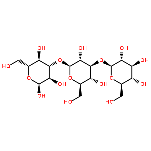
DOI:10.1016/j.carbpol.2010.05.027
A salt-tolerant strain Agrobacterium sp. ZX09 produced a high molecular mass, water-soluble extracellular polysaccharide (EPS) composed of d-glucose. By examining periodate oxidation, 1H NMR and 13C NMR spectra, it was proven that the EPS consisted of the following repeating unit: →3)-β-d-Glcp-(1 → 3)-[β-d-Glcp-(1 → 3)-β-d-Glcp-(1 → 3)]3-α-d-Glcp-(1 → 3)-α-d-Glcp-(1→. In vitro the EPS was indigestible by α-amylase, and strongly inhibited pancreatic α-amylase activity. Fasting mice fed on the EPS failed to increase blood glucose levels. Experimental diets containing the EPS suppressed steep increase in blood glucose concentration. These results suggest that the novel water-soluble glucan could be potentially useful for decreasing absorption of dietary carbohydrate and postprandial blood glucose level.
Co-reporter:Xiamusiya Kakan, Peng Chen, Jianfa Zhang
Experimental and Toxicologic Pathology (September 2011) Volume 63(Issue 6) pp:581-585
Publication Date(Web):1 September 2011
DOI:10.1016/j.etp.2010.04.011
The circadian clock gene Period2 (Per2) plays important roles in many physiologic and pathologic processes in mammals. In the previous study, we have reported the protective role of mPer2 against carbon tetrachloride induced hepatotoxicity. Here, we further explore the contribution of this gene to acetaminophen (APAP) induced liver injury in mice. It is reported that the hepatotoxicity induced by APAP exhibited a circadian rhythm in which the peak sensitive injection time is 20:00 while when the administration time becomes to 08:00, it caused markedly decreased liver damage. Thus, we injected APAP into wide type (WT) and mPer2 null mice at the dose of 300 mg/kg at both 08:00 and 20:00. Interestingly, the liver damage showed no significant difference between WT and mPer2 null mice when administered at 08:00, however, liver injury occurred in mPer2 null mice displayed less severe than WT at 20:00. In addition, Cyp1a2, one of the most important cytochrome P450 isoforms responsible for APAP bioactivation, decreased mRNA level at 20:00 in mPer2 null mice while its expression was not different in both strain mice at 08:00. Coincidently, the hepatic circadian rhythm expression of Per2 revealed that its mRNA level was weak at 08:00 but reached peak expression during 24 h at 20:00 in WT mice. Therefore, it is speculated that clock gene mPer2 may function in diurnal variation of APAP induced hepatotoxicity via modulating Cyp1a2 expression in mice.
Co-reporter:Peng Chen, Xiamusiya Kakan, Shiming Wang, Wei Dong, Aiqun Jia, Chun Cai, Jianfa Zhang
Experimental and Toxicologic Pathology (May 2013) Volume 65(Issue 4) pp:427-432
Publication Date(Web):1 May 2013
DOI:10.1016/j.etp.2011.12.007
The Period 2 (Per2) gene is an important component of the circadian system and is thought to modulate many physiological and pathological processes in mammals. In the previous study, we have disclosed the protective role of Per2 against carbon tetrachloride induced liver injury and fibrosis. Here we further assess the effect of Per2 deficiency on cholestatic hepatic injury and fibrosis. Cholestasis was induced by bile duct ligation (BDL) for 10 days in wild-type (WT) and Per2−/− mice. Masson trichrome staining and analysis of α-SMA immunohistochemistry were performed to show the collagen accumulation and the HSC activation, respectively. The mRNA levels of fibrosis-related genes were monitored by quantitative real-time PCR. Following BDL, livers from Per2−/− mice exhibited markedly increased extent of bile infarct and extracellular matrix (ECM) deposition compared with WT mice. Furthermore, the expressions of fibrosis-related genes like TNF-α, TGF-β1, Col1α1 and TIMP-1 were dramatically elevated in Per2−/− cholestatic liver. Our observations indicated that clock gene Per2 plays a protective role in mediating liver injury and fibrosis during cholestasis.
Co-reporter:Ping Yang, Zhongqiu Wang, Yibei Zhan, Tao Wang, Mengyi Zhou, Lin Xia, Xiao Yang, Jianfa Zhang
Toxicology (5 July 2013) Volume 309() pp:100-106
Publication Date(Web):5 July 2013
DOI:10.1016/j.tox.2013.05.003
•A1AR deficiency aggravates ethanol-induced oxidative stress and liver damage.•Loss of A1AR promotes lipid accumulation by upregulating lipogenic genes.•DPCPX pretreatment exacerbates liver damage and early steatosis induced by ethanol.Previous studies have indicated a critical role of adenosine and its receptors in the pathogenesis of liver diseases. The aim of this study was to determine the contribution of A1 adenosine receptor (A1AR) to acute ethanol-induced hepatotoxicity. Wild-type (WT) and A1AR−/− mice were intragastrically administered with ethanol (5 g/kg), and hepatic injury was evaluated 6 h thereafter. Mice lacking A1AR were more susceptible to ethanol-induced liver damage than WT mice, as evidenced by higher serum transaminase levels and increased extent of histopathological changes. Ethanol induced triglycerides accumulation in the serum and liver, and this accumulation was augmented in A1AR−/− mice. Analysis of gene expression in the liver revealed up-regulated mRNA levels of genes related to lipogenesis (including: FAS, SCD1, ACC1, DGAT2, and PPARγ) in A1AR−/− mice after ethanol treatment. In addition, lack of A1AR aggravated lipid peroxidation and superoxide dismutase depletion caused by acute ethanol exposure. A subsequent study revealed that, pretreatment with A1AR antagonist DPCPX increases the sensitivity of mice to ethanol-induced liver injury. In conclusion, these results indicated that endogenous A1AR activation protects mice against acute ethanol-induced liver injury by reducing oxidative stress and decreasing lipid accumulation.
Co-reporter:Peng Chen, Xiamusiya Kakan, Jianfa Zhang
FEBS Letters (16 April 2010) Volume 584(Issue 8) pp:1597-1601
Publication Date(Web):16 April 2010
DOI:10.1016/j.febslet.2010.03.019
Disruption in circadian rhythms either by mutation in mice or by shiftwork in people, is associated with an increased risk for the development of multiple organ diseases. In turn, organ disease may influence the function of clock genes and peripheral circadian systems. Here we showed that hepatic fibrosis induced by carbon tetrachloride in mice leads to alterations in the circadian rhythms of hepatic clock genes. Especially, we found an impaired daily Cry2 rhythm in the fibrotic livers, with markedly decreased levels during the day time while compared with control livers. Associatively, the expressions of two important clock-regulated genes peroxisome proliferator-activated receptor alpha and cytochrome P450 oxidoreductase lost circadian rhythm with significantly decreased levels during the light–dark (12/12 h) cycle in fibrotic livers.
Co-reporter:Tao Wang, Ping Yang, Yibei Zhan, Lin Xia, Zichun Hua, Jianfa Zhang
Toxicology (15 December 2013) Volume 314(Issues 2–3) pp:193-201
Publication Date(Web):15 December 2013
DOI:10.1016/j.tox.2013.09.009
The severity of ethanol-induced liver injury is associated with oxidative stress and lipid accumulation in the liver. Core circadian clock is known to mediate antioxidative enzyme activity and lipid metabolism. However, the link between circadian clock and ethanol-induced hepatotoxicity remains unclear. Here we showed that extents of acute ethanol-induced liver injury and steatosis in mice exhibit circadian variations consistent with hepatic expression of Period (Per) genes. Mice lacking clock gene Per1 displayed less susceptible to ethanol-induced liver injury, as evidenced by lower serum transaminase activity and less severe histopathological changes. Ethanol-induced lipid peroxidation was alleviated in Per1−/− mice. However, Per1 deletion had no effect on antioxidants depletion caused by ethanol administration. Ethanol-induced triglycerides (TG) accumulation in the serum and liver was significantly decreased in Per1−/− mice compared with that in wild-type (WT) mice. Analysis of gene expression in the liver revealed peroxisome proliferators activated receptor-gamma (PPARγ) and its target genes related to TG synthesis are remarkably down-regulated in Per1−/− mice. HepG2 cells were treated with ethanol at 150 mM for 3 days. Per1 overexpression augmented lipid accumulation after treatment with ethanol in HepG2 cells, but had no effect on ethanol-induced oxidative stress. Expression of genes related to lipogenesis, including PPARγ and its target genes, was up-regulated in cells overexpressing Per1. In conclusion, these results indicated that circadian rhythms of ethanol-induced hepatotoxicity are controlled by clock gene Per1, and deletion of Per1 protected mice from ethanol-induced liver injury by decreasing hepatic lipid accumulation.