Co-reporter:Baohua Huang, James Otis, Melvin Joice, Alina Kotlyar, and Thommey P. Thomas
Biomacromolecules 2014 Volume 15(Issue 3) pp:
Publication Date(Web):January 6, 2014
DOI:10.1021/bm401777w
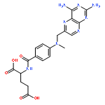
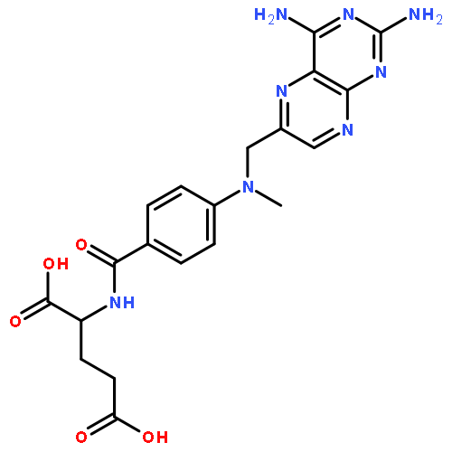
One of the important criteria for achieving efficient nanoparticle-based targeted drug delivery is that the drug is not prematurely released at off-target sites. Here we report the preclinical evaluation of a serum-stable dendrimer-based drug conjugate capable of actively targeting into prostate cancer (PC) cells, delivered through the prostate-specific membrane antigen (PSMA). Multiple molecules of PSMA-binding small molecule glutamate urea (GLA; targeting agent) and the drug methotrexate (MTX) were conjugated to generation 5 PAMAM dendrimer (G5) through Cu-free “click” chemistry. The GLA was conjugated through a stable amide bond, and the MTX was conjugated either through ester (Es)- or amide (Am)-coupling, to generate G5-GLAm-(Es)MTXn and G5-GLAm-(Am)MTXn, respectively. In serum-containing medium, free MTX was slowly released from “G5-GLAm-(Es)MTXn”, with ∼8% MTX released from the dendrimer in 72 h, whereas the MTX on G5-GLAm-(Am)MTXn was completely stable. The G5-GLAm-(Am)MTXn bound and internalized into PSMA-expressing LNCaP cells, but not into PSMA-negative PC3 cells. The conjugate-inhibited recombinant dihydrofolate reductase and induced potent cytotoxicity in the LNCaP cells, but not in the PC3 cells. Similar to the action of free GLA, stable amide-linked dendrimer-GLA was capable of inhibiting the enzyme N-acetylated α-linked acidic dipeptidase (NAALADase) activity of PSMA. The G5-GLAm-MTXn may serve as a serum-stable nanoparticle conjugate to specifically and effectively target and treat PSMA-overexpressing prostate tumors.
Co-reporter:Thommey P. Thomas, Baohua Huang, Seok Ki Choi, Justin E. Silpe, Alina Kotlyar, Ankur M. Desai, Hong Zong, Jeremy Gam, Melvin Joice, and James R. Baker Jr.
Molecular Pharmaceutics 2012 Volume 9(Issue 9) pp:2669-2676
Publication Date(Web):July 24, 2012
DOI:10.1021/mp3002232
Our previous studies have demonstrated that a generation 5 dendrimer (G5) conjugated with both folic acid (FA) and methotrexate (MTX) has a higher chemotherapeutic index than MTX alone. Despite this, batch-to-batch inconsistencies in the number of FA and MTX molecules linked to each dendrimer led to conjugate batches with varying biological activity, especially when scaleup synthesis was attempted. Since the MTX is conjugated through an ester linkage, there were concerns that biological inconsistency could also result from serum esterase activity and differential bioavailability of the targeted conjugate. In order to resolve these problems, we undertook a novel approach to synthesize a polyvalent G5–MTXn conjugate through click chemistry, attaching the MTX to the dendrimer through an esterase-stable amide linkage. Surface plasmon resonance binding studies show that a G5–MTX10 conjugate synthesized in this manner binds to the FA receptor (FR) through polyvalent interaction showing 4300-fold higher affinity than free MTX. The conjugate inhibits dihydrofolate reductase, and induces cytotoxicity in FR-expressing KB cells through FR-specific cellular internalization. Thus, the polyvalent MTX on the dendrimer serves the dual role as a targeting molecule as well as a chemotherapeutic drug. The newly synthesized G5–MTXn conjugate may serve as a FR-targeted chemotherapeutic with potential for cancer therapy.Keywords: cancer; dendrimer; drug delivery; methotrexate; nanoparticle;
Co-reporter:Thommey P. Thomas, Baohua Huang, Ankur Desai, Hong Zong, Xue-min Cheng, Alina Kotlyar, Pascale R. Leroueil, Thomas Dunham, Abraham van der Spek, Brent B. Ward, James R. Baker Jr.
Bioorganic & Medicinal Chemistry Letters 2010 Volume 20(Issue 21) pp:6250-6253
Publication Date(Web):1 November 2010
DOI:10.1016/j.bmcl.2010.08.098
Two morphine prodrugs (‘PDA’ and ‘PDB’) were synthesized and the kinetics of esterase-mediated morphine release from these prodrugs were determined when incubated with plasma from different animal species. Morphine was rapidly released from PDA by all species plasma with the maximum reached within 5–10 min; the released morphine was biologically active as determined by an in vitro cAMP assay. The morphine was released from PDB at a slower and species-dependent rate (mouse > rat > guinea pig > human). Morphine’s release from PDB appeared to be mediated by carboxyl esterases as the release was inhibited by the carboxyl esterase inhibitor benzil. PDA nor PDB induce cytotoxicity in the neuronal cell lines SK-NSH and SH-SY5Y. The carboxyl and amino functional moieties present on the linker portions of PDA and PDB, respectively, may facilitate their conjugation to nanoparticles to tailor morphine pharmacokinetics and specific targeting. These studies suggest the potential clinical utility of these prodrugs for morphine release at desired rates by administration of their mixture at selected ratios.
Co-reporter:Thommey P. Thomas, Rameshwer Shukla, Alina Kotlyar, Jola Kukowska-Latallo, James R. Baker Jr.
Bioorganic & Medicinal Chemistry Letters 2010 Volume 20(Issue 2) pp:700-703
Publication Date(Web):15 January 2010
DOI:10.1016/j.bmcl.2009.11.065
Fibroblast Growth Factor Receptor (FGFR) is overexpressed in a wide variety of tumors, and therefore is an attractive target for drug delivery. Recombinant FGF-1 was purified and attached to a fifth-generation (G5) polyamidoamine dendrimer. The specific binding and internalization of this conjugate labeled with FITC was demonstrated by flow cytometry as well as by confocal microscopic analysis in cell lines expressing FGFR. The binding and uptake of FGF-conjugated dendrimers was completely blocked by excess nonconjugated FGF-1. Confocal microscopic analysis showed cytosolic as well as nuclear localization. Multivalent G5-FGF nanoparticles may serve as a platform for cytosolic as well as nuclear drug delivery in tumor cells, and as an FGF delivery agent for angiogenesis and wound healing. Our study shows for the first time the applicability of a dendrimer nanodevice for tumor cell targeting through FGFR.The specific tumor cell-targeting of a dendrimer–FGF conjugate ‘G5-FI-FGF’ is reported.
Co-reporter:Thommey P. Thomas, Istvan Majoros, Alina Kotlyar, Douglas Mullen, Mark M. Banaszak Holl and James R. Baker Jr.
Biomacromolecules 2009 Volume 10(Issue 12) pp:
Publication Date(Web):November 19, 2009
DOI:10.1021/bm900683r
Poly(amidoamine) (PAMAM) dendrimers carrying different amounts of surface amino groups were synthesized and tested for their effects on cellular cytotoxicity, lysosomal pH, and mitochondria-dependent apoptosis. In KB cells, the PAMAM dendrimers were taken up into the lysosomal compartment, and they increased the lysosomal pH and cytotoxicity as a function of the number of surface amino groups on the dendrimer. PAMAM dendrimers that were surface-neutralized by acetylation of >80% of the surface amino groups failed to show any cytotoxicity. The positively charged, amine-terminated PAMAM dendrimer induced cellular apoptosis, as demonstrated by mitochondrial membrane potential changes and caspase activity measurements. These results suggest that PAMAM dendrimers are endocytosed into the KB cells through a lysosomal pathway, leading to lysosomal alkalinization and induction of mitochondria-mediated apoptosis.

![Thiourea, N-(3-azidopropyl)-N'-(3',6'-dihydroxy-3-oxospiro[isobenzofuran-1(3H),9'-[9H]xanthen]-5-yl)-](/data/chemimg/3722800/908007-15-8.png)
![Thiourea, N-(3-azidopropyl)-N'-(3',6'-dihydroxy-3-oxospiro[isobenzofuran-1(3H),9'-[9H]xanthen]-5-yl)-](/data/chemimg/3722800/908007-15-8_b.png)


![Carbamic acid, [3-[(bromoacetyl)amino]propyl]-, 1,1-dimethylethyl ester](http://img.cochemist.com/ccimg/180100/180066-87-9.png)
![Carbamic acid, [3-[(bromoacetyl)amino]propyl]-, 1,1-dimethylethyl ester](http://img.cochemist.com/ccimg/180100/180066-87-9_b.png)