Co-reporter:Hana Tawarahara, Isao Kuraoka, Shigenori Iwai
Analytical Biochemistry 2017 Volume 526(Volume 526) pp:
Publication Date(Web):1 June 2017
DOI:10.1016/j.ab.2017.03.023
We previously developed a method to detect the cellular ability of nucleotide excision repair, which functions to remove UV-induced lesions in DNA, using a plasmid-type fluorescent probe. A drawback to the popular use of this method was that the oligonucleotide containing the (6–4) photoproduct, which was used as a primer in the plasmid preparation, must be synthesized chemically. In this study, we prepared the probe using a post-synthetically UV-irradiated oligonucleotide as the primer. Transfection of cells demonstrated that this probe detected the repair ability of the cells in the same manner as the original probe.
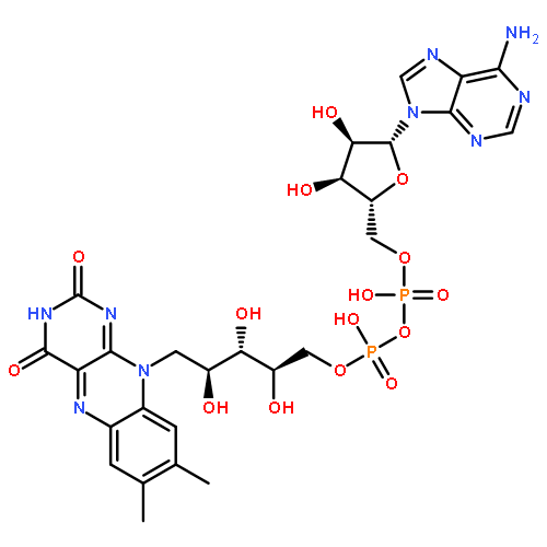
Co-reporter:Jing Zhao, Satoshi Katsube, Junpei Yamamoto, Kazuhiko Yamasaki, Makoto Miyagishi and Shigenori Iwai
Analyst 2015 vol. 140(Issue 17) pp:5881-5884
Publication Date(Web):16 Jul 2015
DOI:10.1039/C5AN01347J
Imidazole was tethered to the C5 position of thymine in an ATP-binding DNA aptamer with two types of linkers, and the affinities of each aptamer for ATP and AMP were determined by surface plasmon resonance measurements. The imidazole-tethered aptamers exhibited higher affinity for ATP, almost independently of the linker structure or the modification site.
Co-reporter:Norihito Arichi, Junpei Yamamoto, Chiaki Takahata, Emi Sano, Yuji Masuda, Isao Kuraoka and Shigenori Iwai
Organic & Biomolecular Chemistry 2013 vol. 11(Issue 21) pp:3526-3534
Publication Date(Web):28 Mar 2013
DOI:10.1039/C3OB00012E
The (6–4) photoproduct is one of the major UV-induced lesions in DNA. We previously showed that hydrolytic ring opening of the 5′ base and subsequent hydrolysis of the glycosidic bond of the 3′ component occurred when this photoproduct was treated with aqueous NaOH. In this study, we found that another product was obtained when the (6–4) photoproduct was heated at 90 °C for 6 h, in a 0.1 M solution of N,N′-dimethyl-1,2-ethanediamine adjusted to pH 7.4 with acetic acid. An analysis of the chemical structure of this product revealed that the 5′ base was intact, whereas the glycosidic bond at the 3′ component was hydrolyzed in the same manner. The strand break was detected for a 30-mer oligonucleotide containing the (6–4) photoproduct upon treatment with the above solution or other pH 7.4 solutions containing biogenic amines, such as spermidine and spermine. In the case of spermidine, the rate constant was calculated to be 1.4 × 10⁻⁸ s−1 at 37 °C. The strand break occurred even when the oligonucleotide was heated at 90 °C in 0.1 M sodium phosphate (pH 7.0), although this treatment produced several types of 5′ fragments. The Dewar valence isomer was inert to this reaction. The product obtained from the (6–4) photoproduct-containing 30-mer was used to investigate the enzymatic processing of the 3′ end bearing the damaged base and a phosphate. The ERCC1–XPF complex removed several nucleotides containing the damaged base, in the presence of replication protein A.
Co-reporter:Tatsuya Toga, Isao Kuraoka, Akira Yasui, Shigenori Iwai
Analytical Biochemistry 2013 Volume 440(Issue 1) pp:9-11
Publication Date(Web):1 September 2013
DOI:10.1016/j.ab.2013.04.027
Abstract
We previously developed a molecular beacon-type probe to detect the strand scission in cellular base excision repair and found that the phosphodiester linkages in the fluorophore/quencher linkers were cleaved. This reaction was applied to a transfection reporter, which contained the unmodified phosphodiester in the linker to another type of fluorophore. After cotransfection of cells with the probe and the reporter, the signals were used to detect the incision and to confirm the proper transfection, respectively. This method will contribute to the prevention of false-negative results in experiments using molecular beacon-type probes.
Co-reporter:Norihito Arichi, Aki Inase, Sachise Eto, Toshimi Mizukoshi, Junpei Yamamoto and Shigenori Iwai
Organic & Biomolecular Chemistry 2012 vol. 10(Issue 11) pp:2318-2325
Publication Date(Web):20 Dec 2011
DOI:10.1039/C2OB06966K
The (6–4) photoproduct is one of the major damaged bases produced by ultraviolet light in DNA. This lesion is known to be alkali-labile, and strand breaks occur at its sites when UV-irradiated DNA is treated with hot alkali. We have analyzed the product obtained by the alkali treatment of a dinucleoside monophosphate containing the (6–4) photoproduct, by HPLC, NMR spectroscopy, and mass spectrometry. We previously found that the N3–C4 bond of the 5′ component was hydrolyzed by a mild alkali treatment, and the present study revealed that the following reaction was the hydrolysis of the glycosidic bond at the 3′ component. The sugar moiety of this component was lost, even when a 3′-flanking nucleotide was not present. Glycosidic bond hydrolysis was also observed for a dimer and a trimer containing 5-methyl-2-pyrimidinone, which was used as an analog of the 3′ component of the (6–4) photoproduct, and its mechanism was elucidated. Finally, the alkali treatment of a tetramer, d(GT(6–4)TC), yielded 2′-deoxycytidine 5′-monophosphate, while 2′-deoxyguanosine 3′-monophosphate was not detected. This result demonstrated the hydrolysis of the glycosidic bond at the 3′ component of the (6–4) photoproduct and the subsequent strand break by β-elimination. It was also shown that the glycosidic bond at the 3′ component of the Dewar valence isomer was more alkali-labile than that of the (6–4) photoproduct.
Co-reporter:Junpei Yamamoto, Yoshiyuki Tanaka and Shigenori Iwai
Organic & Biomolecular Chemistry 2009 vol. 7(Issue 1) pp:161-166
Publication Date(Web):10 Nov 2008
DOI:10.1039/B815458A
We synthesized a dinucleoside monophosphate of the 15N-labeled (6–4) photoproduct, which is one of the major UV-induced lesions in DNA, to investigate the (6–4) photolyase repair mechanism, and characterized its protonation state by measuring 15N NMR spectra as a function of pH. We expected that chemical-shift changes of the pyrimidone15N3, due to protonation, would be observed at pH 3, as observed for the 15N-labeled 5-methylpyrimidin-2-one nucleoside. Interestingly, however, the changes were observed only in alkaline solutions. In UV absorption spectroscopy and HPLC analyses under acidic conditions, a change in the maximum absorption wavelength, due to the protonation-induced hydrolysis, was observed at and below pH 1, but not at pH 2, whereas the protonation of 5-methylpyrimidin-2-one occurred at pH values between 2 and 3. These results indicated that the pKa value for this N3 is remarkably lower than that of a normal pyrimidone ring, and strongly suggest that an intramolecular hydrogen bond is formed between the N3 of the 3′ base and the 5-OH of the 5′ base under physiological conditions. The results of this study have implications not only for the recognition and reaction mechanisms of (6–4) photolyase, but also for the chemical nature of the (6–4) photoproduct.
Co-reporter:Junpei Yamamoto, Kenichi Hitomi, Ryosuke Hayashi, Elizabeth D. Getzoff and Shigenori Iwai
Biochemistry 2009 Volume 48(Issue 39) pp:
Publication Date(Web):August 28, 2009
DOI:10.1021/bi900956p
The (6−4) photoproduct, which is one of the major UV-induced DNA lesions, causes carcinogenesis with high frequency. The (6−4) photolyase is a flavoprotein that can restore this lesion to the original bases, but its repair mechanism has not been elucidated. In this study, we focused on the interaction between the enzyme and the 3′ pyrimidone component of the (6−4) photoproduct and prepared a substrate analogue in which the carbonyl group, a hydrogen-bond acceptor, was replaced with an imine, a hydrogen-bond donor, to investigate the involvement of this carbonyl group in the (6−4) photolyase reaction. UV irradiation of oligodeoxyribonucleotides containing a single thymine−5-methylisocytosine site yielded products with absorption bands at wavelengths longer than 300 nm, similar to those obtained from the conversion of the TT site to the (6−4) photoproduct. Nuclease digestion, MALDI-TOF mass spectrometry, and the instability of the products indicated the formation of the 2-iminopyrimidine-type photoproduct. Analyses of the reaction and the binding of the (6−4) photolyase using these oligonucleotides revealed that this imine analogue of the (6−4) photoproduct was not repaired by the (6−4) photolyase, although the enzyme bound to the oligonucleotide with considerable affinity. These results indicate that the carbonyl group of the 3′ pyrimidone ring plays an important role in the (6−4) photolyase reaction. On the basis of these results, we discuss the repair mechanism.
Co-reporter:Tatsuya Toga, Junpei Yamamoto, Shigenori Iwai
Tetrahedron Letters 2009 50(6) pp: 723-726
Publication Date(Web):
DOI:10.1016/j.tetlet.2008.12.001
Co-reporter:Aki Inase-Hashimoto, Shinya Yoshikawa, Yusuke Kawasaki, Takashi S. Kodama, Shigenori Iwai
Bioorganic & Medicinal Chemistry 2008 Volume 16(Issue 1) pp:164-170
Publication Date(Web):1 January 2008
DOI:10.1016/j.bmc.2007.10.002
We previously reported that distamycin A, a natural antibiotic known as a minor groove binder, could bind to DNA duplexes containing the (6–4) photoproduct formed at its target site, whereas the binding was not observed for duplexes containing the cis-syn cyclobutane pyrimidine dimer in the same sequence context. In this study, we have further analyzed the binding of this drug to lesion-containing duplexes to elucidate its damaged-DNA recognition mechanism. Surface plasmon resonance measurements using various types of DNA showed that distamycin A could bind to several types of lesion-containing DNA. Curve fitting of the CD titration data revealed that the complex formation occurred with Kd values around 10−6 and a stoichiometry of 1:1. The results obtained in this study suggested that distamycin A binds to damaged DNA in the same way as to the normal target site, by recognizing the chemical structure of the minor groove.The binding of distamycin A to various lesion-containing oligonucleotide duplexes was analysed. The results suggested that this drug recognized the chemical structure of the minor groove at the lesion site.
Co-reporter:Junpei Yamamoto, Yoshiyuki Tanaka and Shigenori Iwai
Organic & Biomolecular Chemistry 2009 - vol. 7(Issue 1) pp:NaN166-166
Publication Date(Web):2008/11/10
DOI:10.1039/B815458A
We synthesized a dinucleoside monophosphate of the 15N-labeled (6–4) photoproduct, which is one of the major UV-induced lesions in DNA, to investigate the (6–4) photolyase repair mechanism, and characterized its protonation state by measuring 15N NMR spectra as a function of pH. We expected that chemical-shift changes of the pyrimidone15N3, due to protonation, would be observed at pH 3, as observed for the 15N-labeled 5-methylpyrimidin-2-one nucleoside. Interestingly, however, the changes were observed only in alkaline solutions. In UV absorption spectroscopy and HPLC analyses under acidic conditions, a change in the maximum absorption wavelength, due to the protonation-induced hydrolysis, was observed at and below pH 1, but not at pH 2, whereas the protonation of 5-methylpyrimidin-2-one occurred at pH values between 2 and 3. These results indicated that the pKa value for this N3 is remarkably lower than that of a normal pyrimidone ring, and strongly suggest that an intramolecular hydrogen bond is formed between the N3 of the 3′ base and the 5-OH of the 5′ base under physiological conditions. The results of this study have implications not only for the recognition and reaction mechanisms of (6–4) photolyase, but also for the chemical nature of the (6–4) photoproduct.
Co-reporter:Norihito Arichi, Aki Inase, Sachise Eto, Toshimi Mizukoshi, Junpei Yamamoto and Shigenori Iwai
Organic & Biomolecular Chemistry 2012 - vol. 10(Issue 11) pp:NaN2325-2325
Publication Date(Web):2011/12/20
DOI:10.1039/C2OB06966K
The (6–4) photoproduct is one of the major damaged bases produced by ultraviolet light in DNA. This lesion is known to be alkali-labile, and strand breaks occur at its sites when UV-irradiated DNA is treated with hot alkali. We have analyzed the product obtained by the alkali treatment of a dinucleoside monophosphate containing the (6–4) photoproduct, by HPLC, NMR spectroscopy, and mass spectrometry. We previously found that the N3–C4 bond of the 5′ component was hydrolyzed by a mild alkali treatment, and the present study revealed that the following reaction was the hydrolysis of the glycosidic bond at the 3′ component. The sugar moiety of this component was lost, even when a 3′-flanking nucleotide was not present. Glycosidic bond hydrolysis was also observed for a dimer and a trimer containing 5-methyl-2-pyrimidinone, which was used as an analog of the 3′ component of the (6–4) photoproduct, and its mechanism was elucidated. Finally, the alkali treatment of a tetramer, d(GT(6–4)TC), yielded 2′-deoxycytidine 5′-monophosphate, while 2′-deoxyguanosine 3′-monophosphate was not detected. This result demonstrated the hydrolysis of the glycosidic bond at the 3′ component of the (6–4) photoproduct and the subsequent strand break by β-elimination. It was also shown that the glycosidic bond at the 3′ component of the Dewar valence isomer was more alkali-labile than that of the (6–4) photoproduct.
Co-reporter:Norihito Arichi, Junpei Yamamoto, Chiaki Takahata, Emi Sano, Yuji Masuda, Isao Kuraoka and Shigenori Iwai
Organic & Biomolecular Chemistry 2013 - vol. 11(Issue 21) pp:NaN3534-3534
Publication Date(Web):2013/03/28
DOI:10.1039/C3OB00012E
The (6–4) photoproduct is one of the major UV-induced lesions in DNA. We previously showed that hydrolytic ring opening of the 5′ base and subsequent hydrolysis of the glycosidic bond of the 3′ component occurred when this photoproduct was treated with aqueous NaOH. In this study, we found that another product was obtained when the (6–4) photoproduct was heated at 90 °C for 6 h, in a 0.1 M solution of N,N′-dimethyl-1,2-ethanediamine adjusted to pH 7.4 with acetic acid. An analysis of the chemical structure of this product revealed that the 5′ base was intact, whereas the glycosidic bond at the 3′ component was hydrolyzed in the same manner. The strand break was detected for a 30-mer oligonucleotide containing the (6–4) photoproduct upon treatment with the above solution or other pH 7.4 solutions containing biogenic amines, such as spermidine and spermine. In the case of spermidine, the rate constant was calculated to be 1.4 × 10⁻⁸ s−1 at 37 °C. The strand break occurred even when the oligonucleotide was heated at 90 °C in 0.1 M sodium phosphate (pH 7.0), although this treatment produced several types of 5′ fragments. The Dewar valence isomer was inert to this reaction. The product obtained from the (6–4) photoproduct-containing 30-mer was used to investigate the enzymatic processing of the 3′ end bearing the damaged base and a phosphate. The ERCC1–XPF complex removed several nucleotides containing the damaged base, in the presence of replication protein A.