Co-reporter:Brian Stapleton, Lawrence R. Walker, and Timothy M. Logan
Biochemistry 2013 Volume 52(Issue 11) pp:
Publication Date(Web):February 22, 2013
DOI:10.1021/bi301608p
Thermodynamic measurements of Fe(II) binding and activation of repressor function in the iron-dependent repressor from Mycobacterium tuberculosis (IdeR) are reported. IdeR, a member of the diphtheria toxin repressor family of proteins, regulates iron homeostasis and contributes to the virulence response in M. tuberculosis. Although iron is the physiological ligand, this is the first detailed analysis of iron binding and activation in this protein. The results showed that IdeR binds 2 equiv of Fe(II) with dissociation constants that differ by a factor of 25. The high- and low-affinity iron binding sites were assigned to physical binding sites I and II, respectively, using metal binding site mutants. IdeR was also found to contain a high-affinity Zn(II) binding site that was assigned to physical metal binding site II through the use of binding site mutants and metal competition assays. Fe(II) binding was modestly weaker in the presence of Zn(II), but the coupled metal binding–DNA binding affinity was significantly stronger, requiring 30-fold less Fe(II) to activate DNA binding compared to Fe(II) alone. Together, these results suggest that IdeR is a mixed-metal repressor, where Zn(II) acts as a structural metal and Fe(II) acts to trigger the physiologically relevant promoter binding. This new model for IdeR activation provides a better understanding of IdeR and the biology of iron homeostasis in M. tuberculosis.
Co-reporter:Joshua M. Kogot, Alex M. Parker, Jihun Lee, Michael Blaber, Geoffrey F. Strouse and Timothy M. Logan
Bioconjugate Chemistry 2009 Volume 20(Issue 11) pp:2106
Publication Date(Web):October 7, 2009
DOI:10.1021/bc900224d
Whether assembling proteins onto nanoscale, mesoscopic, or macroscropic material surfaces, maintaining a protein’s structure and function when conjugated to a surface is complicated by the high propensity for electrostatic or hydrophobic surface interactions and the possibility of direct metal coordination of protein functional groups. In this study, the assembly of a 1.5 nm CAAKA passivated gold nanoparticle (AuNP) onto FGF1 (human acidic fibroblast growth factor) using an amino terminal His6 tag is analyzed. The impact of structure and time-dependent changes in the structural elements in FGF1and FGF1-heparin in the presence of the AuNP is probed by a molecular beacon fluorescence assay, circular dichroism, and NMR spectroscopy. Analysis of the results indicates that a time-dependent evolution of the protein structure without loss of FGF1 heparin binding occurs following the formation of the initial FGF1-AuNP complex. The time-dependent changes are believed to reflect protein sampling of the AuNP surface to minimize the free energy of the AuNP-FGF1 complex without impacting FGF1 function.
Co-reporter:Nathalie Muñoz, Junho Kim, Yijun Liu, Timothy M. Logan, Teng Ma
Journal of Biotechnology (10 January 2014) Volume 169() pp:95-102
Publication Date(Web):10 January 2014
DOI:10.1016/j.jbiotec.2013.11.010
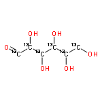
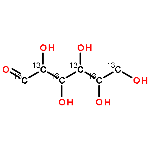
•GC–MS was used to investigate glucose metabolism in hMSC and hMSC-derived osteoblasts (hMSC-OS) cultured at 2% or 20% O2.•hMSC and hMSC-OS exhibited different metabolic responses to low oxygen tension.•Hypoxia-induced inhibition of PDH was more pronounced in hMSC-OS compared to hMSC.•Hypoxia increased the apparent activity of the malate aspartate shuttle in hMSC, but not in hMSC-OS.Bone marrow derived human mesenchymal stem cells (hMSC) are the primary cell type in bone tissue engineering, and their life span during osteogenic differentiation is associated with changes in oxygen tension. As a ubiquitous regulator of cellular metabolic activity, oxygen tension influences the profiles of metabolites in the entire metabolic network and plays an important role in hMSC survival, function, and osteogenic differentiation. In the current study, we hypothesize that hMSC have a metabolic phenotype that supports growth in low oxygen environments and that this phenotype changes upon differentiation, leading to differential responses to oxygen tension. We developed a gas chromatography–mass spectrometry (GC–MS) based metabolic profiling approach to analyze the metabolic fate of 13C-glucose in glycolysis and the tricarboxylic acid cycle (TCA) in undifferentiated hMSC and hMSC-derived osteoblasts (hMSC-OS) in response to perturbation in oxygen tension; specifically we compared changes induced by culture under 20% vs. 2% O2. The isotope enrichments in the metabolites were calculated and used to infer activities of specific metabolic enzymes and the associated pathways. The results revealed contrasting metabolic profiles for hMSC and the hMSC-OS in both 20% and 2% O2 states, with the most significant differences involving coupling of glycolysis to the TCA cycle, glutaminolysis, and the malate-aspartate shuttle. The results have important implications in defining the optimal culture conditions for hMSC expansion and osteogenic differentiation.
.jpg)


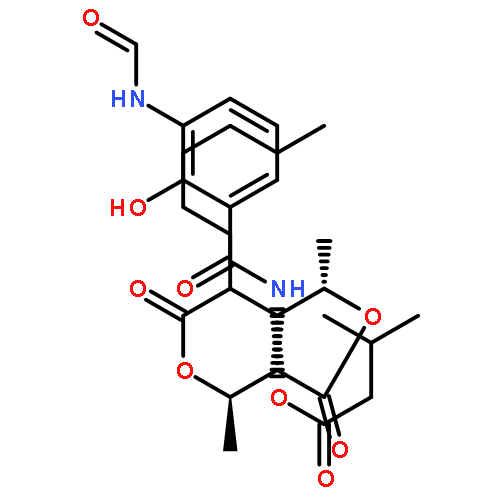


![[D-MeAla]3-[EtVal]4-cyclosporin A](http://img.cochemist.com/ccimg/254500/254435-95-5.png)
![[D-MeAla]3-[EtVal]4-cyclosporin A](http://img.cochemist.com/ccimg/254500/254435-95-5_b.png)