Co-reporter:Wei Zhang, Huiwen Xiong, Miaomiao Chen, Xiuhua Zhang, Shengfu Wang
Biosensors and Bioelectronics 2017 Volume 96(Volume 96) pp:
Publication Date(Web):15 October 2017
DOI:10.1016/j.bios.2017.04.035
•A novel molecularly imprinted electrochemical luminescence (MIP-ECL) sensor based on Ru(bpy)32+-doped silica nanoparticles (Ru@SiO2 NPs) was employed to determine fumonisin B1 (FB1).•The localized surface plasmon resonance (LSPR) and electrochemical effect of gold-nanoparticles (AuNPs) was used to enhance the ECL intensity of Ru@SiO2 NPs.•FB1 with an amino group was used both as the target molecule and coreactant of Ru@SiO2 in this method.A novel molecularly imprinted electrochemiluminescence (MIP-ECL) sensor based on Ru(bpy)32+-doped silica nanoparticles (Ru@SiO2 NPs) is developed for highly sensitive detection of fumonisin B1 (FB1). Gold-nanoparticles (AuNPs), Ru@SiO2 NPs with chitosan (CS) composites and a molecularly imprinted polymer (MIP) are assembled on a glassy carbon electrode (GCE) to fabricate an ECL platform step by step. AuNPs could greatly promote the ECL intensity and improve the analytical sensitivity according to the localized surface plasmon resonance (LSPR) and the electrochemical effect. In this surface-enhanced electrochemiluminescence (SEECL) system, AuNPs work as the LSPR source to improve the ECL intensity and Ru@SiO2 NPs are used as ECL luminophores. In the phosphate buffer solution (PBS), the evident anodic ECL of Ru@SiO2 on the above working electrode is observed in the presence of the template molecule fumonisin B1 (FB1), which could act as the coreactant of Ru@SiO2 NPs due to the amino group of FB1. When the template molecules were eluted from the MIP, little coreactant was left, resulting in an apparent decrease of ECL signal. After the MIP-ECL sensor was incubated in FB1 solution, the template molecules rebound to the MIP surface, leading to the enhancement of ECL signal again. On the basis of these results, a facile MIP-ECL sensor has been successfully fabricated, which exhibited a linear range from 0.001 to 100 ng mL−1 with a detection limit of 0.35 pg mL−1 for FB1. Moreover, the proposed MIP-ECL sensor displayed an excellent application in real samples.
Co-reporter:Fan Zhang;Miaomiao Chen;Haiqing Zhang;Huayu Xiong;Wei Wen;Shengfu Wang
Analytical Methods (2009-Present) 2017 vol. 9(Issue 6) pp:929-936
Publication Date(Web):2017/02/09
DOI:10.1039/C6AY02711C
A sensitive, convenient and rapid fluorescent sensor for effectively and directly detecting propranolol was proposed based on the fluorescence suppression of 3-mercaptopropionic acid stabilized CdTe quantum dots. The fluorescence suppression mechanism between CdTe QDs and propranolol was studied by time-resolved fluorescence spectroscopy, UV-Vis spectroscopy and fluorescence spectroscopy, and it was attributed to the simultaneous action of the inner filter effect (IFE) and dynamic quenching effect (DQE). The effects of the concentration of quantum dots, pH value of buffer solution and incubation time on the fluorescence signal of the propranolol/CdTe quantum dot system were also investigated. The results indicated that, when the experiment was conducted under optimal experimental conditions, the linear range of this assay was 0.1 μM to 2 μM and 5 μM to 200 μM, with a corresponding detection limit of 0.055 μM. Moreover, some common drug excipients and analogues of propranolol and stimulants had no apparent effect on the determination of propranolol. The proposed fluorescent sensor could be readily used for effectively and directly detecting propranolol in biological samples.
Co-reporter:Yan Xiao;Yuhao Sheng;Jie Zhou;Miaomiao Chen;Wei Wen;Shengfu Wang
Analyst (1876-Present) 2017 vol. 142(Issue 14) pp:2617-2623
Publication Date(Web):2017/07/10
DOI:10.1039/C7AN00553A
Graphitic carbon nitride (g-C3N4) nanosheets are a type of two-dimensional nanomaterial with stable photoluminescence and large specific surface areas that make them an ideal candidate for fluorescence sensing. However, fluorescence sensing based on g-C3N4 nanosheets for disease-specific biomolecules is still limited. Moreover, current g-C3N4 nanosheet-based fluorescence sensors for nucleic acid require pre-labeling which is time-consuming and cost-ineffective. In this work, a novel label-free strategy for HBV gene determination based on metal ion binding-induced fluorescence quenching of g-C3N4 nanosheets was designed. Under the optimized conditions, this bioassay provided high sensitivity for HBV gene determination with the limit of detection as low as 1.0 nM, and has been successfully applied for the detection of HBV gene in human serum with satisfactory results. By embedding different recognition sequences of the probes, this method may be conveniently used for versatile detection of diverse disease markers. This novel design may provide a simple, flexible, sensitive and low-cost strategy for g-C3N4 nanosheet-based disease-specific biomolecule sensing.
Co-reporter:Weiming Li, Miaomiao Chen, Huayu Xiong, Wei Wen, Hanping He, Xiuhua Zhang and Shengfu Wang
New Journal of Chemistry 2016 vol. 40(Issue 1) pp:564-570
Publication Date(Web):05 Nov 2015
DOI:10.1039/C5NJ02879E
Molecular imprinting for the detection of protein has gained great interest in recent years. For this purpose, we prepared magnetic molecularly imprinted polymers (MIPs) for the recognition of bovine hemoglobin (BHb) through the surface imprinting technique with two-stage core–sell sol–gel polymerization on the surface of silica modified Fe3O4 nanospheres. 3-Aminopropyltriethoxylsilane and octyltrimethoxysilane were chosen as monomers to construct the MIP layer. The morphology and structure property of prepared nanoparticles were characterized by TEM, X-ray diffraction, Fourier transform infrared spectrometry, and the vibrating sample magnetometer. The obtained magnetic MIPs with high saturation magnetization (60 emu g−1) made it easy to separate the target protein from solution by an external magnetic field. The adsorption and recognition performance of this magnetic MIPs was discussed through adsorption kinetics, adsorption isotherms, special selectivity, reusability and reproducibility tests. It turned out that the magnetic MIPs showed a relatively high adsorption capacity of 124.86 mg g−1 and excellent selectivity towards BHb with a separation factor of 1.99. Moreover, the adsorption capacity of magnetic MIPs was not significantly reduced after three continuous adsorption and elution processes, which indicated their good reusability for at least three repeated cycles.
Co-reporter:Qingling Wang, Miaomiao Chen, Haiqing Zhang, Wei Wen, Xiuhua Zhang, Shengfu Wang
Biosensors and Bioelectronics 2016 Volume 79() pp:561-567
Publication Date(Web):15 May 2016
DOI:10.1016/j.bios.2015.12.098
•This paper developed the RuSi NPs/CdTe QDs ECL-ET system.•This method has high sensitivity and good selectivity for detection of ochratoxin A.•The method has been applied in real samples with good sensitivity and selectivity.This paper develops a new approach to enhance the electrochemiluminescence (ECL) emission of the Ru(bpy)32+-tripropyl amine (TPrA) system for ultrasensitive determination of ochratoxin A (OTA). Ru(bpy)32+-doped silica nanoparticles (RuSi NPs) act as ECL materials, which are immobilized on the surface of electrode by chitosan to fabricate a solid-state ECL sensor. CdTe quantum dots (QDs) can enhance the ECL emission of the Ru(bpy)32+-TPrA ECL system by energy transfer. This strategy can improve the sensitivity of the sensor. In this assay, we combine the ECL with molecular imprinting technique to improve the selectivity of this sensor. The template molecule could be eluted from the molecularly imprinted polymer (MIP), and the formed cavities could then selectively recognize the target. The cavities could also work as the tunnel for the transfer of coreactant TPrA to produce responsive signal. With the increase of the concentration of OTA in samples, more cavities were filled because of the rebinding of OTA to the MIP surface, resulting in a gradual decrease in ECL intensity. The results showed that the ECL decrease value depended linearly on the logarithm of the OTA concentration in the range from 1.00×10⁻⁵ to 11.13 ng mL−1 with lower detection limit of 3.0 fg mL−1 (S/N=3). This ECL sensor has also been applied to detect OTA concentration in the real samples with satisfied results, and the recoveries range from 85.1% to 107.9%.
Co-reporter:Qingling Wang, Miaomiao Chen, Haiqing Zhang, Wei Wen, Xiuhua Zhang, Shengfu Wang
Sensors and Actuators B: Chemical 2016 Volume 222() pp:264-269
Publication Date(Web):January 2016
DOI:10.1016/j.snb.2015.08.057
•A simple and convenient sensor for OTA was prepared.•The electrochemiluminescence sensor based on Ru(bpy)32+-doped silica nanoparticles has high sensitivity.•The electrochemiluminescence sensor combines with desirable selectivity of molecularly imprinted technology.•The sensor showed good sensitivity and stability for OTA in corn sample.An electrochemiluminescence (ECL) sensor based on Ru(bpy)32+-doped silica nanoparticles (RuSi NPs) combined with molecularly imprinted polymer (MIP) has been developed for the determination of ochratoxin A (OTA). The sensor was fabricated by remodification of RuSi NPs with a thin film of molecularly imprinted polymer, which provided the specific binding sites for OTA. The process of template elution and rebinding acted as a gate to control the flux of probes, which passed through the cavities and reacted with Ru(bpy)32+ immobilized on the electrode surface to emit ECL signal. The ECL intensity decreased with the OTA molecules rebound in MIP. The as-prepared sensor exhibited a very high sensitivity and excellent selectivity to the target molecule. The ΔIECL depended linearly on the logarithm of the OTA concentration in the range from 0.1 pg/mL to 14.76 ng/mL with lower detection limit of 0.027 pg/mL (S/N = 3). The MIP-ECL sensor enabled the successful application of this method to determine OTA in corn.Schematic diagram of the molecularly imprinted polymer electrochemiluminescence sensor for the detecting of ochratoxin A.
Co-reporter:Q. L. Wang, M. M. Chen, H. Q. Zhang, W. Wen, X. H. Zhang and S. F. Wang
Analytical Methods 2015 vol. 7(Issue 24) pp:10224-10228
Publication Date(Web):02 Nov 2015
DOI:10.1039/C5AY02327K
This paper reports a new molecularly imprinted electrochemical luminescence (MIP-ECL) sensor for the determination of ochratoxin A (OTA) with high selectivity and sensitivity. Ru(bpy)32+ was immobilized on the electrode surface as the luminescent material by using Nafion. The process of template elution and rebinding acted as a gate to control the flux of probes, which passed through the cavities and reacted on the electrode surface. When the imprinted film was rebound with OTA, the ECL signal decreased. Under optimal conditions, the MIP-ECL sensor showed a wide linear range from 0.1 ng mL−1 to 10 ng mL−1 with a detection limit (S/N = 3) of 0.03 ng mL−1. Corn samples were assayed by using this sensor, and recoveries ranging from 95.2% to 102.7% were obtained.
Co-reporter:Huayu Xiong, Chunhui Guo, Ping Liu, Wei Xu, Xiuhua Zhang, and Shengfu Wang
Analytical Chemistry 2014 Volume 86(Issue 10) pp:4729
Publication Date(Web):May 1, 2014
DOI:10.1021/ac5009139
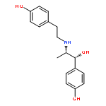
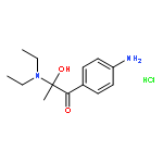
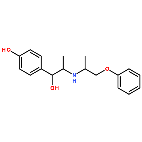
The fast, visual discrimination of β2-agonist drugs is needed for the on-site screening of various types of β2-agonists in blood and urine samples. We developed a simple, rapid, one-step colorimetric method to detect phenolic β2-agonists by use of a tyrosinase catalytic reaction, which involved the oxidation of the phenol group on the benzene rings of β2-agonists. The enzymatic oxidation products of β2-agonists with phenolic groups exhibited different color transitions based on the different substituent groups on the aromatic ring, whereas β2-agonists with the aniline group or the resorcinol group remained colorless. This visual color discrepancy has been used to intuitively and conveniently differentiate the phenolic group β2-agonists, such as ractopamine, isoxsuprine, ritodrine, and fenoterol. The oxidation products of these compounds have been identified using mass spectrometry, and the possible reaction mechanisms between β2-agonists and tyrosinase have been deduced. The parameters that govern the analytical performance of the reaction product, including the pH of the buffer solution, the concentration of tyrosinase, and the incubation time, have been studied and optimized using ultraviolet–visible (UV–vis) spectroscopy and electrochemical methods. Under the optimal experimental conditions, the absorbance intensity and electrochemical signal were found to increase proportionally to the concentrations of the phenolic group β2-agonists, which gave a quantitative description of the β2-agonists in solution.
Co-reporter:Wenying Cao, Huayu Xiong, Xing Gao, Xiuhua Zhang and Shengfu Wang
Analytical Methods 2014 vol. 6(Issue 7) pp:2349-2355
Publication Date(Web):21 Jan 2014
DOI:10.1039/C3AY42282H
A novel strategy for preparing highly sensitive molecularly imprinted sensors based on the electro-polymerization of o-phenylenediamine (o-PD) on a columnar-structured platinum (CSPt) electrode was proposed for β2-agonist determination. The CSPt electrode was used as a working electrode to increase the specific surface area and to extend the linear range. The sensor surface morphology was characterized by scanning electron microscopy. The preparation process of the sensor was characterized by an electrochemical quartz crystal microbalance. The electrochemical performance of the sensor was investigated by electrochemical impedance spectroscopy and cyclic voltammetry. Additionally, sodium dodecyl sulfonate, which was used in the electro-polymerization reaction of o-PD, prevented the hydrolysis degradation of poly-o-phenylenediamine (PoPD) and enhanced the stability of the PoPD film in the anionic micellar media. The recognition and determination of the sensor were carried out by measuring the changes of the amperometric response of [Fe(CN)6]3−/4−. The proposed sensor was successfully applied to detect β2-agonists in real human serum samples.
Co-reporter:Wenying Cao;Huayu Xiong;Xing Gao;Shengfu Wang
Analytical Methods (2009-Present) 2014 - vol. 6(Issue 7) pp:NaN2355-2355
Publication Date(Web):2014/03/13
DOI:10.1039/C3AY42282H
A novel strategy for preparing highly sensitive molecularly imprinted sensors based on the electro-polymerization of o-phenylenediamine (o-PD) on a columnar-structured platinum (CSPt) electrode was proposed for β2-agonist determination. The CSPt electrode was used as a working electrode to increase the specific surface area and to extend the linear range. The sensor surface morphology was characterized by scanning electron microscopy. The preparation process of the sensor was characterized by an electrochemical quartz crystal microbalance. The electrochemical performance of the sensor was investigated by electrochemical impedance spectroscopy and cyclic voltammetry. Additionally, sodium dodecyl sulfonate, which was used in the electro-polymerization reaction of o-PD, prevented the hydrolysis degradation of poly-o-phenylenediamine (PoPD) and enhanced the stability of the PoPD film in the anionic micellar media. The recognition and determination of the sensor were carried out by measuring the changes of the amperometric response of [Fe(CN)6]3−/4−. The proposed sensor was successfully applied to detect β2-agonists in real human serum samples.
Co-reporter:
Analytical Methods (2009-Present) 2015 - vol. 7(Issue 24) pp:NaN10228-10228
Publication Date(Web):2015/11/02
DOI:10.1039/C5AY02327K
This paper reports a new molecularly imprinted electrochemical luminescence (MIP-ECL) sensor for the determination of ochratoxin A (OTA) with high selectivity and sensitivity. Ru(bpy)32+ was immobilized on the electrode surface as the luminescent material by using Nafion. The process of template elution and rebinding acted as a gate to control the flux of probes, which passed through the cavities and reacted on the electrode surface. When the imprinted film was rebound with OTA, the ECL signal decreased. Under optimal conditions, the MIP-ECL sensor showed a wide linear range from 0.1 ng mL−1 to 10 ng mL−1 with a detection limit (S/N = 3) of 0.03 ng mL−1. Corn samples were assayed by using this sensor, and recoveries ranging from 95.2% to 102.7% were obtained.


![L-Phenylalanine,N-[[(3R)-5-chloro-3,4-dihydro-8-hydroxy-3-methyl-1-oxo-1H-2-benzopyran-7-yl]carbonyl]-](http://img.cochemist.com/ccimg/400/303-47-9.png)
![L-Phenylalanine,N-[[(3R)-5-chloro-3,4-dihydro-8-hydroxy-3-methyl-1-oxo-1H-2-benzopyran-7-yl]carbonyl]-](http://img.cochemist.com/ccimg/400/303-47-9_b.png)


![Benzenemethanol, 4-amino-3,5-dichloro-α-[[(1,1-dimethylethyl)amino]methyl]-](/data/chemimg/568900/37148-27-9.png)
![Benzenemethanol, 4-amino-3,5-dichloro-α-[[(1,1-dimethylethyl)amino]methyl]-](/data/chemimg/568900/37148-27-9_b.png)


