Co-reporter:Lifu Xiao, Karen A. Bailey, Hao Wang, and Zachary D. Schultz
Analytical Chemistry September 5, 2017 Volume 89(Issue 17) pp:9091-9091
Publication Date(Web):August 14, 2017
DOI:10.1021/acs.analchem.7b01796
The specific interaction between a ligand and a protein is a key component in minimizing off-target effects in drug discovery. Investigating these interactions with membrane protein receptors can be quite challenging. In this report, we show how spectral variance observed in surface-enhanced Raman scattering (SERS) and tip-enhanced Raman scattering (TERS) can be correlated with ligand specificity in affinity-based assays. Variations in the enhanced Raman spectra of three peptide ligands (i.e., cyclic-RGDFC, cyclic-isoDGRFC, and CisoDGRC), which have different binding affinity to αvβ3 integrin, are reported from isolated proteins and from receptors in intact cancer cell membranes. The SERS signal from the purified proteins provides basis spectra to analyze the signals in cells. Differences in the spectral variance within the SERS and TERS data for each ligand indicate larger variance for nonspecific ligand–receptor interactions. The SERS and TERS results are correlated with single particle tracking experiments of the ligand-functionalized nanoparticles with purified receptors on glass surfaces and living cells. These results demonstrate the ability to elucidate protein–ligand recognition using the observed vibrational spectra and provide perspective on binding specificity for small-molecule ligands in intact cell membranes, demonstrating a new approach for investigating drug specificity.
Co-reporter:James M. Marr and Zachary D. Schultz
The Journal of Physical Chemistry Letters October 3, 2013 Volume 4(Issue 19) pp:
Publication Date(Web):September 16, 2013
DOI:10.1021/jz401551u
Electric fields associated with Raman enhancements are typically inferred from changes in the observed scattering intensity. Here, we use the vibrational Stark effect from a nitrile reporter to determine the electric-field-dependent frequency shift of cyanide (CN) on a gold (Au) surface. Electroplated Au surfaces with surface-enhanced Raman (SERS) activity exhibit larger Stark shifts near the edge and in areas with large roughness. The Stark shift is observed to correlate with the intensity of a coadsorbed thiophenol molecule. Gap-mode tip-enhanced Raman scattering (TERS), using a Au nanoparticle tip, shows dramatic shifts in the CN stretch that correlate to enhancement factors of 1013 in the gap region. The observed peak widths indicate that the largest fields are highly localized. Changes in the nitrile stretch frequency provide a direct measurement of the electric fields in SERS and TERS experiments.Keywords: vibrational Stark effect; nitrile; plasmonics; Raman; SERS; TERS;
Co-reporter:Zhi-Cong Zeng, Hao Wang, Paul Johns, Gregory V. Hartland, and Zachary D. Schultz
The Journal of Physical Chemistry C June 1, 2017 Volume 121(Issue 21) pp:11623-11623
Publication Date(Web):May 5, 2017
DOI:10.1021/acs.jpcc.7b01220
The optical properties of plasmonic nanoparticles are strongly dependent on interactions with other nanoparticles, which complicates analysis for systems larger than a few particles. In this work we examined heat dissipation in aggregated nanoparticles and its influence on surface-enhanced Raman scattering (SERS) through correlated photothermal heterodyne imaging, electron microscopy, and SERS measurements. For dimers the per particle absorption cross sections show evidence of interparticle coupling; however, the effects are much smaller than those for the field enhancements that are important for SERS. For larger aggregates the total absorption was observed to be simply proportional to aggregate volume. This observation allows us to model light absorption and heating in the aggregates by assuming that the particles act as independent heat sources. The heat dissipation calculations show that very high temperatures can be created at the nanoparticle surface and that the temperature decreases with increasing thermal conductivity of the surroundings. This is in agreement with the SERS measurements that show faster signal degradation for air compared to water environments.
Co-reporter:Ju-Young Kim, Zhi-Cong Zeng, Lifu Xiao, and Zachary D. Schultz
Analytical Chemistry December 19, 2017 Volume 89(Issue 24) pp:13074-13074
Publication Date(Web):November 14, 2017
DOI:10.1021/acs.analchem.7b04246
The ability to distinguish between specific and nonspecific binding is important for assessing the interactions between protein receptors and ligands. Surface plasmon resonance (SPR) spectroscopy is an advanced tool to measure binding events, yet the ability to distinguish between specific and nonspecific binding remains a limitation. To address this problem, we use SPR spectroscopy correlated with surface enhanced Raman scattering (SERS). The chemical information present in SERS spectra provides insight into the molecular interactions between functionalized nanoparticles and proteins, which are not detectable by SPR alone. Using a custom instrument with the Kretschmann configuration, we successfully demonstrate simultaneous affinity and the chemical characterization of streptavidin-functionalized gold nanoparticles (STV-NPs) binding to biotin immobilized on a gold film in both air and flowing phosphate buffered saline (PBS). The SPR performance is consistent with that of previous reports. The association constant (KA) for streptavidin/biotin and STV-NPs/biotin interactions observed (2 ± 1 × 10⁷ M–1 and 2.4 ± 0.3 × 10¹⁰ M–1, respectively) agree with literature values and show a strong avidity effect associated with the STV-NPs. The SERS scattering from STV-NPs is excited by the surface plasmon polariton and collected from an objective lens mounted over the fluidic channel. The SERS spectra are recorded simultaneously with the SPR sensorgram, and the detected Raman bands provide chemical insight into the binding event. Multivariate curve resolution analysis of the spectra can differentiate specific from nonspecific binding. This label-free, real time, and surface sensitive detection method provides chemical information to protein/ligand binding affinity measurements.
Co-reporter:Hao Wang;Kun Yao;John A. Parkhill
Physical Chemistry Chemical Physics 2017 vol. 19(Issue 8) pp:5786-5796
Publication Date(Web):2017/02/23
DOI:10.1039/C6CP08168A
The significant electric field enhancements that occur in plasmonic nanogap junctions are instrumental in boosting the performance of spectroscopy, optoelectronics and catalysis. Electron tunneling, associated with quantum effects in small junctions, is reported to limit the electric field enhancement. However, observing and quantitatively determining how tunneling alters the electric fields within small gaps is challenging due to the nanoscale dimensions and heterogeneity present experimentally. Here, we report the use of a nitrile probe placed in the nanoparticle–film gap junctions to demonstrate that the change in the nitrile stretching band associated with the vibrational Stark effect can be directly correlated with the local electric field environment modulated by gap size variations. The emergence of Stark shifts correlates with plasmon resonance shifts associated with electron tunneling across the gap junction. Time dependent changes in the nitrile band with extended illumination further support a build up of charge associated with optical rectification in the coupled plasmon system. Computational models agree with our experimental observations that the frequency shifts arise from a vibrational Stark effect. Large local electric fields associated with the smallest gap junctions give rise to significant Stark shifts. These results indicate that nitrile Stark probes can measure the local field strengths in plasmonic junctions and monitor the subtle changes in the local electric fields resulting from electron tunneling.
Co-reporter:Lifu Xiao, Hao Wang, and Zachary D. Schultz
Analytical Chemistry 2016 Volume 88(Issue 12) pp:6547
Publication Date(Web):May 18, 2016
DOI:10.1021/acs.analchem.6b01344
Ligand–receptor interactions play important roles in many biological processes. Cyclic arginine–glycine–aspartic acid (RGD) containing peptides are known to mimic the binding domain of extracellular matrix protein fibronectin and selectively bind to a subset of integrin receptors. Here we report the tip enhanced Raman scattering (TERS) detection of RGD-functionalized nanoparticles bound to integrins produces a Raman scattering signal specific to the bound protein. These results demonstrate that this method can detect and differentiate between two different integrins (α5β1 and αvβ3) bound to RGD-conjugated gold nanoparticles both on surfaces and in a cancer cell membrane. In situ measurements of RGD nanoparticles bound to purified α5β1 and αvβ3 receptors attached to a glass surface provide reference spectra for a multivariate regression model. The TERS spectra observed from nanoparticles bound to cell membranes are analyzed using this regression model and the identity of the receptor can be determined. The ability to distinguish between receptors in the cell membrane provides a new tool to chemically characterize ligand–receptor recognition at molecular level and provide chemical perspective on the molecular recognition of membrane receptors.
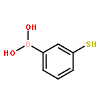
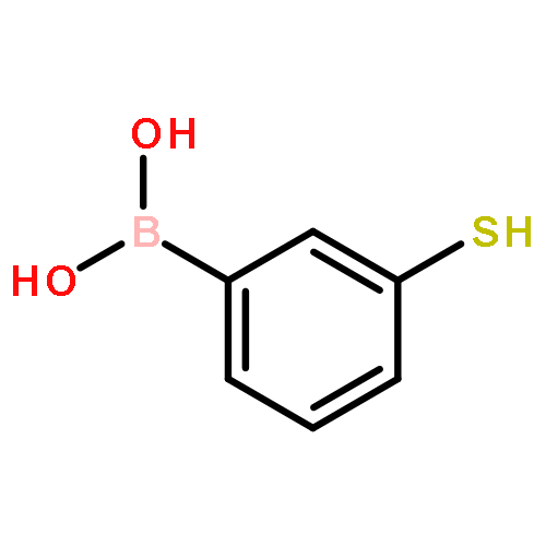
Co-reporter:Xin Gu, Hao Wang, Zachary D. Schultz, and Jon P. Camden
Analytical Chemistry 2016 Volume 88(Issue 14) pp:7191
Publication Date(Web):June 29, 2016
DOI:10.1021/acs.analchem.6b01378
Hydrogen peroxide (H2O2) is known as a key molecule in a variety of biological processes, as well as a crucial byproduct in many enzymatic reactions. Therefore, being able to selectively and sensitively detect H2O2 is not only important in monitoring, estimating, and decoding H2O2 relevant physiological pathways but also very helpful in developing enzymatic-based biosensors for other analytes of interest. Herein, we report a plasmonic probe for H2O2 based on 3-mercaptophenylboronic acid (3-MPBA) modified gold nanoparticles (AuNPs) which is coupled with surface-enhanced Raman scattering (SERS) to yield a limit of detection (LOD) of 70 nM. Our probe quantifies both exogenous and endogenous H2O2 levels in living cells and can further be coupled with glucose oxidase (GOx) to achieve quantitative and selective detection of glucose in artificial urine and human serum.
Co-reporter:Matthew R. Bailey and Zachary D. Schultz
Analyst 2016 vol. 141(Issue 17) pp:5078-5087
Publication Date(Web):08 Jun 2016
DOI:10.1039/C6AN01054G
The reduction and oxidation of the flavin system is an important electron transfer reaction in biological systems. Several reaction pathways exist to connect oxidized to fully reduced riboflavin, each with unique intermediates including a semi-quinone radical. By performing surface-enhanced Raman scattering (SERS) with simultaneous electrochemical detection of riboflavin at different pH values, we are able to correlate reversible changes in spectral features to the current changes observed in the cyclic voltammetry. Multivariate curve resolution analysis of the SERS spectra indicates that three distinct components were present at the SERS electrode at each pH during the potential sweep. To verify and better understand the variations in Raman bands across the voltammogram, density functional theory (DFT) calculations were performed to model the effect of pH and oxidation state on the riboflavin Raman spectrum. The calculated spectra show qualitative agreement with the species identified in the chemometric analysis. This combination of results indicates the presence of the oxidized, semi-quinone, and reduced forms of riboflavin and provides insight into the mechanism of the flavin redox system.
Co-reporter:Anh Nguyen and Zachary D. Schultz
Analyst 2016 vol. 141(Issue 12) pp:3630-3635
Publication Date(Web):05 Apr 2016
DOI:10.1039/C6AN00155F
Sheath-flow surface-enhanced Raman spectroscopy (SERS) was used for online detection and quantification of small molecules separated by liquid chromatography. A mixture of model metabolites (thiamine, folic acid, and riboflavin) was separated and characterized by UV-Vis and SERS detectors connected in series. Acetonitrile in the mobile phase provided an internal standard enabling quantitative detection across SERS experiments.
Co-reporter:Karen A. Bailey and Zachary D. Schultz
The Journal of Physical Chemistry B 2016 Volume 120(Issue 27) pp:6819-6828
Publication Date(Web):June 20, 2016
DOI:10.1021/acs.jpcb.6b04304
Multiplex coherent anti-Stokes Raman scattering correlation spectroscopy (CARS-CS) is shown as a label-free, chemically specific approach for monitoring the molecular mobility of particles in solution and at interfaces on the millisecond time scale. The CARS spectral range afforded by broadband excitation facilitates a quantitative measurement for the number of particles in the focal volume, whereas the autocorrelation of spectral data elucidates dynamic events, such as diffusion. The measured diffusion coefficients for polymer beads ranging from 100 nm to 1.1 μm in diameter are on the order of 10–8–10–9 cm2/s, in good agreement with predicted Stokes–Einstein values. Diffusion at different interfaces shows particles are fastest in bulk medium, marginally slower at the liquid/glass interface, and 1.5–2 times slower rate at the air/liquid interface. Multivariate curve resolution analysis of distinct spectral features in multiplex CARS measurement distinguishes different composition lipid vesicles in a mixture diffusing through the focal volume. The observed diffusion is consistent with results obtained from single particle tracking experiments. This work demonstrates the utility of multiplex CARS correlation spectroscopy for monitoring particle diffusion from different chemical species across diverse interfaces.
Co-reporter:Matthew R. Bailey, R. Scott Martin, and Zachary D. Schultz
The Journal of Physical Chemistry C 2016 Volume 120(Issue 37) pp:20624-20633
Publication Date(Web):March 17, 2016
DOI:10.1021/acs.jpcc.6b01196
The strength of the analyte–substrate interaction is a key component when evaluating the observed enhancements in surface-enhanced Raman scattering (SERS) detection. By performing Raman and electrochemical measurements on a series of neurotransmitters, including dopamine, serotonin, norepinephrine, and epinephrine, as well as catechol as it allows us to examine the diol moiety without the side chains present, we were able to correlate surface chemistry with the measured SERS signal and examine the oxidation mechanism of each analyte. Finite element simulations of fluid flow, mass transport, and Langmuir adsorption to a surface in a microchannel were used to expand on the experiments. By holding kads constant and changing kdes, Keq was varied systematically to elucidate how the adsorption kinetics change for different molecular adsorbates. The modeling indicates that the largest surface concentration is observed from the analyte with the strongest affinity for the surface in both the continuous flow and time dependent injection scenarios. The COMSOL model of varying surface concentration explains differences observed in integrated current during amperometry and signal intensities in SERS measurements. This combination of results indicates that molecular structure and surface affinity influence the sensitivity in SERS such that the species with the strongest affinity for the surface has the highest signal-to-noise in the SERS experiments in flowing solutions.
Co-reporter:Daniel T. Kwasnieski, Hao Wang and Zachary D. Schultz
Chemical Science 2015 vol. 6(Issue 8) pp:4484-4494
Publication Date(Web):04 Jun 2015
DOI:10.1039/C5SC01265A
Vibrational Stark shifts observed from mercaptoalkyl monolayers on surface enhanced Raman (SERS) active materials are reported to provide a direct measurement of the local electric field around plasmonic nanostructures. Adlayers of CN−, p-mercaptobenzonitrile, and n-mercaptobutylnitrile were adsorbed to a heterogeneous nanostructured Ag surface. The frequency of the CN moiety was observed to shift in a correlated fashion with the SERS intensity. These shifts are attributed to a vibrational Stark shift arising from rectification of the optical field, which gives rise to a DC potential on the surface. All three molecules showed CN Stark shifts on the plasmonic surfaces. p-Mercaptobenzonitrile is observed to be a well-behaved probe of the electric field, providing a narrow spectral line, suggesting a more uniform orientation on the surface. The utility of p-mercaptobenzonitrile was demonstrated by successfully assessing the electric field between gold nanoparticles adsorbed to a monolayer of the nitrile on a flat gold surface. A model is presented where the Stark shift of the alkyl-nitrile probe can be correlated to optical field, providing an intensity independent measurement of the local electric field environment.
Co-reporter:Kevin T. Jacobs and Zachary D. Schultz
Analytical Chemistry 2015 Volume 87(Issue 16) pp:8090
Publication Date(Web):July 13, 2015
DOI:10.1021/acs.analchem.5b02055
Improved surface-enhanced Raman scattering (SERS) measurements of a flowing aqueous sample are accomplished by combining line focus optics with sheath-flow SERS detection. The straightforward introduction of a cylindrical lens into the optical path of the Raman excitation laser increases the efficiency of SERS detection and the reproducibility of SERS signals at low concentrations. The width of the line focus is matched to the width of the sample capillary from which the analyte elutes under hydrodynamic focusing conditions, allowing for increased collection across the SERS substrate while maintaining the power density below the damage threshold at any specific point. We show that a 4× increase in power spread across the line increases the signal-to-noise ratio by a factor of 2 for a variety of analytes, such as rhodamine 6G, amino acids, and lipid vesicles, without any detectable photodamage. COMSOL simulations and Raman maps elucidate the hydrodynamic focusing properties of the flow cell, providing a clearer picture of the confinement effects at the surface where the sample exits the capillary. The lipid vesicle results suggest that the combination of hydrodynamic focusing and increased optical collection enables the reproducible detection of rare events, in this case individual lipid vesicles.
Co-reporter:Matthew R. Bailey, Amber M. Pentecost, Asmira Selimovic, R. Scott Martin, and Zachary D. Schultz
Analytical Chemistry 2015 Volume 87(Issue 8) pp:4347
Publication Date(Web):March 27, 2015
DOI:10.1021/acs.analchem.5b00075
The combination of hydrodynamic focusing with embedded capillaries in a microfluidic device is shown to enable both surface enhanced Raman scattering (SERS) and electrochemical characterization of analytes at nanomolar concentrations in flow. The approach utilizes a versatile polystyrene device that contains an encapsulated microelectrode and fluidic tubing, which is shown to enable straightforward hydrodynamic focusing onto the electrode surface to improve detection. A polydimethyslsiloxane (PDMS) microchannel positioned over both the embedded tubing and SERS active electrode (aligned ∼200 μm from each other) generates a sheath flow that confines the analyte molecules eluting from the embedded tubing over the SERS electrode, increasing the interaction between the Riboflavin (vitamin B2) and the SERS active electrode. The microfluidic device was characterized using finite element simulations, amperometry, and Raman experiments. This device shows a SERS and amperometric detection limit near 1 and 100 nM, respectively. This combination of SERS and amperometry in a single device provides an improved method to identify and quantify electroactive analytes over either technique independently.
Co-reporter:Pierre Negri, Scott A. Sarver, Nicole M. Schiavone, Norman J. Dovichi and Zachary D. Schultz
Analyst 2015 vol. 140(Issue 5) pp:1516-1522
Publication Date(Web):05 Jan 2015
DOI:10.1039/C4AN01980F
There is a need for low cost, sensitive and chemical specific detectors for routine characterization of biomolecules. In this study, we utilize sheath-flow surface-enhanced Raman scattering (SERS) to analyze a mixture of eight biologically-active peptides separated by capillary zone electrophoresis (CZE). Analysis of the SERS electropherogram resulting from online detection resolves the characteristic Raman bands attributed to the amino acid constituents of each peptide, which enables identification. The detection limit by SERS was found to be 10−8 M. Our results suggest that the structural information obtained from the detected vibrational modes provides complementary characterization to other chemically specific detectors like mass spectrometry and improved chemical identification over other commonly used optical-based post-chromatographic detection methods. In addition, the sheath-flow SERS detection results in band narrowing in the observed electropherogram that enables distinction of closely migrating species. The results presented here indicate that this platform can provide fast, robust, reproducible, and chemical specific detection to facilitate the characterization of peptides.
Co-reporter:Steven M. Asiala, James M. Marr, Gediminas Gervinskas, Saulius Juodkazis and Zachary D. Schultz
Physical Chemistry Chemical Physics 2015 vol. 17(Issue 45) pp:30461-30467
Publication Date(Web):19 Oct 2015
DOI:10.1039/C5CP04506A
Red-Green-Blue (RGB) dark-field imaging can direct the choice of laser excitation for Raman enhancements on nanostructured plasmonic surfaces. Here we demonstrate that black silicon (b-Si) is a structured surface that has been shown to effectively absorb broad wavelengths of light, but also enables surface enhanced Raman scattering (SERS) when coated with silver (Ag). Coating b-Si with increasing amounts of Ag results in increased dark-field scattering at discrete frequencies associated with localized plasmon resonances. The dark-field scattering was monitored by collecting a far-field image with an inexpensive complementary metal oxide semiconductor (CMOS) camera, similar to what is available on most mobile phones. Color analysis of the RGB pixel intensities correlates with the observed SERS intensity obtained with either green (532 nm) or red (633 nm) laser excitation in SERS experiments. Of particular note, the SERS response at 633 nm showed low spectral variation and a lack of background scattering compared to SERS at 532 nm. The difference in background suggests sub-radiant (dark or Fano resonances) may be associated with the SERS response at 633 nm and a non-resonant character of SERS. These results indicate that b-Si serves a template where Ag nucleates during physical vapor deposition. Increased deposition causes the deposits to coalesce, and at larger Ag thicknesses, bulk scattering is observed. Comparison with a high enhancement Ag SERS substrate further illustrates that a high density of plasmonic junctions, or hotspots, is important for maximizing the SERS response. The randomness of the b-Si substrate and the corresponding Ag nano-features contributes to a broadband spectral response and enhancement in SERS. Metal-coated b-Si is a promising SERS substrate due to its performance and facile fabrication.
Co-reporter:Pierre Negri, Ryan J. Flaherty, Oluwatosin O. Dada and Zachary D. Schultz
Chemical Communications 2014 vol. 50(Issue 21) pp:2707-2710
Publication Date(Web):02 Jan 2014
DOI:10.1039/C3CC49030K
A mixture of structural isomers was separated and identified at nanomolar concentrations (∼100000 molecules) by incorporating capillary zone electrophoresis (CZE) with a sheath flow surface-enhanced Raman scattering (SERS) detector. Baseline resolution was obtained from three structural isomers of rhodamine using a planar silver SERS substrate, demonstrating the utility of this approach for trace chemical analysis.
Co-reporter:Karen A. Antonio and Zachary D. Schultz
Analytical Chemistry 2014 Volume 86(Issue 1) pp:30
Publication Date(Web):November 12, 2013
DOI:10.1021/ac403640f
Co-reporter:Steven M. Asiala and Zachary D. Schultz
Analytical Chemistry 2014 Volume 86(Issue 5) pp:2625
Publication Date(Web):February 6, 2014
DOI:10.1021/ac403882h
Surface enhanced Raman correlation spectroscopy (SERCS) is shown as a label-free, chemically specific method for monitoring individual polymer beads and lipid vesicles interacting with a 2-D planar surface enhanced Raman (SERS) substrate in solution. The enhancement afforded by the SERS substrate allows for spectral data to be acquired in series at rates between 31 and 83 Hz. Auto- and cross-correlation of spectral data facilitates the measurement of diffusion constants for particles ranging in radius from 50 to 500 nm while discriminating signal associated with the target analyte from extraneous fluctuations. The measured diffusion coefficients are on the order of 10–10–10–11 cm2/s, a factor of 40 times slower than predicted from the Stokes–Einstein equation, suggesting that particles are experiencing hindered diffusion at the surface. The enhanced signals appear to originate from particles less than 5 nm of the SERS substrate, consistent with adsorption to the surface. This work provides a means to measure and monitor surface interactions and demonstrates the utility and limits of SERS detection in solution over planar SERS substrates.
Co-reporter:Pierre Negri and Zachary D. Schultz
Analyst 2014 vol. 139(Issue 22) pp:5989-5998
Publication Date(Web):15 Sep 2014
DOI:10.1039/C4AN01177E
A sheath-flow surface-enhanced Raman scattering (SERS) detector is demonstrated to provide chemical information enabling identification of the 20 proteinogenic L-amino acids separated by capillary zone electrophoresis (CZE). Amino acids were used to illustrate the chemical specificity of SERS detection from structurally related molecules. Analysis of the SERS electropherograms obtained from the separation and sequential online detection of six groups of structurally related amino acids shows that our sheath-flow SERS detector is able to resolve the characteristic Raman bands attributed to the amine, carboxyl, and side chain constituents. The results demonstrate the chemical information available from our detector and also provide insight into the nature of the analyte interaction with the silver SERS substrate. The spectra extracted from the SERS electropherogram of a mixture containing the 20 proteinogenic L-amino acids show unique signatures characteristic to each amino acid, thus enabling identification. The results presented here demonstrate the potential of this sheath-flow SERS detector as a general purpose method for high throughput characterization and identification following separations of complex biomolecular mixtures.
Co-reporter:Hao Wang ;Dr. Zachary D. Schultz
ChemPhysChem 2014 Volume 15( Issue 18) pp:3944-3949
Publication Date(Web):
DOI:10.1002/cphc.201402466
Abstract
Integrins are important membrane receptors that form focal adhesions with the extracellular matrix and are transmembrane signaling proteins. We demonstrate that nanoparticles functionalized with c-RGDfC ligands bind to intact cell membranes and selectively enhance the amino acid signals of the integrin receptor when coupled with tip-enhanced Raman scattering (TERS) detection. Controlling the plasmonic interaction between the functionalized nanoparticle and the TERS tip provides a clear Raman signal from αVβ3 integrins in the cell membrane that matches the signal of the purified integrin receptor. Random aggregation of nanoparticles on the cell does not provide the same spectral information. Chemical characterization of membrane receptors in intact cellular membranes is important for understanding membrane signaling and drug targeting. These results provide a new method to investigate the chemical interactions associated with ligand binding to membrane receptors in cells.
Co-reporter:Zachary D. Schultz
Analytical and Bioanalytical Chemistry 2014 Volume 406( Issue 25) pp:6083-6084
Publication Date(Web):2014 October
DOI:10.1007/s00216-014-8072-5
Co-reporter:Steven M. Asiala and Zachary D. Schultz
Chemical Communications 2013 vol. 49(Issue 39) pp:4340-4342
Publication Date(Web):17 Oct 2012
DOI:10.1039/C2CC37268A
We demonstrate label-free detection of lipid vesicles and polystyrene beads freely diffusing in aqueous solution using surface enhanced Raman scattering (SERS). The signals observed enable real-time identification and monitoring of individual particles interacting with the SERS substrate. SERS is demonstrated as a label-free method capable of monitoring transient species in solution on the millisecond time scale.
Co-reporter:Pierre Negri, Kevin T. Jacobs, Oluwatosin O. Dada, and Zachary D. Schultz
Analytical Chemistry 2013 Volume 85(Issue 21) pp:10159
Publication Date(Web):September 29, 2013
DOI:10.1021/ac401537k
Label-free, chemical specific detection in flow is important for high throughput characterization of analytes in applications such as flow injection analysis, electrophoresis, and chromatography. We have developed a surface-enhanced Raman scattering (SERS) flow detector capable of ultrasensitive optical detection on the millisecond time scale. The device employs hydrodynamic focusing to improve SERS detection in a flow channel where a sheath flow confines analyte molecules eluted from a fused silica capillary over a planar SERS-active substrate. Increased analyte interactions with the SERS substrate significantly improve detection sensitivity. The performance of this flow detector was investigated using a combination of finite element simulations, fluorescence imaging, and Raman experiments. Computational fluid dynamics based on finite element analysis was used to optimize the flow conditions. The modeling indicates that a number of factors, such as the capillary dimensions and the ratio of the sheath flow to analyte flow rates, are critical for obtaining optimal results. Sample confinement resulting from the flow dynamics was confirmed using wide-field fluorescence imaging of rhodamine 6G (R6G). Raman experiments at different sheath flow rates showed increased sensitivity compared with the modeling predictions, suggesting increased adsorption. Using a 50 ms acquisition, a sheath flow rate of 180 μL/min, and a sample flow rate of 5 μL/min, a linear dynamic range from nanomolar to micromolar concentrations of R6G with a limit of detection (LOD) of 1 nM is observed. At low analyte concentrations, rapid analyte desorption is observed, enabling repeated and high-throughput SERS detection. The flow detector offers substantial advantages over conventional SERS-based assays such as minimal sample volumes and high detection efficiency.
Co-reporter:Hao Wang and Zachary D. Schultz
Analyst 2013 vol. 138(Issue 11) pp:3150-3157
Publication Date(Web):11 Feb 2013
DOI:10.1039/C3AN36898J
Here we present results that investigate the origins of signals observed in tip-enhanced Raman (TERS) measurements of functionalized nanoparticles. Surface enhanced Raman scattering (SERS) is known to give the largest enhancements in gap junctions. Similarly, gap-mode TERS also produces significant enhancements. The methodology developed here provides gap-mode like enhancements in TERS measurements without the need for a metal surface. Using a combination of aggregated nanoparticle SERS and TERS detection of functionalized nanoparticles, we assess the chemical origins of the observed peaks and show that molecules outside of gap junctions are also enhanced using our methodology. Our experiments use biotin and streptavidin as a model system for protein–ligand binding. Different size functionalized nanoparticles (20, 50, 80 nm) show changes in intensity in both SERS and TERS measurements. SERS measurements indicate that streptavidin has a larger Raman cross-section than biotin and is preferentially observed. The specific streptavidin peaks observed by TERS vary depending on whether streptavidin is attached to the nanoparticle and located in the gap or bound to the substrate surface. This methodology suggests a route to enhancing TERS signals associated with protein receptors in biological systems that cannot be isolated to a metallic surface.
Co-reporter:Kristen D. Alexander and Zachary D. Schultz
Analytical Chemistry 2012 Volume 84(Issue 17) pp:7408
Publication Date(Web):August 10, 2012
DOI:10.1021/ac301739k
Tip enhanced Raman scattering (TERS) microscopy is used to image antibody conjugated nanoparticles on intact cellular membranes. The combination of plasmonic coupling and the resultant electric field obtained from intermediate focusing of a radially polarized source gives rise to Raman images with spatial resolution below 50 nm. Finite element method calculations are used to explain the origins of the observed image resolution and spectroscopic signals. The observed Raman scattering provides information about the biomolecules present near the nanoparticle probes. The results show that aggregates of nanoparticles produce spectroscopic results similar to those reported from other surface enhanced Raman spectroscopies, e.g., shell isolated nanoparticle enhanced Raman spectroscopy (SHINERS) and aggregated nanoparticles; however, TERS enables the detection of isolated nanoparticles on cell membranes where the observed spectra provide information about the interaction of the specific biomolecule conjugated to the nanoparticle probe. These measurements present a new technique for exploring biomolecular interactions on the surface of cells and tissue.
Co-reporter:James M. Marr, Frank Li, Alexandra R. Petlick, Robert Schafer, Ching-Ting Hwang, Adrienne Chabot, Steven T. Ruggiero, Carol E. Tanner, and Zachary D. Schultz
Langmuir 2012 Volume 28(Issue 32) pp:11874-11880
Publication Date(Web):July 16, 2012
DOI:10.1021/la301976s
We assess the role of lateral tension in rupturing anionic dipalmitoylphosphatidyserine (DPPS), neutral dipalmitoylphosphatidylcholine (DPPC), and mixed DPPS–DPPC vesicles. Binding of Ca2+ is known to have a significant impact on the effective size of DPPS lipids and little effect on the size of DPPC lipids in bilayer structures. In the present work we utilized laser transmission spectroscopy (LTS) to assess the effect of Ca2+-induced stress on the stability of the DPPS and DPPC vesicles. The high sensitivity and resolution of LTS has permitted the determination of the size and shape of liposomes in solution. The results indicate a critical size after which DPPS single shell vesicles are no longer stable. Our measurements indicate Ca2+ promotes bilayer fusion up to a maximum diameter of ca. 320 nm. These observations are consistent with a straightforward free-energy-based model of vesicle rupture involving lateral tension between lipids regulated by the binding of Ca2+. Our results support a critical role of lateral interactions within lipid bilayers for controlling such processes as the formation of supported bilayer membranes and pore formation in vesicle fusion. Using this free energy model we are able to infer a lower bound for the area dilation modulus for DPPS (252 pN/nm) and demonstrate a substantial free energy increase associated with vesicle rupture.
Co-reporter:Stacey L. Carrier, Corey M. Kownacki and Zachary D. Schultz
Chemical Communications 2011 vol. 47(Issue 7) pp:2065-2067
Publication Date(Web):04 Jan 2011
DOI:10.1039/C0CC05059H
We report TERS imaging of individual 50 nm, biotin-labeled gold nanoparticles bound to a streptavidin-derivatized glass slide. Individual gold nanoparticles detected by a nanoparticle TERS tip generate Raman enhancements in both the biotin and streptavidin signals. These results indicate that nanoparticles are capable of investigating nanoscale spatial and chemical environments with non-resonant Raman enhancements.
Co-reporter:Steven M. Asiala and Zachary D. Schultz
Analyst 2011 vol. 136(Issue 21) pp:4472-4479
Publication Date(Web):22 Sep 2011
DOI:10.1039/C1AN15432J
Vapor deposition of silver and gold onto a porous anodized aluminum oxide template is shown to produce a SERS substrate with an average surface enhancement factor of 107–108. The high level of enhancement is explored using a combination of dark-field Rayleigh scattering and Raman spectroscopy and imaging. The scattering spectrum of the surface indicates a Plasmon resonance at 633 nm and dark-field imaging shows a relatively uniform scattering intensity at this wavelength. These measurements are consistent with the uniform enhanced Raman intensity observed in Raman maps of the substrate. Scanning electron microscopy shows the surface exhibits heterogeneous nanostructures with diameters of approximately 100 nm, the size of the pores in the template. Our measurements indicate that interactions between adjacent structures forming junctions and crevices likely give rise to a high density of hotspots, which provide the extraordinary SERS enhancement. The advantage of substrates prepared in this way is the reproducibly dense distribution of hotspots across the surface, increasing the likelihood that an analyte will experience the largest enhancement.
Co-reporter:Steven M. Asiala, James M. Marr, Gediminas Gervinskas, Saulius Juodkazis and Zachary D. Schultz
Physical Chemistry Chemical Physics 2015 - vol. 17(Issue 45) pp:NaN30467-30467
Publication Date(Web):2015/10/19
DOI:10.1039/C5CP04506A
Red-Green-Blue (RGB) dark-field imaging can direct the choice of laser excitation for Raman enhancements on nanostructured plasmonic surfaces. Here we demonstrate that black silicon (b-Si) is a structured surface that has been shown to effectively absorb broad wavelengths of light, but also enables surface enhanced Raman scattering (SERS) when coated with silver (Ag). Coating b-Si with increasing amounts of Ag results in increased dark-field scattering at discrete frequencies associated with localized plasmon resonances. The dark-field scattering was monitored by collecting a far-field image with an inexpensive complementary metal oxide semiconductor (CMOS) camera, similar to what is available on most mobile phones. Color analysis of the RGB pixel intensities correlates with the observed SERS intensity obtained with either green (532 nm) or red (633 nm) laser excitation in SERS experiments. Of particular note, the SERS response at 633 nm showed low spectral variation and a lack of background scattering compared to SERS at 532 nm. The difference in background suggests sub-radiant (dark or Fano resonances) may be associated with the SERS response at 633 nm and a non-resonant character of SERS. These results indicate that b-Si serves a template where Ag nucleates during physical vapor deposition. Increased deposition causes the deposits to coalesce, and at larger Ag thicknesses, bulk scattering is observed. Comparison with a high enhancement Ag SERS substrate further illustrates that a high density of plasmonic junctions, or hotspots, is important for maximizing the SERS response. The randomness of the b-Si substrate and the corresponding Ag nano-features contributes to a broadband spectral response and enhancement in SERS. Metal-coated b-Si is a promising SERS substrate due to its performance and facile fabrication.
Co-reporter:Pierre Negri, Ryan J. Flaherty, Oluwatosin O. Dada and Zachary D. Schultz
Chemical Communications 2014 - vol. 50(Issue 21) pp:NaN2710-2710
Publication Date(Web):2014/01/02
DOI:10.1039/C3CC49030K
A mixture of structural isomers was separated and identified at nanomolar concentrations (∼100000 molecules) by incorporating capillary zone electrophoresis (CZE) with a sheath flow surface-enhanced Raman scattering (SERS) detector. Baseline resolution was obtained from three structural isomers of rhodamine using a planar silver SERS substrate, demonstrating the utility of this approach for trace chemical analysis.
Co-reporter:Daniel T. Kwasnieski, Hao Wang and Zachary D. Schultz
Chemical Science (2010-Present) 2015 - vol. 6(Issue 8) pp:NaN4494-4494
Publication Date(Web):2015/06/04
DOI:10.1039/C5SC01265A
Vibrational Stark shifts observed from mercaptoalkyl monolayers on surface enhanced Raman (SERS) active materials are reported to provide a direct measurement of the local electric field around plasmonic nanostructures. Adlayers of CN−, p-mercaptobenzonitrile, and n-mercaptobutylnitrile were adsorbed to a heterogeneous nanostructured Ag surface. The frequency of the CN moiety was observed to shift in a correlated fashion with the SERS intensity. These shifts are attributed to a vibrational Stark shift arising from rectification of the optical field, which gives rise to a DC potential on the surface. All three molecules showed CN Stark shifts on the plasmonic surfaces. p-Mercaptobenzonitrile is observed to be a well-behaved probe of the electric field, providing a narrow spectral line, suggesting a more uniform orientation on the surface. The utility of p-mercaptobenzonitrile was demonstrated by successfully assessing the electric field between gold nanoparticles adsorbed to a monolayer of the nitrile on a flat gold surface. A model is presented where the Stark shift of the alkyl-nitrile probe can be correlated to optical field, providing an intensity independent measurement of the local electric field environment.
Co-reporter:Stacey L. Carrier, Corey M. Kownacki and Zachary D. Schultz
Chemical Communications 2011 - vol. 47(Issue 7) pp:NaN2067-2067
Publication Date(Web):2011/01/04
DOI:10.1039/C0CC05059H
We report TERS imaging of individual 50 nm, biotin-labeled gold nanoparticles bound to a streptavidin-derivatized glass slide. Individual gold nanoparticles detected by a nanoparticle TERS tip generate Raman enhancements in both the biotin and streptavidin signals. These results indicate that nanoparticles are capable of investigating nanoscale spatial and chemical environments with non-resonant Raman enhancements.
Co-reporter:Steven M. Asiala and Zachary D. Schultz
Chemical Communications 2013 - vol. 49(Issue 39) pp:NaN4342-4342
Publication Date(Web):2012/10/17
DOI:10.1039/C2CC37268A
We demonstrate label-free detection of lipid vesicles and polystyrene beads freely diffusing in aqueous solution using surface enhanced Raman scattering (SERS). The signals observed enable real-time identification and monitoring of individual particles interacting with the SERS substrate. SERS is demonstrated as a label-free method capable of monitoring transient species in solution on the millisecond time scale.
Co-reporter:Hao Wang, Kun Yao, John A. Parkhill and Zachary D. Schultz
Physical Chemistry Chemical Physics 2017 - vol. 19(Issue 8) pp:NaN5796-5796
Publication Date(Web):2017/01/20
DOI:10.1039/C6CP08168A
The significant electric field enhancements that occur in plasmonic nanogap junctions are instrumental in boosting the performance of spectroscopy, optoelectronics and catalysis. Electron tunneling, associated with quantum effects in small junctions, is reported to limit the electric field enhancement. However, observing and quantitatively determining how tunneling alters the electric fields within small gaps is challenging due to the nanoscale dimensions and heterogeneity present experimentally. Here, we report the use of a nitrile probe placed in the nanoparticle–film gap junctions to demonstrate that the change in the nitrile stretching band associated with the vibrational Stark effect can be directly correlated with the local electric field environment modulated by gap size variations. The emergence of Stark shifts correlates with plasmon resonance shifts associated with electron tunneling across the gap junction. Time dependent changes in the nitrile band with extended illumination further support a build up of charge associated with optical rectification in the coupled plasmon system. Computational models agree with our experimental observations that the frequency shifts arise from a vibrational Stark effect. Large local electric fields associated with the smallest gap junctions give rise to significant Stark shifts. These results indicate that nitrile Stark probes can measure the local field strengths in plasmonic junctions and monitor the subtle changes in the local electric fields resulting from electron tunneling.
.jpg)


![3,5,9-Trioxa-4-phosphaheptacos-18-en-1-aminium,4-hydroxy-N,N,N-trimethyl-10-oxo-7-[[(9Z)-1-oxo-9-octadecen-1-yl]oxy]-, innersalt, 4-oxide, (7R,18Z)-](http://img.cochemist.com/ccimg/4300/4235-95-4.png)
![3,5,9-Trioxa-4-phosphaheptacos-18-en-1-aminium,4-hydroxy-N,N,N-trimethyl-10-oxo-7-[[(9Z)-1-oxo-9-octadecen-1-yl]oxy]-, innersalt, 4-oxide, (7R,18Z)-](http://img.cochemist.com/ccimg/4300/4235-95-4_b.png)


![3,5,9-Trioxa-4-phosphapentacosan-1-aminium,4-hydroxy-N,N,N-trimethyl-10-oxo-7-[(1-oxohexadecyl)oxy]-, inner salt, 4-oxide](http://img.cochemist.com/ccimg/2700/2644-64-6.png)
![3,5,9-Trioxa-4-phosphapentacosan-1-aminium,4-hydroxy-N,N,N-trimethyl-10-oxo-7-[(1-oxohexadecyl)oxy]-, inner salt, 4-oxide](http://img.cochemist.com/ccimg/2700/2644-64-6_b.png)


![(4S)-4,9-DIHYDROXY-2,2-DIMETHYL-3,4-DIHYDRO-2H-BENZO[G]CHROMENE-5<WBR />,10-DIONE](http://img.cochemist.com/ccimg/3100/3036-82-6.png)
![(4S)-4,9-DIHYDROXY-2,2-DIMETHYL-3,4-DIHYDRO-2H-BENZO[G]CHROMENE-5<WBR />,10-DIONE](http://img.cochemist.com/ccimg/3100/3036-82-6_b.png)

