Co-reporter:Zhipeng Qi, Guohua Hu, Chang Liu, Lei Li, Binfeng Yun, Ruohu Zhang, Yiping Cui
Optics Communications 2017 Volume 385() pp:130-135
Publication Date(Web):15 February 2017
DOI:10.1016/j.optcom.2016.10.050
•A heterogeneous structure combining Si with PLZT is firstly proposed here.•The proposed waveguide structure has a strong mode confinement capacity.•Such microring modulator exhibits excellent performance charisteristics.•The modulator can also be used as a tunable polarizer.A silicon (Si) and lanthanum-doped lead zirconium titanate (PLZT) hybrid microring modulator based on silicon-on-insulator (SOI) platform is designed theoretically and investigated numerically in this paper. The heterogeneous integration of PLZT film with Si material enables the waveguide to acquire both excellent electro-optical property and strong mode confinement capacity. Such hybrid microring modulator (100 μm in radius) has a PLZT rib-loaded cladding and is integrated with optimized tuning electrodes. The simulation results demonstrated that the Si/PLZT hybrid microring modulator could operate at 14 GHz with a relative high modulation efficiency (<0.8 V cm), which is much better than the other proposed Si/ferroelectric modulators. Meanwhile, under a driving voltage of 20 V, our modulator exhibits an extinction ratio of 32 dB at 1550.22 nm wavelength and a resonant wavelength tunability of 25 pm/V for TE mode. With these outstanding performances, the Si/PLZT hybrid microring modulator holds a great potential as a reliable on-chip device for optical communications and links.
Co-reporter:Guangguang Huang;Chunlei Wang;Shuhong Xu;Shenfei Zong;Ju Lu;Zhuyuan Wang;Changgui Lu;Yiping Cui
Advanced Materials 2017 Volume 29(Issue 29) pp:
Publication Date(Web):2017/08/01
DOI:10.1002/adma.201700095
Unlike widely used postsynthetic halide exchange for CsPbX3 (X is halide) perovskite nanocrystals (NCs), cation exchange of Pb is of a great challenge due to the rigid nature of the Pb cationic sublattice. Actually, cation exchange has more potential for rendering NCs with peculiar properties. Herein, a novel halide exchange-driven cation exchange (HEDCE) strategy is developed to prepare dually emitting Mn-doped CsPb(Cl/Br)3 NCs via postsynthetic replacement of partial Pb in preformed perovskite NCs. The basic idea for HEDCE is that the partial cation exchange of Pb by Mn has a large probability to occur as a concomitant result for opening the rigid halide octahedron structure around Pb during halide exchange. Compared to traditional ionic exchange, HEDCE is featured by proceeding of halide exchange and cation exchange at the same time and lattice site. The time and space requirements make only MnCl2 molecules (rather than mixture of Mn and Cl ions) capable of doping into perovskite NCs. This special molecular doping nature results in a series of unusual phenomenon, including long reaction time, core–shell structured mid states with triple emission bands, and dopant molecules composition-dependent doping process. As-prepared dual-emitting Mn-doped CsPb(Cl/Br)3 NCs are available for ratiometric temperature sensing.
Co-reporter:Zhipeng Qi;Guohua Hu;Pengfei Zheng;Ruigong Su;Wenhua Shi;Binfeng Yun;Ruohu Zhang;Yiping Cui
Advanced Optical Materials 2017 Volume 5(Issue 24) pp:
Publication Date(Web):2017/12/01
DOI:10.1002/adom.201700304
AbstractA novel on-chip Fano device is experimentally demonstrated operating at telecom wavelengths, which consists of an ultracompact plasmonic nanocavity integrated with two Si waveguides on a silicon-on-insulator substrate. It is observed that an Au symmetric split ring with a large linewidth can be coupled to an embedded Au nanorod with a narrow linewidth via excitation of an Si waveguide, contributing to a Fano resonance phenomenon. In addition, the relative precise control of the resonant properties, including the depth, lineshape, and central wavelength, is realized by varying the rotation angle of the Au nanorod. For further investigations, a coupled Lorentz oscillator model is applied to study the transmission peak arising within an absorption region in the nanocavity. Slow-light effects and sensing characteristics are also verified with finite difference time domain simulations and numerical calculations. This device has achieved Fano resonance in integrated photonic–plasmonic hybrid circuits, which may find utility in optical communications, buffering, and sensing.
Co-reporter:Pengfei Zheng, Huimin Yang, Linsen Jiao, Meiyong Fan, Binfeng Yun, Yiping Cui
Optics Communications 2017 Volume 396(Volume 396) pp:
Publication Date(Web):1 August 2017
DOI:10.1016/j.optcom.2017.03.067
•Proposed a PIT system constructed by the trapezoid cavity coupled with MIM stub.•The physical mechanisms of the appearance and disappearance of PIT was given.•The slow light effects of the proposed PIT system were analyzed and optimized.A plasmonic induced transparency system constructed by a metal-insulate-metal stub coupled with a trapezoid cavity resonator was proposed. The results show that the spectra of different narrow modes in the trapezoid resonator can overlap with the broad stub mode and induce the plasmonic induced transparency effect. However, some of them cannot produce a plasmonic induced transparency effect because there is hardly any near field overlap between the trapezoid cavity mode and the stub mode, which was proved by the mode field distributions in the coupled resonator system. The “disappeared” plasmonic induced transparency can be reproduced by changing the relative position between the stub and trapezoid resonator. Also the coupling strength can be modulated by this method to manipulate the plasmonic induced transparency and slow light effect.
Co-reporter:Shenfei Zong;Chen Chen;Yizhi Zhang;Lang Li;Zhuyuan Wang;Yiping Cui
RSC Advances (2011-Present) 2017 vol. 7(Issue 63) pp:39977-39988
Publication Date(Web):2017/08/11
DOI:10.1039/C7RA06949A
When performing immunofluorescent labeling of microtubules, Triton X-100 (TX100) is commonly used as the cell membrane permeabilization agent to improve the accessibility of antigens. Usually, before immunofluorescent labeling, cells are fixed first by aldehydes, followed by permeabilization with TX100. Here, we report an innovative immunofluorescent labeling strategy for microtubules with a meaningful alteration, that is, to treat cells with TX100 first and fix with aldehydes later. We proved that this subtle change can greatly improve the specificity of microtubular immunolabeling. However, treating cells first with TX100 can also severely disrupt the integrity of microtubules if an excessive amount of TX100 is used. Hence, TX100 is a “double-edged sword” in immunofluorescent labeling of microtubules and elaborative control of its dosage is required. In the experiment, we compared different immunofluorescent labeling protocols using various cell lines and found that treating cells with 0.02% TX100 before fixation is an optimal solution. Confocal laser scanning microscopy (CLSM), atomic force microscopy (AFM) and single molecule localization microscopy (SMLM) are utilized to verify the immunofluorescent labeling results performed via the presented unusual protocol. It is possible that such a modified immunofluorescent labeling protocol of microtubules can be generalized as a universal strategy.
Co-reporter:Jia Li;Shenfei Zong;Zhuyuan Wang;Yiping Cui
RSC Advances (2011-Present) 2017 vol. 7(Issue 77) pp:48738-48744
Publication Date(Web):2017/10/16
DOI:10.1039/C7RA08156A
Fluorophores for single molecule localization microscopy (SMLM) must exhibit continuous signal fluctuations (i.e. blinking). Here, we present a strategy to make silica nanoparticles (NPs) blink using Alexa Fluor 647 (A647), an organic dye widely used in SMLM. We find that encapsulating A647 inside the silica NPs produces non-blinking silica NPs, while decorating A647 onto the outer surface of silica NPs generates blinking silica NPs. The morphology and fluorescence properties of the blinking silica NPs are studied. Besides, by attaching cancer specific antibodies, the blinking silica NPs can specifically target the cancer cells. Cells incorporated with the blinking silica NPs are imaged using confocal laser scanning microscopy and SMLM. With the optical super resolution imaging ability, this kind of cancer specific blinking silica NPs can have a great potential in detailed investigation of the interaction between cancer cells and NPs.
Co-reporter:Lei Wu;Zhuyuan Wang;Yizhi Zhang;Jiayuan Fei;Hui Chen;Shenfei Zong
Nano Research 2017 Volume 10( Issue 2) pp:584-594
Publication Date(Web):2017 February
DOI:10.1007/s12274-016-1316-2
Discovering novel drugs for cancer immunotherapy requires a robust in vitro drug screening platform that allows for straightforward probing of cell–cell communications. Here, we combined surface-enhanced Raman scattering (SERS) nanoprobes with microfluidic networks to monitor in situ the cancer–immune system intercellular communications. The microfluidic platform links up immune cells with cancer cells, where the cancer-cell secretions act as signaling mediators. First, gold@silver core–shell nanorods were employed to fabricate SERS immunoprobes for analysis of the signaling molecules. Multiple cancer secretions in a tumor microenvironment were quantitatively analyzed by a SERS-assisted three-dimensional (3D) barcode immunoassay with high sensitivity (1 ng/mL). Second, in an on-chip cell proliferation assay, multiple immunosuppressive proteins secreted by cancer cells were found to inhibit activation of immune cells, indicating that the platform simulates the physiological process of cancer–immune system communications. Furthermore, potential drug candidates were tested on this platform. A quantitative SERS immunoassay was performed to evaluate drug efficacy at regulating the secretion behavior of cancer cells and the activity of immune cells. This assay showed the suitability of this platform for in vitro drug screening. It is expected that the fully integrated and highly automated SERS-microfluidic platform will become a powerful analytical tool for probing intercellular communications and should accelerate the discovery and clinical validation of novel drugs.
Co-reporter:Chunlei Wang;Jingkun Xu;Yanbin Wang;Shuhong Xu;Zhengqing Qi;Changgui Lu ;Yiping Cui
Advanced Functional Materials 2016 Volume 26( Issue 24) pp:4274-4282
Publication Date(Web):
DOI:10.1002/adfm.201505172
In the past few decades, trap emission was always believed hardly manipulated to luminescent quantum dots (QDs). Actually, not all trap emissions are useless. This work shows that the interface between MnSe dopant and ZnSe host could be used for manipulating irradiative defects with a controllable manner. This study focuses on three basic challenges for manipulating interface defects, including (i) how to introduce irradiative defects at the dopant–host interface, (ii) how to control the intensity of the interface trap emission, and (iii) how to tune QD emission color via the interface trap emission. Finally, this study shows the application of dopant–host interface defects in ratiometric optical thermometry.
Co-reporter:Chen Chen, Shenfei Zong, Zhuyuan Wang, Ju Lu, Dan Zhu, Yizhi Zhang, and Yiping Cui
ACS Applied Materials & Interfaces 2016 Volume 8(Issue 39) pp:25825
Publication Date(Web):September 12, 2016
DOI:10.1021/acsami.6b09442
Exosomes are small membrane vesicles secreted by cells and enriched with plenty of proteins. Considering their significant roles in different physical activities and potential value for diagnostic drug delivery, researchers have put great efforts in in vitro tracking and content analysis of exosomes. Recently, the emergence of different kinds of super-resolution microscopy provides powerful tools for exosome study. Here, we demonstrate the application of single-molecule localization based super-resolution imaging technique in the imaging and tracking of cancer-derived exosomes. In the experiment, first, cancer-derived exosomes are extracted from the culture media of tumor cells. Then the exosome membrane receptors are labeled with photoswitchable probes, which allow super-resolution imaging of these membrane receptors via photoactivated localization microscopy (PALM) or stochastic optical reconstruction microscopy (STORM). By using human breast cancer cell-derived exosomes, we demonstrated simultaneous dual-color PALM/STORM imaging of two kinds of membrane receptors on the exosome membrane. Moreover, the successful labeling and imaging of exosomes make it possible to observe the interaction between cancer-derived exosomes and normal cells. Meanwhile, we realized the colocalization of cancer-derived exosomes and lysosomes in recipient cells with PALM/STORM imaging. Since exosomes play a vital role in intercellular communications, we anticipate that the presented PALM/STORM-based imaging and tracking of exosomes holds a great potential in the investigation of the mechanism of exosome-mediated cancer metastasis.Keywords: exosomes; intracellular tracking; membrane receptors; PALM/STORM; super-resolution imaging
Co-reporter:Jinhua Du, Shuhong Xu, Chunlei Wang, Jiangyong Pan, Jing Chen, Li Zhu, Changgui Lv and Yiping Cui
RSC Advances 2016 vol. 6(Issue 81) pp:77963-77967
Publication Date(Web):01 Aug 2016
DOI:10.1039/C6RA15558H
The first aqueous red CdTe QD-LED was fabricated with the structure of ITO/PEDOT:PSS/PVK/CdTe QDs/ZnO/Al. The device has been considered to be an advanced aqueous QD-LED, which demonstrated a turn-on voltage of about 5 V. Further, its current density and brightness would also reach 92 mA cm−2 and 58 cd m−2 at the voltage of 10 V. Moreover, we found that there were two essential factors for fabricating an aqueous QD-LED successfully: (1) adding wet agent to the QD solution, (2) removing the composite ions remaining in the QD solution. Meanwhile, the EL intensity of the aqueous QD-LEDs will perform better if the solution of QDs is acidic. These results offer a promising approach to the further development of aqueous QD-LEDs.
Co-reporter:Shenfei Zong, Le Wang, Chen Chen, Ju Lu, Dan Zhu, Yizhi Zhang, Zhuyuan Wang and Yiping Cui
Analytical Methods 2016 vol. 8(Issue 25) pp:5001-5008
Publication Date(Web):26 May 2016
DOI:10.1039/C6AY00406G
Exosomes play an important role in intercellular communications. Here, we present a surface-enhanced Raman scattering (SERS) based detection method for tumor-derived exosomes using SERS nanoprobes and magnetic nanobeads. The SERS nanoprobe has a core–shell structure, with gold core–silver shell nanorods (Au@Ag NRs) as the SERS active core, Raman molecules as the SERS reporter and a silica layer as the protecting shell. The outmost surface of the SERS nanoprobe is decorated with exosome-specific antibodies. Each magnetic nanobead is fabricated by coating Fe3O4 nanoparticles (NPs) with a silica shell and attaching specific antibodies to the silica shell. With target exosomes present, the magnetic nanobeads and SERS nanoprobes can capture the exosomes by forming a sandwich-type immunocomplex. The immunocomplex (as well as the SERS nanoprobes) can be precipitated with a magnet, and thus SERS signals can be detected in the precipitates. With no target exosomes present, no immunocomplex can be formed, and thus quite weak SERS signals will be detected in the precipitates. Hence, the SERS signal of the final magnetic separation product can be used to detect exosomes. In the experiment, using one kind of tumor cells and one kind of normal cells, we proved that the presented method can be used for both qualitative and quantitative detection of tumor-derived exosomes.
Co-reporter:Shuhong Xu;Xiaojing Xu;Chunlei Wang;Zengxia Zhao;Zhuyuan Wang ;Yiping Cui
Luminescence 2016 Volume 31( Issue 2) pp:312-316
Publication Date(Web):
DOI:10.1002/bio.3056
Abstract
The optical and bonding characteristics of doping ZnSe quantum dots (QDs) were investigated. Cd-, Mn-, Ag- and Cu-doped ZnSe were synthesized in aqueous solution. Theoretically, the intensity of the Cd–Se bond was similar to that of the Zn–Se bond, which illustrates that Cd can be doped into ZnSe materials at any ratio. We found that Mn–Se bonding was stronger than Zn–Se bonding. Ag-doped ZnSe nanoclusters show the same bonding and configuration as Cu-doped ZnSe. Moreover, Cd can be doped into ZnSe using both the substitution- and vacancy-doping method. For Mn-doped ZnSe clusters, small amounts of Mn impurity lead to stronger bonding with Se, but larger amounts of Mn impurity led to the formation of a Mn–Mn metal bond. The theoretical results show that it is difficult to form a vacancy-doping cluster for Mn-doped ZnSe materials. In experiments, the absorption and photoluminescence (PL) spectral wavelengths of Mn-doped ZnSe nanocrystals were the same as those of pure ZnSe nanocrystals, showing that the Mn impurity is not doped into ZnSe nanocrystals. Ag- and Cu-doped ZnSe nanocrystals have the same PL characteristics. The doping of an impurity is related to the solubility product, and not the bonding intensity. Copyright © 2015 John Wiley & Sons, Ltd.
Co-reporter:Peng Chen, Zhuyuan Wang, Shenfei Zong, Dan Zhu, Hui Chen, Yizhi Zhang, Lei Wu, Yiping Cui
Biosensors and Bioelectronics 2016 Volume 75() pp:446-451
Publication Date(Web):15 January 2016
DOI:10.1016/j.bios.2015.09.002
•A pH-sensitive nanocarrier based on carbon manotubes (fMWCNTs) for tracing drug release is presented.•The endocytosis pathway of fMWCNTs can be observed by pH and fluorescence imaging.•Drugs release from the nanocarrier is specifically triggered by pH variation.We fabricate a multifunctional nanocarrier based on multi-walled carbon nanotubes (MWCNTs) decorated with gold/silver core–shell nanoparticles (Au@Ag NPs) and fluorescein isothiocyanate (FITC) for tracking the intracellular drug release process. In the demonstrated nanocarrier, the Au@Ag NPs adsorbed on the surface of MWCNTs were labeled with the pH-dependent SERS reporter 4-Mercaptobenzoic acid (4MBA) for SERS based pH sensing. FITC was conjugated on MWCNTs to provide fluorescence signal for tracing the MWCNTs. Fluorescent doxorubicin (DOX) was used as the model drug which can be loaded onto MWCNTs via π–π stacking and released from the MWCNTs under acidic condition. By detecting the SERS spectrum of 4MBA, the pH value around the nanocarrier could be monitored. Besides, by tracing the fluorescence of FITC and DOX, we can also investigate the drug release process in cells. Experimental results show that the proposed nanocarrier retained a well pH-sensitive performance in living cells, and the DOX detached from MWCNTs inside the lysosomes and entered into the cytoplasm with the MWCNTs being left in lysosomes. To further investigate the drug release dynamics, 2-D color-gradient pH mapping were plotted, which were calculated from the SERS spectra of 4MBA. The detailed release process and carrier distribution have been recorded as environmental pH changes during cell endocytosis. Furthermore, we also confirmed that the proposed nanocarrier has a good biocompatibility. It indicates that the designed nanocarrier have a great potential in intraceable drug delivery, cancer cells imaging and pH monitoring.
Co-reporter:Shenfei Zong, Chen Chen, Zhuyuan Wang, Yizhi Zhang, and Yiping Cui
ACS Nano 2016 Volume 10(Issue 2) pp:2950
Publication Date(Web):January 26, 2016
DOI:10.1021/acsnano.6b00198
Assessing telomere length is of vital importance since telomere length is closely related with several fatal diseases such as atherosclerosis and cancer. Here, we present a strategy to assess/measure telomere length, that is, surface enhanced Raman scattering (SERS) based in situ hybridization (SISH). The SISH method uses two kinds of SERS nanoprobes to hybridize in situ with telomeres and centromeres, respectively. The telomere specific SERS nanoprobe is called the Telo-probe, while the centromere specific SERS nanoprobe is called the Centro-probe. They are composed of metal nanoparticles (NPs), Raman reporter molecules and specially designed DNA strands. With longer telomeres, more Telo-probes will hybridize with them, resulting in a stronger SERS signal. To exclude possible influence of the SERS intensity by external factors (such as the nanoprobe concentration, the cell number or different batches of nanoprobes), centromeres are used as the inner control, which can be recognized by Centro-probes. Telomere length is evaluated using a redefined telomere-to-centromere ratio (T/C ratio). The calculation method for T/C ratio in SISH method is more reliable than that in fluorescent in situ hybridization (FISH). In addition, unlike FISH method, the SISH method is insensitive to autofluorescence. Moreover, SISH method can be used to analyze single telomeres. These features make SISH an excellent alternative strategy for telomere length measurement.Keywords: in situ hybridization; nanoprobes; surface enhanced Raman scattering; T/C ratio; telomere length;
Co-reporter:Jinhua Du;Chunlei Wang;Xiaojing Xu;Zhuyuan Wang;Shuhong Xu;Yiping Cui
Luminescence 2016 Volume 31( Issue 2) pp:419-422
Publication Date(Web):
DOI:10.1002/bio.2976
Abstract
Stable photoluminescence QD light-emitting diodes (QD-LEDs) were made based on hydrophilic CdTe quantum dots (QDs). A quantum dot-inorganic nanocomposite (hydrophilic CdTe QDs incorporating dehydrated silica gel) was prepared by two methods (rotary evaporation and freeze drying). Taking advantage of its viscosity, plasticity and transparency, dehydrated silica gel could be coated on the surface of ultraviolet (UV) light LEDs to make photoluminescence QD-LEDs. This new photoluminescence QD-LED, which is stable, environmentally non-toxic, easy to operate and low cost, could expand the applications of hydrophilic CdTe QDs in photoluminescence. Copyright © 2015 John Wiley & Sons, Ltd.
Co-reporter:Hui Chen, Zhuyuan Wang, Shenfei Zong, Peng Chen, Dan Zhu, Lei Wu and Yiping Cui
Nanoscale 2015 vol. 7(Issue 37) pp:15477-15486
Publication Date(Web):24 Aug 2015
DOI:10.1039/C5NR03454J
A graphene quantum dot-based FRET system is demonstrated for nuclear-targeted drug delivery, which allows for real-time monitoring of the drug release process through FRET signals. In such a system, graphene quantum dots (GQDs) simultaneously serve as the carriers of drugs and donors of FRET pairs. Additionally, a peptide TAT as the nuclear localization signal is conjugated to GQDs, which facilitates the transportation of the delivery system to the nucleus. We have demonstrated that: (a) both the conjugated TAT and small size of GQDs contribute to targeting the nucleus, which results in a significantly enhanced intranuclear accumulation of drugs; (b) FRET signals being extremely sensitive to the distance between donors and acceptors are capable of real-time monitoring of the separation process of drugs and GQDs, which is more versatile in tracking the drug release dynamics. Our strategy for the assembly of a FRET-based drug delivery system may be unique and universal for monitoring the dynamic release process. This study may give more exciting new opportunities for improving the therapeutic efficacy and tracking precision.
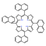
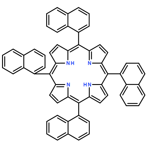
Co-reporter:Ning Sheng, Shenfei Zong, Wei Cao, Jianzhuang Jiang, Zhuyuan Wang, and Yiping Cui
ACS Applied Materials & Interfaces 2015 Volume 7(Issue 35) pp:19718
Publication Date(Web):August 20, 2015
DOI:10.1021/acsami.5b05256
The hydrophobility of most porphyrin and porphyrin derivatives has limited their applications in medicine and biology. Herein, we developed a novel and general strategy for the design of porphyrin nanospheres with good biocompatibility and water dispersibility for biological applications using hydrophobic porphyrins. In order to display the generality of the method, we used two hydrophobic porphyrin isomers as starting material which have different structures confirmed by an X-ray technique. The porphyrin nanospheres were fabricated through two main steps. First, the uniform porphyrin nanospheres stabilized by surfactant were prepared by an interfacially driven microemulsion method, and then the layer-by-layer method was used for the synthesis of polyelectrolyte-coated porphyrin nanospheres to reduce the toxicity of the surfactant as well as improve the biocompatibility of the nanospheres. The newly fabricated porphyrin nanospheres were characterized by TEM techniques, the electronic absorption spectra, photoluminescence emission spectra, dynamic light scattering, and cytotoxicity examination. The resulting nanospheres demonstrated good biocompatibility, excellent water dispersibility and low toxicity. In order to show their application in biophotonics, these porphyrin nanospheres were successfully applied in targeted living cancer cell imaging. The results showed an effective method had been explored to prepare water dispersible and highly stable porphyrin nanomaterial for biophotonics applications using hydrophobic porphyrin. The approach we reported shows obvious flexibility because the surfactants and polyelectrolytes can be optionally selected in accordance with the characteristics of the hydrophobic material. This strategy will expand the applications of hydrophobic porphyrins owning excellent properties in medicine and biology.Keywords: biophotonics applications; hydrophobic porphyrin; layer-by-layer method; microemulsion method; polyelectrolyte; surfactant
Co-reporter:Ning Sheng, Dahui Liu, Bing Gu, Jun He, Yiping Cui
Dyes and Pigments 2015 Volume 122() pp:346-350
Publication Date(Web):November 2015
DOI:10.1016/j.dyepig.2015.07.011
•The 2PA properties of a series of sandwich type complexes were investigated.•All complexes exhibit large 2PA cross-sections.•The unique π–π interaction in these complexes is effective to improve their 2PA properties.The third-order nonlinear optical properties of a series of homoleptic substituted phthalocyaninato rare earth double-decker complexes were experimentally investigated by the Z-scan technique at the wavelength of 800 nm. All complexes were revealed to exhibit large two-photon absorption cross-sections. The measured two-photon absorption values of these complexes remain constant at different laser intensities, indicating nonlinearity is originated from pure two-photon absorption behavior. In addition, the two-photon absorption cross-section of these complexes increases along with the decrease of the rare earth atomic radius, suggesting that intense intramolecular π–π interaction between the phthalocyaninato macrocycles improves the third-order nonlinear optical properties. The optical limiting behavior of these complexes in chloroform was also investigated by using femtosecond laser pulses. The excellent two-photon absorption properties and optical limiting performance indicate their good application potential in photonic and optoelectronic devices.
Co-reporter:Ning Sheng, Dahui Liu, Jialu Wu, Bing Gu, Zhuyuan Wang, Yiping Cui
Dyes and Pigments 2015 Volume 119() pp:116-121
Publication Date(Web):August 2015
DOI:10.1016/j.dyepig.2015.03.033
•Both D-π-A type porphyrins exhibit large 2PA cross sections.•The 2PA properties of them were investigated using femtosecond-pulsed Z-scan technique.•Intramolecular energy transfer takes place in the newly prepared asymmetric porphyrin.The third-order nonlinear optical properties of 5-(4-pyrenylalkynylphenyl)-10,15,20-tris(4-octyloxyphenyl)porphyrin (1) and 5-(4-iodophenyl)-10,15,20-tris(4-octyloxyphenyl)porphyrin (2) have been comparatively investigated with symmetrical 5,10,15,20-tetra(4-octyloxyphenyl)porphyrin (3) and pyrene (4) as reference using femtosecond-pulsed Z-scan technique. Due to the D–π–A structure, both A3B type asymmetric porphyrins 1 and 2 were revealed to exhibit larger two-photon absorption (2PA) cross sections over the symmetrical analogue 3 with the largest value of 6.1 × 10⁵ GM revealed for 1 because of the intramolecular energy transfer from the excited pyrene moiety to the porphyrin moiety as indicated by the fluorescence spectroscopic result. The good 2PA properties of these A3B type porphyrins with ultrafast response indicate their good application potential in nonlinear photonic devices.
Co-reporter:Zhao-Chong Wang, Shu-Hong Xu, Chun-lei Wang, Li Zhu, Fan Bo, Xiao-Yan Lin, Zhu-Yuan Wang and Yi-Ping Cui
RSC Advances 2015 vol. 5(Issue 57) pp:46186-46191
Publication Date(Web):07 May 2015
DOI:10.1039/C5RA04242A
We report the assembly of non-toxic Ag-doped ZnInSe (AZIS) quantum dot (QD)-sensitized solar cells (QDSSC) with a conversion efficiency of 0.89% at 1 sun. The QDs were directly adsorbed on a TiO2 film with the addition of a surface-active agent (Triton X-100). ZnS treatment was performed on QDs by the successive ionic layer adsorption and reaction method to inhibit the recombination current in QDSSCs. The results demonstrated that Triton X-100 increased the QD loading on mesoporous TiO2. The conversion efficiency of an aqueous, non-toxic AZIS QDSSC containing Triton X-100, a ZnS passivating layer and a Cu2S counter electrode reached 0.89% at 1 sun.
Co-reporter:Yuan Jiang, Shuhong Xu, Zengxia Zhao, Liang Zheng, Zhuyuan Wang, Chunlei Wang and Yiping Cui
RSC Advances 2015 vol. 5(Issue 24) pp:18379-18383
Publication Date(Web):06 Feb 2015
DOI:10.1039/C4RA14134B
In this work, the record for a photoluminescence (PL) quantum yield (QY) of positively-charged CdTe nanocrystals (NCs) was brought up to 31%, by using a water–ethanol mixture as a new medium for NC synthesis. A series of water–ethanol feed ratios were investigated, with the maximal PL QY achieved in 50% ethanol solvent mixtures. A photoluminescence lifetime measurement and element analysis demonstrated an improved NC surface modification via the synthesis in the water–ethanol solvent mixtures.
Co-reporter:Xiaojing Xu, Zhengqing Qi, Zengxia Zhao, Chunlei Wang, Changgui Lu, Shuhong Xu and Yiping Cui
New Journal of Chemistry 2015 vol. 39(Issue 11) pp:8818-8824
Publication Date(Web):03 Sep 2015
DOI:10.1039/C5NJ01725D
In this work, water-dispersible Mn:ZnSe/ZnO core/shell quantum dots (QDs) with pure dopant emission were prepared via a two-step method. Compared with the traditional organometallic approach, this two-step method has many advantages, such as using low-cost and non-toxic chemicals, low reaction temperature and simple operation. Moreover, the obtained Mn:ZnSe/ZnO core/shell QDs also showed superior properties to those prepared by the aqueous solution synthesis, such as narrow emission and good monodispersity. The influence of the storage time of Mn:ZnSe QDs in organic medium, the amount of MPA and temperature during the period of ZnO shell deposition on the photo-luminescence (PL) properties was investigated. Under optimal conditions, MPA-capped Mn:ZnSe/ZnO QDs with an average diameter of 2.6 nm and a dopant PL quantum yield (QY) of 30% in water were produced.
Co-reporter:Shuhong Xu, Zhaochong Wang, Chunlei Wang, Zhuyuan Wang and Yiping Cui
New Journal of Chemistry 2015 vol. 39(Issue 4) pp:3105-3108
Publication Date(Web):09 Feb 2015
DOI:10.1039/C5NJ00102A
Configurations and spectra of Ag7 clusters have been investigated using DFT/B3LYP. First, four stable structures of Ag7 clusters have been obtained, which are composed of pentagons and triangles. The most stable one is the Ag7-1 cluster that has one pentagon and ten triangles. The Wiberg bond index values of Natural Bond Orbital analysis and Raman spectra have verified the configurations of four clusters and the existence of pentagons and triangles. Second, Ag7 nano-clusters have been synthesized in an experiment. From comparison of the absorbance spectra from the experiment and the data from calculations, we have confirmed that Ag7-2 and Ag7-4 molecules were the possible configurations of experimental products.
Co-reporter:Yun Binfeng, Hu Guohua, Zhang Ruohu, Zhou Juan, Cui Yiping
Optics Communications 2015 Volume 354() pp:89-93
Publication Date(Web):1 November 2015
DOI:10.1016/j.optcom.2015.05.045
•The polymer sampled waveguide Bragg grating is proposed and fabricated.•Third order Bragg grating were used to lower the fabrication cost.•Low cost polymers were chosen to lower the material cost.•Special processes were introduced to optimize the contact lithography.A low cost polymer sampled waveguide Bragg grating which can be fabricated by using the simple contact lithography is proposed. By using the contact lithography and reactive ion etching, the sampled Bragg grating structures were fabricated on the waveguide bottom cladding, where a channel waveguide was fabricated on top of it as the waveguide core to form the polymer sampled waveguide Bragg grating working at around 1550 nm. The measured transmission and reflection spectra of the fabricated polymer sampled waveguide Bragg grating exhibit multiple dips and peaks, respectively. And the wavelength interval among the peaks is 0.376 nm, which shows good agreement with the theoretical predictions.
Co-reporter:Binfeng Yun, Guohua Hu, Ruohu Zhang, Yiping Cui
Optics Communications 2015 Volume 336() pp:30-33
Publication Date(Web):1 February 2015
DOI:10.1016/j.optcom.2014.09.048
A tunable erbium-doped fiber ring laser incorporating a tunable thermo-optic polymer waveguide Bragg grating is proposed. Nano-imprinting technique was used to fabricate the polymer waveguide Bragg grating whose reflectivity and 3 dB bandwidth are 25 dB and 0.8 nm, respectively. The polymer waveguide Bragg grating was used as wavelength selective element in the ring laser system and the center wavelength can be tuned by controlling the electrical power on the micro-heater. The fabricated tunable laser exhibits an output of 4.1 dBm, side-mode suppression ratio of 55 dB, 3 dB bandwidth less than 16 pm, and wavelength tuning range of 8.4 nm.
Co-reporter:Chunlei Wang;Shuhong Xu;Zengxia Zhao;Zhuyuan Wang;Yiping Cui
Journal of Fluorescence 2015 Volume 25( Issue 1) pp:41-48
Publication Date(Web):2015 January
DOI:10.1007/s10895-014-1476-y
We reported the synthesis of water-soluble and nontoxic Ag2Se/ZnSe Quantum Dots (QDs) using for fluorescence sensors. The influences of various experimental conditions including the synthesis pH, types of ligand, feed ratios, and the refluxed time on the growth process and fluorescence of QDs were investigated in detail. Under optimal conditions, Ag2Se/ZnSe QDs show a single emission peak around 490 nm with the maximal photoluminescence (PL) quantum yield (QYs) of 13.7 %. As-prepared Ag2Se/ZnSe QDs can be used for detection of Ag(II) and Cu(II). The detection limits are 1 × 10⁻⁶ mol/L to 5 × 10⁻⁵ mol/L for Ag (I), and 2 × 10⁻⁶ mol/L to 1.10 × 10⁻⁴ mol/L for Cu(II).
Co-reporter:Zhuyuan Wang, Xueqin Ma, Shenfei Zong, Yuzhong Wang, Hui Chen, Yiping Cui
Talanta 2015 Volume 131() pp:259-265
Publication Date(Web):January 2015
DOI:10.1016/j.talanta.2014.07.088
•A magnetic fluorescent nano-thermometer is proposed.•The nano-thermometer relies on iron oxide nanoparticles and N-isopropylacrylamide copolymers.•The nano-thermometer can detect the temperature in the range from 10 °C to 45 °C.•With the aid of an external magnetic field, targeted temperature sensing in living cells can be achieved.A magnetic fluorescent nano-thermometer is presented. To fabricate the nano-thermometer, magnetic nanoparticles (Fe3O4) were first encapsulated with a silica layer. Then a poly (N-isopropylacrylamide) (pNIPAM) copolymer shell with Rhodamine B isothiocyanate (RhBITC) embedded inside was further coated, which was denoted as the pNIPAM-co-RhBITC shell. Finally, gold nanoparticles were introduced onto the copolymer shell by in-situ growth method and the nano-thermometer (denoted as Fe3O4@SiO2@(pNIPAM-co-RhBITC)/Au) was obtained. The nano-thermometer shows dual responses to both magnetism and temperature. Specifically, the fluorescence intensity of the nano-thermometer decreases as the temperature increases, which makes the nano-thermometer suitable for intracellular temperature sensing. Using this nano-thermometer, temperature changes in live HeLa cells can be successfully detected. Moreover, due to the Fe3O4 component, magnetic field guided targeting can be realized, thus targeted temperature sensing can be achieved for living cells. Cellular temperature changes can be easily detected using the proposed nano-thermometer in the range of 26 °C to 41 °C with a sensitivity of −4.84% °C−1.A magnetic fluorescent nano-thermometer have been proposed which can achieve targeted temperature sensing in live cells.
Co-reporter:Haibao Shao;Chunlei Wang;Shuhong Xu;Zhuyuan Wang;Haihong Yin
Journal of Fluorescence 2015 Volume 25( Issue 2) pp:305-310
Publication Date(Web):2015 March
DOI:10.1007/s10895-015-1509-1
Controllable doping is an effective way of tuning the properties of semiconductor nanocrystals (NCs). In this work, a simple strategy of fast doping Cu ions into ZnSe NCs under ambient conditions was proposed. The principle of doping is based on hydrazine (N2H4) promoted cation exchange reaction. By direct addition of Cu ion stock solution into the preformed ZnSe NCs, Cu doped ZnSe NCs can be obtained. Furthermore, the emission of doped NCs can be tuned by changing the amount of impurity ion addition. The cation exchange reaction is facilitated by three factors: 1) N2H4 addition, 2) fast impurity ions, and 3) partial stabilizer removal. The proposed cation exchange reaction in aqueous solution could be an alternate route for NC doping as well as synthesis of ionic NCs.
Co-reporter:Shenfei Zong, Zhuyuan Wang, Hui Chen, Guohua Hu, Min Liu, Peng Chen and Yiping Cui
Nanoscale 2014 vol. 6(Issue 3) pp:1808-1816
Publication Date(Web):13 Nov 2013
DOI:10.1039/C3NR04942F
As an important biomarker and therapeutic target, telomerase has attracted considerable attention concerning its detection and monitoring. Here, we present a colorimetry and surface enhanced Raman scattering (SERS) dual-mode telomerase activity detection method, which has several distinctive advantages. First, colorimetric functionality allows rapid preliminary discrimination of telomerase activity by the naked eye. Second, the employment of SERS technique results in greatly improved detection sensitivity. Third, the combination of colorimetry and SERS into one detection system can ensure highly efficacious and sensitive screening of numerous samples. Besides, the avoidance of polymerase chain reaction (PCR) procedures further guarantees fine reliability and simplicity. Generally, the presented method is realized by an “elongate and capture” procedure. To be specific, gold nanoparticles modified with Raman molecules and telomeric repeat complementary oligonucleotide are employed as the colorimetric-SERS bifunctional reporting nanotag, while magnetic nanoparticles functionalized with telomerase substrate oligonucleotide are used as the capturing substrate. Telomerase can synthesize and elongate telomeric repeats onto the capturing substrate. The elongated telomeric repeats subsequently facilitate capturing of the reporting nanotag via hybridization between telomeric repeat and its complementary strand. The captured nanotags can cause a significant difference in the color and SERS intensity of the magnetically separated sediments. Thus both the color and SERS can be used as indicators of the telomerase activity. With fast screening ability and outstanding sensitivity, we anticipate that this method would greatly promote practical application of telomerase-based early-stage cancer diagnosis.
Co-reporter:Chunlei Wang, Shuhong Xu, Yanbin Wang, Zhuyuan Wang and Yiping Cui
Journal of Materials Chemistry A 2014 vol. 2(Issue 4) pp:660-666
Publication Date(Web):29 Oct 2013
DOI:10.1039/C3TC31602E
In this work, we prepared white-emitting multilayer Mn:ZnSe/Cu:ZnS quantum dots (QDs) in an aqueous solution. The core–shell multilayer structure provides a unique opportunity to dope different types of impurities into each host layer. White-emitting Mn:ZnSe/Cu:ZnS QDs have two emission bands at around 477 nm and 585 nm. The latter emission band around 585 nm is assigned to Mn-induced emission. The first emission band around 477 nm is caused by both the trap emission in the ZnSe core and Cu-doping emission from the trap state in the ZnS shell to the Cu+ energy level. Due to doping of Cu and Mn in QDs, internally doped Mn:ZnSe/Cu:ZnS QDs have good stability against UV irradiation or alteration due to surroundings during blending with polymers, making as-prepared QDs available for white light-emitting diodes.
Co-reporter:Dan Zhu, Zhuyuan Wang, Shenfei Zong, Hui Chen, Xin Wu, Yuwei Pei, Peng Chen, Xueqin Ma and Yiping Cui
Nanoscale 2014 vol. 6(Issue 14) pp:8155-8161
Publication Date(Web):16 Apr 2014
DOI:10.1039/C4NR00557K
A liposome–Ag nanohybrid has been demonstrated as a SERS traceable intracellular drug nanocarrier. Liposomes have been introduced for their special qualities in drug delivery systems. In essence, 4-aminothiophenol (4ATP) tagged Ag nanoparticles (Ag@4ATP) were adsorbed onto the surfaces of liposomes via electrostatic interactions, in which 4ATP was used as a SERS reporter. In such a nanohybrid, the locations of the carrier can be tracked by SERS signals while those of the drugs can be monitored through their fluorescence, allowing the simultaneous investigation of the intracellular distribution of both the carriers and the drugs. Our experimental results suggest that the reported liposomal system has substantial potential for intracellular drug delivery.
Co-reporter:Fan Bo, Chufan Zhang, Chunlei Wang, Shuhong Xu, Zhuyuan Wang and Yiping Cui
Journal of Materials Chemistry A 2014 vol. 2(Issue 35) pp:14585-14592
Publication Date(Web):20 Jun 2014
DOI:10.1039/C4TA02545H
A novel route using selenium has been developed to fabricate highly catalytic metal selenide counter electrodes (CE) for sensitized solar cells. To demonstrate the facility of this method, copper selenide is fabricated and characterized in this article. Basically, chemically active red selenium has been deposited firmly on FTO through an ultra facile route by disproportionation of Na2SeSO3 aqueous solution with an ethanol spray. Then the red selenium is manipulated as both template and reaction center to fabricate a cuprous embedded CuxSe composite (Cu:Se = 1.66:1) CE upon selenium self redox reaction with copper ions. For device performance, CdSeS quantum-dot-sensitized solar cells coupled with the CuxSe composite CE exhibit a much higher photovoltaic performance than their platinum counterparts (3.80% vs. 1.07%) and even better performance (3.80% vs. 3.72%) and photocurrent stability (<5% drop vs. 40% drop) than the optimized cuprous sulfide cathode made from etched brass under consecutive 1 sun illumination. This alternative from selenium could greatly facilitate the counter electrode fabricating process under rigorous conditions and is promising to fabricate other metal selenide functional counter electrodes, such as PbSe, Ag2Se, etc.
Co-reporter:Min Liu, Zhuyuan Wang, Shenfei Zong, Hui Chen, Dan Zhu, Lei Wu, Guohua Hu, and Yiping Cui
ACS Applied Materials & Interfaces 2014 Volume 6(Issue 10) pp:7371
Publication Date(Web):April 16, 2014
DOI:10.1021/am5006282
Heavy metal ions, such as Hg2+ and Ag+, pose severe risks in human health and the environment. For sensitive detection and selective removal of Hg2+ and Ag+ ions, here, we demonstrate a surface-enhanced Raman scattering (SERS)-active platform by employing the oligonucleotide-functionalized magnetic silica sphere (MSS)@Au nanoparticles (NPs). This system exploits mismatched T–Hg–T and C–Ag–C bridges to capture Hg2+ and Ag+ ions, exhibiting excellent responses for Hg2+ ions in the range of 0.1–1000 nM and for Ag+ in the range of 10–1000 nM. The assay is highly selective for the target ions and does not respond to other metal ions. Additionally, the Hg2+ and Ag+ ions in this system can be effectively removed from surrounding solutions by an external magnetic field or through spontaneous precipitation. Moreover, more than 80% of the MSS@Au NPs can be easily recycled with the help of cysteine. We anticipate that the designed strategy could be extended to other analytes that can bind to DNA molecules with a high affinity, and can be used in many potential applications such as environmental renovation, toxin detection, and groundwater analysis.Keywords: heavy metal ions; magnetic silica nanospheres; surface-enhanced Raman scattering; T−Hg−T bridges;
Co-reporter:Hui Chen, Zhuyuan Wang, Shenfei Zong, Lei Wu, Peng Chen, Dan Zhu, Chunlei Wang, Shuhong Xu, and Yiping Cui
ACS Applied Materials & Interfaces 2014 Volume 6(Issue 20) pp:17526
Publication Date(Web):October 1, 2014
DOI:10.1021/am505160v
A redox-responsive drug carrier based on nanoscale graphene oxide (NGO) loaded with Ag nanoparticles, whose intracellular release behavior can be investigated by SERS-fluorescence combined spectroscopy, is presented. In this demonstrated drug carrier, to make the carrier integrated with the redox responsive property, we utilized disulfide linkages to load drug molecules to the surfaces of NGO directly, which can be cleaved by glutathione (GSH). Covalent drug loading and GSH-responsive release strategy can reduce the influence of the surface diffusion barriers introduced by multifunctionalization. Interestingly, the intracellular real-time drug release dynamics can be monitored by the combined SERS-fluorescence signals of the drugs, while the distribution of the drug carrier can simultaneously be tracked by the intrinsic SERS signals of NGO in the whole process. Our results show that upon the internalization of doxorubicin (DOX)-loaded nanocarriers into living cells, DOX was efficiently released under a GSH regulated reducing environment. Because tumor cells generally exhibit a higher concentration of GSH than normal ones, this drug carrier should have potential in the field of tumor therapy.Keywords: glutathione; graphene oxide; stimuli−drug delivery; surface enhanced Raman scattering; tumor cells
Co-reporter:Zhuyuan Wang;Shenfei Zong;Hui Chen;Chunlei Wang;Shuhong Xu ;Yiping Cui
Advanced Healthcare Materials 2014 Volume 3( Issue 11) pp:1889-1897
Publication Date(Web):
DOI:10.1002/adhm.201400092
A new kind of cancer cell separation method is demonstrated, using surface-enhanced Raman scattering (SERS) and fluorescence dual-encoded magnetic nanoprobes. The designed nanoprobes can realize SERS-fluorescence joint spectral encoding (SFJSE) and greatly improve the multiplexing ability. The nanoprobes have four main components, that is, the magnetic core, SERS generator, fluorescent agent, and targeting antibody. These components are assembled with a multi-layered structure to form the nanoprobes. Specifically, silica-coated magnetic nanobeads (MBs) are used as the inner core. Au core-Ag shell nanorods (Au@Ag NRs) are employed as the SERS generators and attached on the silica-coated MBs. After burying these Au@Ag NRs with another silica layer, CdTe quantum dots (QDs), that is, the fluorescent agent, are anchored onto the silica layer. Finally, antibodies are covalently linked to CdTe QDs. SFJSE is fulfilled by using different Raman molecules and QDs with different emission wavelengths. By utilizing four human cancer cell lines and one normal cell line as the model cells, the nanoprobes can specifically and simultaneously separate target cancer cells from the normal ones. This SFJSE-based method greatly facilitates the multiplex, rapid, and accurate cancer cell separation, and has a prosperous potential in high-throughput analysis and cancer diagnosis.
Co-reporter:Yizhi Zhang, Zhuyuan Wang, Lei Wu, Yuwei Pei, Peng Chen and Yiping Cui
Analyst 2014 vol. 139(Issue 20) pp:5148-5154
Publication Date(Web):08 Jul 2014
DOI:10.1039/C4AN00771A
A rapid and straightforward method has been employed to simultaneously detect two pesticides (thiram and methamidophos (MTD)) on apple surface using surface enhanced Raman scattering (SERS) technique. In the experiment, ethanol was dropped onto the contaminated apple surface for pesticide extraction and then gold@silver core–shell nanorods (Au@Ag NRs) were added to generate the SERS signals of the pesticides. Under a laser excitation at 632.8 nm, prominent SERS peaks of blended contaminants were observed, which were chosen to characterize and quantify their concentration. It was found that the SERS intensity of these two peaks changed as a function of the concentration ratio of thiram to MTD. In addition, a better SERS enhancement performance of Au@Ag NRs was demonstrated compared with that of gold nanorods. Our experimental results show that the lowest detectable concentration on apple surfaces is ∼4.6 × 10⁻⁷ M for thiram and ∼4.4 × 10⁻⁴ M for MTD. This study provides a straightforward method for the simultaneous detection of multiple pesticides on fruit surfaces, which is important for food safety and human health.
Co-reporter:Xiaoyan Lin, Shuhong Xu, Chunlei Wang, Zhuyuan Wang and Yiping Cui
RSC Advances 2014 vol. 4(Issue 10) pp:4993-4997
Publication Date(Web):02 Jan 2014
DOI:10.1039/C3RA44307H
Thiosalicylic acid (TSA) was used as a stabilizer to synthesize CdTe quantum dots (QDs) in aqueous solution. The reaction conditions, such as refluxing time, precursor concentrations, the molar ratio of reactants, and the pH value, were investigated. Under optimized conditions, the photoluminescence quantum yields (PLQY) of TSA-capped CdTe QDs were up to 15%. The morphology and composition of CdTe QDs were characterized by transmission electron microscopy (TEM), X-ray powder diffraction (XRD) and X-ray photoelectron spectroscopy (XPS). Theoretical calculations show that TSA increases electronic coupling through the conjugated π bond structure, thus the TSA-capped CdTe QDs are expected to be applicable in solar cells.
Co-reporter:Chunlei Wang, Shuhong Xu, Shujie Zhou, Zhuyuan Wang and Yiping Cui
Analytical Methods 2014 vol. 6(Issue 24) pp:9596-9600
Publication Date(Web):17 Oct 2014
DOI:10.1039/C4AY02245A
Investigating the chemical reactions at the surface of quantum dots (QDs) is fundamental for applications of QDs in ionic environments. On the basis of our recent report about the discriminative detection of Mn(II) from Cu(II) via QD fluorescence sensing, we herein focused on the QD surface chemical reactions with Mn(II) or Cu(II) at different QD surface modification conditions. Two types of QDs were used. One is well modified QDs with a surface rich in Cd-ligands complexes. Another is moderately modified QDs with both bared Te atoms and Cd-ligand complexes on the surface. Our results indicated that well modified QDs underwent a chemical reaction with Cu(II), namely, the substitution of Cd from QDs by Cu(II). In this case, the results of discriminative detection of Mn(II) from Cu(II) is always believable. For moderately modified QDs in comparison, it appears there is a new reaction manner via combination of bared Te atoms with Cu(II) or Mn(II). This new reaction manner occurs mainly around pH 10.3. Consequently, the results of discriminative detection of Mn(II) from Cu(II) via moderately modified QDs is not believable.
Co-reporter:Hong Wang, Zhuyuan Wang, Minglang Ye, Shenfei Zong, Mingyue Li, Peng Chen, Xueqin Ma, Yiping Cui
Talanta 2014 Volume 119() pp:144-150
Publication Date(Web):15 February 2014
DOI:10.1016/j.talanta.2013.10.057
•A SERS-fluorescence dual-encoded and magnetic nanoprobe is presented.•Single walled carbon nanotubes are used as the building scaffold.•The as-prepared nanoprobe exhibits well fluorescence and SERS performance.•The nanoprobe can fulfill magnetic field guided dual mode optical imaging of live cells.We construct a novel fluorescent, surface enhanced Raman scattering (SERS) encoded and magnetic nanoprobe for live cell imaging. To fabricate this nanoprobe, single walled carbon nanotube (SWNT) is used as the building scaffold while gold nanoparticles (Au NPs), superparamagnetic iron oxide nanoparticles (SPIONs) and quantum dots (QDs) are employed as the building blocks. Here, Au NPs serve as the SERS substrate and QDs act as the fluorescent agent. Au NPs and SPIONs are first adsorbed on the SWNT via electrostatic interactions. Then a silica layer is coated on the SWNT. Finally, QDs are attached on the silica shell. With such a structure, various optical signals can be readily encoded to the nanoprobe simply by using different Raman molecules and QDs with different emission wavelengths. Experimental results show that the as-prepared nanoprobe exhibits well fluorescence and SERS performance. Furthermore, in vitro experiments demonstrate that the nanoprobe can fulfill magnetic field guided fluorescence and SERS dual mode imaging of live cells. As a fascinating optical encoding material and a multifunctional nanoplatform, the presented nanoprobe holds genuine potential in future biosensing applications.
Co-reporter:Lei Wu, Zhuyuan Wang, Shenfei Zong, Yiping Cui
Biosensors and Bioelectronics 2014 Volume 62() pp:13-18
Publication Date(Web):15 December 2014
DOI:10.1016/j.bios.2014.06.026
•A droplet microfluidic chip has been designed and fabricated.•The microfluidic chip was employed for quantitative analysis of thiocyanate (SCN−) using the SERS technique.•The proposed platform greatly reduced the time consumption for the detection of each sample.•The proposed platform significantly improved the reproducibility of the SERS measurements.•The SERS-microfluidic chip was capable to detect SCN− in real human serum and saliva.As thiocyanate (SCN−) acts as an important biomarker in human health assessment, there remains an urgent need to realize rapid and reproducible analysis of SCN− in body fluids. Here, a droplet microfluidic device has been designed and fabricated for SCN− detection in real human serum and saliva using the surface enhanced Raman scattering (SERS) technique. Only a few minutes are needed for the whole detection process which simply cost a few microliters of real sample. Gold@silver core–shell nanorods (Au@Ag NRs) with a large SERS enhancement factor were selected to capture SCN− ions in body fluids. The intensity of SERS peak at around 2100 cm−1, which originates from the –C≡N stretching mode, was used to indicate the concentrations of SCN− ions. Importantly, by generating a droplet environment for mixing reagents and acquiring signals, this microfluidic platform possesses the advantages of an improved reproducibility and reduced time consumption. For practical applications, the SERS-microfluidic system is capable to achieve rapid analysis of SCN− in the presence of human serum, which is very important for realizing the detection in real biological samples. Additionally, SCN− in saliva samples was detected in the SERS-microfluidic chip and the results provide useful information for distinguishing between smokers and nonsmokers.
Co-reporter:Wei Fang, Zhuyuan Wang, Shenfei Zong, Hui Chen, Dan Zhu, Yuan Zhong, Yiping Cui
Biosensors and Bioelectronics 2014 Volume 57() pp:10-15
Publication Date(Web):15 July 2014
DOI:10.1016/j.bios.2014.01.042
•A pH-controllable drug carrier with SERS activity is developed.•Selective drug release in response to different ambient pH is achieved.•SERS-fluorescence dual mode detection is employed to monitor drug release dynamics.•This dual mode detection is more powerful and more accurate.A type of pH-controllable drug carrier is demonstrated based on mesoporous silica nanoparticles and chitosan/poly (methacrylic acid), which can simultaneously serve as the surface enhanced Raman scattering (SERS) traceable drug carriers for targeting cancer cells. The pH-sensitive releasing characteristics can be achieved by coating the nanoparticles with a layer of chitosan/poly (methacrylic acid) (CS-PMAA), while strong SERS signals can be obtained from the SERS reporter tagged Ag nanoparticles in the core. Our experimental results show that doxorubicin (DOX) was effectively encapsulated into the nanocarriers and can be released in response to the ambient pH value. Specifically, an increased amount of DOX release was observed at lower pH value. In addition, the composite nanoparticles were conjugated with transferrin (Tf) to target transferrin receptor (TfR)-overexpressed cancer cells. The targeting ability as well as the intracellular location of the drug carrier was investigated through SERS mapping while the distribution of DOX was monitored by fluorescence images. The results show that the demonstrated drug carrier can simultaneously fulfill the functionalities of pH-responsive drug release, SERS-traceable characteristics and cancer cells targeting, which has a unique potential for the pH-controllable drug delivery nanosystems.
Co-reporter:Jing Yang, Zhuyuan Wang, Shenfei Zong, Hui Chen, Ruohu Zhang, Yiping Cui
Biosensors and Bioelectronics 2014 Volume 51() pp:82-89
Publication Date(Web):15 January 2014
DOI:10.1016/j.bios.2013.07.034
•A dual mode detection method for tumor cell targeting and drug delivery is developed.•SERS and fluorescence signals were used to monitor drug release dynamics.•Targeted drug delivery was realized using mesoporous silica nanoparticles.•This dual mode detection is more powerful and more accurate.We developed a dual-mode detection method for tumor cell specific targeting and intracellular delivery of the chemotherapeutic agent Doxorubicin (DOX) using folic acid functionalized mesoporous silica nanoparticles (FA-MSNs) as carrier systems. In this method, label free surface enhanced Raman scattering (SERS) spectra were utilized to monitor the dynamic release of DOX inside tumor cells in combination with fluorescence images. To investigate the targeting delivery performance of the carrier system, both normal cells (MRC-5) and tumor cells (HeLa) were used as the model cells. The real-time release of DOX from FA-MSNs inside MRC-5 and HeLa cells was monitored. As demonstrated by both fluorescence and SERS results, the DOX loaded FA-MSNs can actively target FA receptor overexpressed tumor cells. Moreover, the releasing behavior of DOX from FA-MSNs in tumor and in normal cells was quantitatively analyzed. Compared with the traditional sole fluorescence or SERS method, this dual-mode detection is more powerful and more accurate, which should have a potential application in drug tracking in living cells.
Co-reporter:Yuwei Pei, Zhuyuan Wang, Shenfei Zong and Yiping Cui
Journal of Materials Chemistry A 2013 vol. 1(Issue 32) pp:3992-3998
Publication Date(Web):06 Jun 2013
DOI:10.1039/C3TB00519D
We have developed a highly sensitive SERS-based sandwich immunoassay by utilizing two derivatives of gold nanostars simultaneously. One is densely packed self-assembled substrates of gold nanostars and the other is labeled immune nanostar aggregates. Taking advantage of the electromagnetic field coupling between tips of adjacent individual nanostars, the self-assembled substrate of gold nanostars exhibited a better SERS performance than that of gold nanoparticles. On the other hand, the immuno-aggregates made of gold stars also showed an improved SERS activity compared to those made of gold nanoparticles. Thus, by combining the self-assembled substrates of gold nanostars and immuno-aggregates of gold nanostars, highly sensitive sandwich immunoassays were obtained. The experimental results show that there existed a linear correlation between the concentration of antigens and the prominent peak intensity of SERS signals. The limit of detection (LOD) is ∼10 fg mL−1, 5 to 6 orders smaller than that of the conventional ELISA protocols. Such a structure will have potential wide-range applications in biological sensing and quantitative biochemical analysis.
Co-reporter:Xin Wu, Zhuyuan Wang, Dan Zhu, Shenfei Zong, Liping Yang, Yuan Zhong, and Yiping Cui
ACS Applied Materials & Interfaces 2013 Volume 5(Issue 21) pp:10895
Publication Date(Web):October 15, 2013
DOI:10.1021/am403092m
A pH and thermo dual-controllable composite structure was developed as a triggerable drug delivery carrier. In such a drug carrier, a mesoporous silica nanoparticle (MSN) acts as the drug loading core, while a layer of copolymer–lipid serves as the dual-responsive gating shell. Specifically, the copolymer–lipid bilayer consists of natural phospholipids (soy phosphatidylcholine, SPC) and the poly(N-isopropylacrylamide-methacrylic acid-octadecyl acrylate) (p(NIPAM-MAA-ODA)) copolymer. With this structure, a high drug loading capacity and a sustained release effect could be provided by the MSN core, while a pH and thermo dual-responsive releasing ability could be offered by the copolymer–lipid bilayer. In addition, the introduction of SPC instead of the traditionally used phospholipids (such as dioleoyl phosphatidylethanolamine (DOPE) or dipalmitoyl phosphatidylcholine (DPPC)) results in a much lower cost and a better serum stability. Using doxorubicin (DOX) as the drug model, our results confirmed that either pH or temperature can trigger the drug release. However, much more drugs could be released by simultaneously controlling the pH and temperature. Furthermore, after being cocultured with cancer cells (MCF-7), the drug carriers transported DOX into the cells and exhibited a pH-sensitive release behavior. Since most tumor sites usually exhibit a more acidic environment or a higher temperature, the pH- and thermo-responsive releasing ability of this drug carrier is particularly useful and important for the targeted release at the tumor region. Thus, due to the powerful controlled releasing ability, the straightforward preparation method, and low cost, the demonstrated nanocarrier will have potential applications in controllable drug delivery and cancer therapy.Keywords: copolymer; drug carrier; liposome; mesoporous silica nanoparticles; pH; stimuli responsive; thermo;
Co-reporter:Lei Wu, Zhuyuan Wang, Shenfei Zong, Hui Chen, Chunlei Wang, Shuhong Xu and Yiping Cui
Analyst 2013 vol. 138(Issue 12) pp:3450-3456
Publication Date(Web):03 Apr 2013
DOI:10.1039/C3AN00181D
Simultaneous detection of tumor suppressor p53 and cyclin-dependent kinase inhibitor p21 has been achieved for the first time with a high sensitivity using surface enhanced Raman scattering (SERS) technique. In this immunoassay, gold@silver core–shell nanorods (Au@Ag NRs) with a high SERS activity and chemical stability were chosen to fabricate immuno-probes, which were conjugated with different antibodies to specifically target each analyte. Qualitative and quantitative analysis of p53 and p21 were achieved by characterizing SERS signals of two different Raman reporters (4MBA and DTNB), which were labeled on the corresponding optical probes. In addition, we investigated the detection of these biomarkers in the medium of blood serum. Our experimental results indicate the excellent specificity, high sensitivity (1 pg mL−1), as well as the great reproducibility of this immunoassay. More importantly, this assay presents a powerful ability to detect multiple analytes due to the great multiplexing capacity of SERS spectroscopy. Since the combined evaluation of p53 and p21 expression level acts as an indicator for early cancer prediction, our demonstrated immunoassay will potentially promote the application of SERS technique to practical clinical diagnoses.
Co-reporter:Chunlei Wang, Shuhong Xu and Yiping Cui
New Journal of Chemistry 2013 vol. 37(Issue 10) pp:3303-3307
Publication Date(Web):12 Aug 2013
DOI:10.1039/C3NJ00778B
The configuration, bonding, molecule orbitals and the wavelength of absorption spectra of HgX (X = O, Te, Se, and S) molecules and their doping clusters have been systematically investigated at B3LYP level with LanL2DZ basis set. The calculated results showed that Hg2O2 had different bonding, orbitals and spectra than other HgO clusters due to the presence of an O–O bond. From analysis of the calculated wavelengths of absorption spectra, we have found that Hg4O4 (a; hexahedron structure) and Hg4O4 (b; planar octagon) had completely different absorption wavelengths. As for doping, vacancy doping changed the HOMO and the LUMO, and the spectra of clusters. In contrast, substitution doping did not influence the orbital compositions of the structures. Obviously, vacancy doping was more difficult than substitution doping. Finally, through comparison of orbitals, spectra and bonding of HgO, HgS, HgSe and HgTe molecules, we have discovered that the other clusters had completely different orbital compositions, spectra, and bonding from of HgO clusters.
Co-reporter:Binfeng Yun;Guohua Hu;Yiping Cui
Plasmonics 2013 Volume 8( Issue 2) pp:267-275
Publication Date(Web):2013 June
DOI:10.1007/s11468-012-9384-y
The resonant mode characteristics of the nanoscale surface plasmon polaritons (SPP) waveguide filter with rectangle cavity are studied theoretically. By using the finite difference time domain method, both the band-stop- and band-pass-type rectangle SPP filters are analyzed. The results show that the whispering gallery mode (WGM) and the Fabry–Perot (FP) mode can be supported by the rectangle SPP resonator. Furthermore, both traveling-wave mode and standing-wave mode can be realized by the WGM, while only standing-wave mode can be introduced by the FP mode. The traveling-wave mode can only be realized by the square-shaped SPP resonator, and the traveling-wave mode is splitted into two standing-wave modes by transforming the cavity shape from square to rectangle. Also, the effects of the cavity shape, cavity size, and coupling gap size on the transmission spectra of the SPP resonators are analyzed in detail. This simple SPP waveguide filter is very promising for the high-density SPP waveguide integrations.
Co-reporter:Chunlei Wang;Shuhong Xu;Yujie Shao;Zhuyuan Wang
Journal of Cluster Science 2013 Volume 24( Issue 2) pp:439-447
Publication Date(Web):2013 June
DOI:10.1007/s10876-013-0567-2
In this paper, conformations of the ternary structures ZnSe(Te), ZnS(Te), ZnS(Se), CdSe(Te), CdS(Te) and CdS(Se) were optimized, and their orbital, spectra have been investigated at the B3LYP/LANL2DZ level. First, we have found some rules which agreed with experimental results. HOMO–LUMO gaps, WBI values and the wavelengths of the absorption peaks of ternary structures changed gradually with the ratio of Te, Se and S atoms in ternary structures. Second, analysis of Raman spectra revealed that doped structures had the spectra of two relevant binary clusters. Namely, Raman spectra of ternary ZnSe(Te) clusters had the Raman peaks of ZnSe and ZnTe. In this way, with the help of the Raman spectra, the formations of the ternary structures can be determined in experiment. This was important to estimate during synthesis progress. Finally, calculated results have proved that ZnS and CdS structures had the shorter wavelengths of the absorption peaks, the higher excited energies of singlets, and good stability.
Co-reporter:Ruohu Zhang;Zhuyuan Wang;Chunyuan Song;Jing Yang;Asma Sadaf
Journal of Fluorescence 2013 Volume 23( Issue 1) pp:71-77
Publication Date(Web):2013 January
DOI:10.1007/s10895-012-1117-2
A novel gap-plasmon-tunable Ag bilayer nanoparticle film for immunoassays is demonstrated. Different from a traditional Ag monolayer nanoparticle film, a desired number of polyelectrolyte (PEL) layers are deposited on the nanoparticles before the self-assembly of a second Ag nanoparticle layer. Interestingly, by controlling the number of the PEL interlayers, the gap plasmon between the two Ag nanoparticle layers can be tuned across the visible spectral range. The ability of the presented Ag bilayer nanoparticle films in fluorescence enhancement has been examined experimentally. A maximal enhancement of around 15.4 fold was achieved when 7 layers of polyelectrolyte were used. When this optimal Ag bilayer nanoparticle film was applied to fluorescence immunoassay, a performance with approximately 3.3-fold enhancement was obtained compared with that performed on a traditional glass substrate. The experimental results suggest that the presented gap-plasmon tunable Ag bilayer nanoparticle films have great potential in fluorescence-based immunoassays. The method of the bilayer-film construction presented here also provides new insights into the rational design of the plasmonic substrates.
Co-reporter:Zhuyuan Wang ; Shenfei Zong ; Wang Li ; Chunlei Wang ; Shuhong Xu ; Hui Chen ;Yiping Cui
Journal of the American Chemical Society 2012 Volume 134(Issue 6) pp:2993-3000
Publication Date(Web):January 9, 2012
DOI:10.1021/ja208154m
A new concept of optical encoding approach, surface enhanced Raman scattering (SERS)-fluorescence joint spectral encoding method (SFJSE), was demonstrated by using organic–metal–quantum dot (QD) hybrid nanoparticles (OMQ NPs) with a nanolayered structure. This method has two distinct characteristics, which make it more feasible to achieve enormous codes in practice, compared with a sole fluorescence- or SERS-based encoding protocol. One of the two characteristics is to use the joint SERS and fluorescence spectra as the encoding elements instead of an individual optical signal, resulting in a broadened optical spectrum range for efficient encoding. The other is to assemble SERS reporters and fluorescent agents onto different layers of OMQ NPs, leading to an easier fabrication protocol when a large number of agents need to be involved into encoding carriers. By conjugating different antibodies to OMQ NPs with varied codes, the potential application of such an encoding system in high-throughput detection has been investigated by multiplex sandwich immunoassays. The high specificity and sensitivity of the assays suggest that the SFJSE method could be developed as a powerful encoding tool for high-throughput bioanalysis with the use of OMQ NPs.
Co-reporter:Shuhong Xu, Chunlei Wang, Haisheng Zhang, Qingfeng Sun, Zhuyuan Wang and Yiping Cui
Journal of Materials Chemistry A 2012 vol. 22(Issue 18) pp:9216-9221
Publication Date(Web):30 Mar 2012
DOI:10.1039/C2JM00046F
In this report, we report a novel pH-controlled recognition method for the discriminative detection of Mn(II) and Cu(II) ions via CdTe QD fluorescence sensing. The detection limits of Mn(II) are 10 nmol L−1 to 5 μmol L−1. Our results indicate that the sensitivity of QD fluorescence sensing to Mn(II) strongly depends on the solution pH, whereas the sensitivity to Cu(II) is subject to a minor impact of pH. Therefore, the recognizable detection of Mn(II) and Cu(II) can be realized by adjusting the solution pH. The discriminative detection of Cu(II) is realized under Mn(II)-insensitive pH, whereas the recognition of Mn(II) is realized after the deduction of the Cu(II) impact by measuring the sample at both Mn(II)-sensitive pH and Mn(II)-insensitive pH. The mechanism for the peculiar pH-controlled sensitivity of Mn(II) is investigated, which is attributed to the pH-dependent diffusion and adsorption of Mn(II) on the surface of 1-thioglycerol (TG)-capped CdTe QDs followed by the energy transfer from CdTe QDs to Mn(II).
Co-reporter:Yuan Jiang, Shuhong Xu, Chunlei Wang, Haibao Shao, Zhuyuan Wang and Yiping Cui
Journal of Materials Chemistry A 2012 vol. 22(Issue 27) pp:13469-13472
Publication Date(Web):28 May 2012
DOI:10.1039/C2JM32225K
A novel separation technique for aqueous nanoparticles (NPs) is developed via a phase transfer based separation. By using anti-charged surfactant in oil phase, small sized NPs can be extracted into the oil phase, leaving large sized NPs in the aqueous phase. Thus, the separation is achieved.
Co-reporter:Chunyuan Song, Justin L. Abell, Yuping He, S. Hunyadi Murph, Yiping Cui and Yiping Zhao
Journal of Materials Chemistry A 2012 vol. 22(Issue 3) pp:1150-1159
Publication Date(Web):15 Nov 2011
DOI:10.1039/C1JM14133C
Only a few remaining technical hurdles currently prevent the implementation of SERS as a mainstream detection technology. Although oblique-angle deposited silver nanorod arrays provide superior analytical figures of merit for SERS sensing, stability issues of silver surfaces can impede their use for real-world sensing applications within certain environments. To circumvent this issue, silver nanorod arrays are modified with a straight-forward, inexpensive Au-coating via a galvanic replacement reaction. The morphological, structural, compositional, and optical properties of the Au-modified Ag nanorod arrays are studied by multiple ex situ morphological characterization techniques and in situ optical absorbance spectroscopy. Depending on the reaction time, the Au coating experiences five different stages of the morphological and compositional changes. The porosity of the Au layer and the content of Ag decrease with reaction time. The optical measurements show that the representative localized plasmon resonance peak of the nanorod red-shifts as the reaction proceeds. The surface enhanced Raman scattering (SERS) intensity, tested using 4-mercaptophenol, decreases exponentially with reaction time, due to the compositional evolution of the nanostructure from pure Ag to a Au–Ag alloy with increasing Au content. We show that the Au-modified Ag nanorod is very stable in NaCl solution compared to the as-deposited Ag nanorod, and the 20 or 30 minute Au-modified Ag nanorod substrate shows an improved SERS sensitivity for air contamination detection. Such an improved SERS substrate can be used in more hostile environments where a pure Ag nanorod substrate cannot be used, and is good for practical sensing applications.
Co-reporter:Haibao Shao, Chunlei Wang, Zhuyuan Wang, Rongqing Li, Qinying Xu, Shuhong Xu, Yuan Jiang, Qingfeng Sun, Fan Bo, Yiping Cui
Journal of Colloid and Interface Science 2012 Volume 383(Issue 1) pp:43-48
Publication Date(Web):1 October 2012
DOI:10.1016/j.jcis.2012.06.016
Colloidal synthesis of metal–semiconductor hybrid nanostructures is mainly achieved in organic solution. In some applications of hybrid nanoparticles relevant in aqueous media, phase transfer of hydrophobic metal–semiconductor hybrid nanostructures is essential. In this work, we present a simple method for direct synthesis of water-soluble gold (Au) decorated Te@CdTe hybrid nanorods (NRs) at room temperature by using aqueous Te@CdTe NRs as templates, which were preformed by using CdTe nanocrystals (NCs) as precursor in the presence of hydrazine hydrate (N2H4). Our results showed that NRs were decorated with Au islands both on tips and along the surface of the NRs. The size and density of Au islands can be controlled by varying the amount of Au precursor (mixture of HAuCl4 and thioglycolic acid (TGA)) and TGA/HAuCl4 ratio. A possible growth mechanism for the Au decoration of Te@CdTe NRs is concluded as three steps: (1) the formation of AuTe1.7 via the substitution reaction of Cd2+ by Au3+, (2) adsorption of Au–TGA complex onto the preformed AuTe1.7 anchors and following reduction by CdTe and N2H4, leading to the formation of small Au NCs, (3) Au NCs grow to bigger ones, followed by reduction of more Au precursor by N2H4.Graphical abstractHighlights► Te@CdTe nanorods were synthesized by using CdTe nanocrystals as precursors. ► Gold decoration onto Te@CdTe nanorods in aqueous solution was realized. ► The size and density of gold decoration could be well controlled. ► A possible mechanism of gold decoration was considered.
Co-reporter:Haibao Shao;Chunlei Wang;Rongqing Li;Shuhong Xu;Haisheng Zhang;Yiping Cui
Chinese Journal of Chemistry 2012 Volume 30( Issue 4) pp:803-808
Publication Date(Web):
DOI:10.1002/cjoc.201100113
Abstract
As one of the most popular nanocrystals (NCs), aqueous CdTe NCs have very weak green emission under conventional synthesis conditions. In this work, we report the first example of blue-emitting CdTe NCs directly synthesized in aqueous solution by slowing down the growth rate after nucleation. The key for the synthesis is the optimization of NC growth conditions, namely pH range of 7.5 to 8.5, TGA/Cd ratio of 3.6, Cd/Te ratio of 10, and Te concentration of 2×10⁻⁵ mol/L, to get a slow growth rate after nucleation. The as-prepared blue-emitting CdTe NCs have small size (as small as 1.9 nm) and bright emission [with 4% photoluminescence quantum yield (PL QY) at 486 nm and 17% PLQY at 500 nm]. Transmission electron microscopy (TEM) images of the as-prepared CdTe show monodispersed NCs which exhibit cubic zinc blend structure. Moreover, time-resolved PL decay and X-ray photoelectron spectroscopy (XPS) results show the as-prepared NCs have better surface modification by ligand, which makes these luminescent small CdTe NCs have higher photoluminescence quantum yield, compared with NCs synthesized under conventional conditions.
Co-reporter:Rongqing Li;Chunlei Wang;Shuhong Xu;Zhuyuan Wang;Haibao Shao;Yiping Cui
Chinese Journal of Chemistry 2012 Volume 30( Issue 7) pp:1490-1496
Publication Date(Web):
DOI:10.1002/cjoc.201200078
Abstract
Metal-enhanced fluorescence of semiconductor nanocrystals (NCs) is the current investigation focus. In this work, we directly observed metal-enhanced fluorescence of CdTe@PAA nanaospheres in aqueous solution. The enhanced magnitude of photoluminescence (PL) was closely related to solution pH values, and the maximal PL enhancement is about 9 times compared with the ones without Au NPs. Furthermore, based on the results of absorptions and fluorescence lifetimes of CdTe@PAA-Au mixed solution at different pH values, we studied the mechanisms and physics processes of pH-dependent enhanced PL induced by Au NPs. The pH-dependent PL of CdTe@PAA-Au mixed solutions are due to the constantly changing distances between Au NPs and CdTe@PAA nanaospheres with pH. In the CdTe@PAA-Au mixed solutions, CdTe@PAA nanospheres in close proximity to the Au NPs are exposed to the increased electric fields in between and around the NPs, effectively resulting in significant increases in their absorption cross section. This lends itself to a subsequent increase in the excitation and eventually in the fluorescence emission from the CdTe@PAA nanospheres.
Co-reporter:Shenfei Zong, Zhuyuan Wang, Jing Yang, Chunlei Wang, Shuhong Xu, Yiping Cui
Talanta 2012 Volume 97() pp:368-375
Publication Date(Web):15 August 2012
DOI:10.1016/j.talanta.2012.04.047
We report a dual mode cancer cell targeting probe based on CdTe quantum dots (QDs) conjugated, silica coated Au@Ag core–shell nanorods (Au@Ag NRs), which can generate both surface enhanced Raman scattering (SERS) and fluorescence signals. In such a probe, folic acid (FA) is used as a targeting ligand for folate receptors (FRs) overexpressed cancer cells. To synthesize the probe, Au@Ag NRs were first prepared to serve as the SERS substrates by coating an Ag shell on the gold nanorods. Then the Au@Ag NRs were labeled with 4-mercaptobenzoic acid (4MBA) to generate SERS signals, followed by being coated with a silica shell through a modified Stöber method. Finally, CdTe QDs and FA were conjugated to the silica coated Au@Ag NRs by the carbodiimide chemistry to yield fluorescence and the targeting ability, respectively. To validate the targeting capability of the probe, in vitro experiments were conducted, using HeLa cells with overexpressed FRs as the model target cells and MRC-5 cells with a low folate receptor expression level as the negative control. Both the fluorescence imaging and the SERS mapping results confirmed that the proposed probe can be used as an efficient cancer cell targeting agent. This kind of multifunctional probe has great potential in the diagnosis and therapeutics of cancerous diseases due to its specific targeting and multiplex imaging abilities, especially in the simultaneous tracking of multiple components in a hybrid bio-system.Highlights► A metal-organic-QD hybrid nanoprobe is synthesized for cancer cell targeting. ► Its SERS and fluorescence can be switched by changing the exciting wavelength. ► It is advantageous for multi-component tracking due to QD's narrow emission bands. ► Au@Ag nanorods exhibit much stronger SERS enhancement than Au nanorods. ► It can target cancer cells with overexpressed folate receptors by two mode images.
Co-reporter:Lei Wu, Zhuyuan Wang, Shenfei Zong, Zhi Huang, Puyang Zhang, Yiping Cui
Biosensors and Bioelectronics 2012 Volume 38(Issue 1) pp:94-99
Publication Date(Web):October–December 2012
DOI:10.1016/j.bios.2012.05.005
An immunoassay based on surface enhanced Raman scattering (SERS) has been developed using immuno-gold/silver core-shell nanorods with a high sensitivity. The features of these nanoparticles were characterized by UV–vis extinction spectra, TEM images, EDX analyses and SERS spectra. It was found that the obtained gold/silver core-shell nanorods showed a much higher SERS activity than uncoated gold nanorods. After the gold/silver core-shell nanorods were modified with antibody and employed in immunoassay, the antigen concentration-dependent SERS spectra and dose-response calibration curves were obtained. By comparison, it can be concluded that the detection limit of gold/silver core-shell nanorods based immunoassay reaches 70 fM, which is 104 times lower than gold nanorods based detection. As a result, SERS probes fabricated with gold/silver core-shell nanorods are demonstrated to be advantageous to those synthesized with gold nanorods due to their highly increased sensitivity in sandwich immunoassay, which also indicates the potential use of these gold/silver core-shell nanorods in other biological sensing applications.Highlights► Gold/silver core-shell nanorods (Au@Ag NRs) based SERS probes were synthesized. ► Au@Ag NRs SERS probes were employed in immunoassay. ► The detection limit of Au@Ag NRs based immunoassay could reach 70 fM. ► Comparative study was made using gold nanorods as a control in immunoassay. ► Au@Ag NRs based immunoassay is much more sensitive than that based on gold nanorods.
Co-reporter:Chunyuan Song, Jun Chen, Justin L. Abell, Yiping Cui, and Yiping Zhao
Langmuir 2012 Volume 28(Issue 2) pp:1488-1495
Publication Date(Web):December 12, 2011
DOI:10.1021/la203772u
Using the hydrolysis of tetraethylorthosilicate, a uniform and conformal layer of porous SiO2 with controlled thickness has been coated onto the oblique angle deposited Ag nanorod (AgNR) array to form an aligned AgNR-SiO2 core–shell array nanostructure. The morphology, optical property, SERS response, and surface wettability of the AgNRs with different SiO2 shell thicknesses have been obtained by multiple characterization techniques. The morphological characterization shows that each AgNR on the array is coated with a uniform and porous silica shell independently and the growth of shell thickness follows a linear function versus the coating time. Thickening of the shell induces a monotonic decrease of the apparent contact angle, red-shift of the transverse mode of the localized surface plasmon resonance peak, and makes the SiO2 shell more compact. The SERS response of 4-Mercaptophenol on these substrates exhibits an exponential decay behavior with the increasing coating time, which is ascribed to the decreasing Ag surface coverage of core–shell nanorods. Under the assumption that the Ag surface coverage is proportional to the SERS intensity, one can estimate the evolution of SiO2 coverage on AgNRs. Such coverage evolution can be used to qualitatively explain the LSPR wavelength change and quantitatively interpret the contact angle change based on a double Cassie’s law.
Co-reporter:Shuhong Xu, Chunlei Wang, Yiping Cui
Journal of Molecular Structure 2012 1010() pp: 26-31
Publication Date(Web):
DOI:10.1016/j.molstruc.2011.10.058
Co-reporter:Dr. Rongqing Li;Dr. Chunlei Wang;Dr. Fan Bo;Dr. Zhuyuan Wang;Dr. Haibao Shao;Dr. Shuhong Xu; Dr. Yiping Cui
ChemPhysChem 2012 Volume 13( Issue 8) pp:2097-2101
Publication Date(Web):
DOI:10.1002/cphc.201101034
Abstract
Fluorescent Ag nanoclusters are of significant interest because they provide the bridge between atomic and nanoparticle behavior in noble metals. Herein, microwave irradiation was originally used for the synthesis of water-soluble fluorescent Ag nanoclusters. As-prepared Ag nanoclusters present red fluorescence emission around 608 nm and a characteristic absorption peak at about 508 nm. Transmission electron microscopy (TEM) shows an average size of 1.6 nm for Ag nanoclusters. The effect of solution pH on the synthesis process and optical properties of Ag nanoclusters was investigated. The pH-dependent present form and adsorption capacity of poly(methacrylic acid, sodium salt) (PMAA) ligands are believed to be the reason for the pH effect.
Co-reporter:Jing Yang;Zhuyuan Wang;Shenfei Zong
Analytical and Bioanalytical Chemistry 2012 Volume 402( Issue 3) pp:1093-1100
Publication Date(Web):2012 January
DOI:10.1007/s00216-011-5577-z
The detection and identification of epidermal growth factor receptor 2 (HER2)-positive breast cancer cells is crucial for the clinic therapy of breast cancer. For the aim of the detection, a novel surface-enhanced Raman scattering (SERS) probe for distinguishing breast cancers at different HER2 statuses is reported in this paper. In such a probe, anti-HER2 antibody-conjugated silver nanoparticles have been synthesized for specific targeting of HER2-positive breast cancer cells. More importantly, different from the previously reported SERS probe for targeting cancer cells, p-mercaptobenzoic acid is utilized as both the Raman reporter and the conjugation agent for attaching antibody molecules, which leads to a much simplified structure. For investigating the ability of such a probe to distinguish breast cancer cells, SKBR3 and MCF7 cells were chosen as two model systems, which are HER2-positive- and HER2-negative-expressing cells, respectively. The experimental results reveal that SKBR3 cells exhibit much stronger SERS signals than MCF7 cells, indicating that the probe could be utilized to distinguish breast cancer cells at different HER2 statuses. This kind of SERS probe holds a potential for a direct detection of living breast cancer cells with the advantages of easy fabrication, high SERS sensitivity, and biocompatibility.
Co-reporter:Shuhong Xu, Chunlei Wang, Runhuan Wang, Zhuyuan Wang, Qinying Xu and Yiping Cui
Journal of Materials Chemistry A 2011 vol. 21(Issue 41) pp:16427-16429
Publication Date(Web):15 Sep 2011
DOI:10.1039/C1JM13345D
One-dimensional (1D) Te nanostructures are a recent focus in various 1D materials due to their interesting properties of photoconductivity, thermoelectric, catalytic activity and strong piezoelectric effect. In this work, we report a new synthesis method for water-dispersible 1D Te@ZnTe core–shell nanoparticles by using ZnTe nanoparticles as precursors. The as-prepared 1D nanoparticles have a crystalline Te core and an amorphous ZnTe shell, which are believed to be available as the cathode for dye-sensitized nanocrystal solar cells and as nanocables in the interconnection of nanodevices. Moreover, the good water dispersibility of the as-prepared 1D nanoparticles facilitated the fabrication of nanodevices via the surface functional groups of the as-prepared 1D nanoparticles.
Co-reporter:Zhuyuan Wang, Hui Wu, Chunlei Wang, Shuhong Xu and Yiping Cui
Journal of Materials Chemistry A 2011 vol. 21(Issue 12) pp:4307-4313
Publication Date(Web):10 Feb 2011
DOI:10.1039/C0JM03884A
A new class of dual-mode image probe has been designed and prepared, which is composed of silica-coated Au aggregates and water-soluble CdTe quantum dots (QDs). In such a probe, the two kinds of optical signals, SERS and fluorescence, can be separately generated and switched by changing the wavelength of the excitation light. Experimentally, the probe exhibited strong fluorescence when excited by light at a wavelength of 458 nm and showed distinct SERS signals while the excitation wavelength changed to 633 nm. The probe was fabricated through two main steps. The first step involves an addition of an SERS reporter, 4-mercaptobenzoic acid (4MBA), to a dispersion of Au nanoparticles to yield 4MBA-tagged Au aggregates for enhanced SERS performance. The aggregates are thereby incorporated as cores into monodisperse silica spheres with a uniform spherical shape using a modified reverse microemulsion procedure. The second step involves adsorption of water-soluble luminescent CdTe QDs onto the surfaces of the amino-functionalized silica nanoparticles, which are finally coated with a layer of poly(allyamine hydrochloride) (PAH) to reduce the toxicity of CdTe QDs. The in vitro experimental results prove that the fluorescence-SERS switchable characteristics of the probe will be maintained after the probe is incorporated into living cells. Such novel composite nanospheres could find wide applications, especially in the fields of biological tracking and verification procedures that simultaneously provide locational and spectroscopic information about biomolecules.
Co-reporter:Shenfei Zong, Zhuyuan Wang, Jing Yang, and Yiping Cui
Analytical Chemistry 2011 Volume 83(Issue 11) pp:4178
Publication Date(Web):April 22, 2011
DOI:10.1021/ac200467z
We report an intracellular pH sensor based on surface enhanced Raman scattering (SERS) using the hydrochloric acid (HCl) treated gold nanorods (GNRs) as the SERS substrates and p-aminothiophenol (pATP) as the Raman reporter. Using the HCl treated GNRs previously reported by us, the biocompatibility and the SERS performance of GNRs have been greatly improved. Meanwhile, the adsorbed reporters are allowed to be directly exposed to the surrounding environments, which is very important for biosensors. It is found that the SERS spectrum of pATP is strongly dependent on the pH value. The intensities of SERS bands at 1142 cm–1, 1390 cm–1, and 1432 cm–1 increased obviously with the pH value varying from 3.0 to 8.0. This pH-dependent SERS performance of pATP-functionalized HCl treated GNRs was well retained after the incorporation of the GNRs into living HeLa cells. Our experimental results indicate that such pATP-functionalized HCl treated GNRs can be used as an effective intracellular pH sensor. Thus, we show a good example that the bioapplications of the normal CTAB-stabilized GNRs can be expanded after the simple HCl treatment.
Co-reporter:Zhuyuan Wang, Wang Li, Zhiqiang Zhou, Yiping Cui
Optical Materials 2011 Volume 33(Issue 6) pp:920-923
Publication Date(Web):April 2011
DOI:10.1016/j.optmat.2011.01.023
We report on the observation of chemical effects in surface enhanced hyper Rayleigh scattering (SEHRS), using silver nanoparticles adsorbed with stilbazolium dyes. The experimental results show that the enhancement factors varied with different donor structures of the dyes, which cannot be explained only according to the electromagnetic effect. Using the extinction spectra and surface enhanced Raman scattering spectra (SERS) to provide the complementary results, we deduce that chemical effects also play an important role in SEHRS in addition to the electromagnetic effect. The chemical effects may be related with the Ag-adsorbate charge transfer transition, similar to the mechanism in SERS.Research highlights► Surface enhanced hyper Rayleigh scattering (SEHRS) was measured for four dyes-silver complexes. ► The enhancement factors varied with different donor structures of the stilbazolium dyes. ► Chemical effects play an important role in SEHRS besides to the electromagnetic effect. ► The chemical effects may be related with the silver-adsorbate charge transfer transition.
Co-reporter:Li Liu, Zhiqiang Zhou, Jieping Shi, Zeyu Liu, Changgui Lu, Weijiang He, Jing Ma, Yiping Cui, Guo-Yuan Lu
Synthetic Metals 2011 Volume 161(9–10) pp:783-788
Publication Date(Web):May 2011
DOI:10.1016/j.synthmet.2011.01.030
The octupolar chromophore TA with a 1,3,5-triazine core and pyrrole electron-donating end-groups was prepared via Knoevenagel coupling. Its photophysical and two-photon absorption (TPA) properties upon the protonation with CF3COOH were investigated by absorption, fluorescence, NMR spectroscopy and TPA cross-section determination. The UV–vis spectroscopic study shows that the absorption band is shifted from 401 nm to 471 nm with the color change from pale-yellow to red upon CF3COOH addition. The fluorescence spectra shows that the emission band is red-shifted from 490 nm to 553 nm accompanied by the distinct emission quench effect caused by the protonation. NMR spectropic study demonstrates the protonation of triazine upon CF3COOH addition, and the protonated molecule retain the C3h symmetry. In addition, the theoretical calculations of the absorption spectra of neutral and protonated TA demonstrate an obvious red-shift of λmax in the absorption after protonation. TPA cross-section of TA measured by nonlinear transmission (NLT) exhibits the obvious TPA cross-section enhancement effect upon protonation, provides a simple way to enhance the TPA cross-section of molecules and makes it attractive for application involving the proton-controlled optical sensor.
Co-reporter:Chunlei Wang;Shuhong Xu ;Yiping Cui
Chinese Journal of Chemistry 2011 Volume 29( Issue 7) pp:1389-1394
Publication Date(Web):
DOI:10.1002/cjoc.201180258
Abstract
During traditional 2-propanol-based purification of aqueous nanocrystals (NCs), it is very difficult to recycle 2-propanol from the aqueous solution, which brings great consumption of 2-propanol during the purification process. A major contribution of this work is to provide a simple way to reduce the consumption via recycling of 2-propanol during the purification process. The recycled 2-propanol is available for precipitating NCs from aqueous solution in a new round of NC purification process. Due to the recycling of 2-propanol, the great consumption of 2-propanol can be avoided, which makes the purification process of aqueous NCs much greener and at a much lower cost.
Co-reporter:Binfeng Yun, Guohua Hu, Yiping Cui
Optics Communications 2011 Volume 284(Issue 1) pp:485-489
Publication Date(Web):1 January 2011
DOI:10.1016/j.optcom.2010.08.080
A compact nanometric surface plasmon polariton Fabry–Perot filter based on three metal-insulator-metal (MIM) waveguides is proposed and studied. The characteristics of this SPP band pass filter are analyzed by the finite difference time domain method and the Fabry–Perot resonance model. The results show that the resonance wavelengths of the pass bands can be linearly changed by the resonant cavity length, and the transmission ratios of the pass bands can be varied by altering the gaps between the MIM waveguides. The metal loss and dispersion effects on the SPP filter's spectra are considered as well. This kind of simple filter is very promising for high density SPP waveguide integrations.
Co-reporter:Chunlei Wang;Shuhong Xu;Lihua Ye;Wei Lei;Yiping Cui
Journal of Molecular Modeling 2011 Volume 17( Issue 5) pp:1075-1080
Publication Date(Web):2011 May
DOI:10.1007/s00894-010-0814-5
Four clusters of ZnO, O–Zn–SR (–SR = ligand) and doping ZnO structures (with Cr, Cu, Al atoms) were investigated using density functional theory at theB3LYP/Lanl2dz level. The characteristics of Zn3O3 and Zn4O4 structures, which are the units of experimental wurtzite and zinc blende structures, were found to be similar to those of experimental ZnO nanocrystals. Moreover, the calculated Raman and IR spectra of ZnO clusters were almost consistent with experimental results. Raman spectra were observed to shift to higher frequencies with decreasing numbers of atoms. Both ligands and solvent make the wavelength of absorption peaks shift to blue. All transitions of absorption peaks for these pure clusters were from d to p orbitals. Finally, doping clusters and experimental doping nanocrystals were similar in character. The doping of metal changed the orbital of ZnO nanocrystals. The transitions in doping clusters (Cr–ZnO, Cu–ZnO) are from d to d orbitals, while Al–ZnO clusters have s–p transitions.
Co-reporter:Zhuyuan Wang, Shenfei Zong, Jing Yang, Jin Li, Yiping Cui
Biosensors and Bioelectronics 2011 Volume 26(Issue 6) pp:2883-2889
Publication Date(Web):15 February 2011
DOI:10.1016/j.bios.2010.11.032
A dual-mode imaging probe for targeting cancer cells has been fabricated based on mesoporous silica coated gold nanorods (MS-GNRs) for the first time. In this probe, fluorescence and surface enhanced Raman scattering (SERS) signals can be generated independently by using different excitation wavelengths. To investigate the targeting performance of the probe, folic acid (FA) is conjugated on the outer surfaces of MS-GNRs as a targeting ligand and HeLa cells were used as model cancer cells because they overexpress folate receptors (FRs). The endocytosis mechanism was verified by competing experiments with free FA through both fluorescence images and SERS mappings. Moreover, the cytotoxicity of the probe was remarkably reduced in comparison with the GNRs without the silica shell as proved by the results of MTT assay. Compared with traditional imaging probes, this new type of nanoprobe has great potential for multiplexed imaging in living cells, which can be easily realized by using fluorescence and SERS signals.
Co-reporter:Dr. Ruohu Zhang; Dr. Zhuyuan Wang;Dr. Chunyuan Song;Dr. Jing Yang;Dr. Jin Li;Dr. Asma Sadaf ; Dr. Yiping Cui
ChemPhysChem 2011 Volume 12( Issue 5) pp:992-998
Publication Date(Web):
DOI:10.1002/cphc.201000849
Abstract
A new and simple procedure to enhance the fluorescence of analytes on the surfaces of a solid substrate is demonstrated based on Ag@SiO2 nanoparticles. Two kinds of silver–silica core–shell nanoparticles with shell thicknesses of around 3 and 15 nm have been prepared and used as enhancing agents, respectively. By simply pipetting drops of the enhancing agents onto substrate surfaces with Rose Bengal monolayers, an enhancement of about 27 times, compared with the control sample, is achieved by using the Ag@SiO2 nanoparticles with shells of about 3 nm, whereas an enhancement of around 11.7 times is obtained when using those with thicker shells. The effects of shell thickness and surface density of the enhancing agents on the enhancement have been investigated experimentally. The results show that this method can be potentially helpful in fluorescence-based surface analysis.
Co-reporter:Chunlei Wang ; Shuhong Xu ; Zhuyuan Wang ;Yiping Cui
The Journal of Physical Chemistry C 2011 Volume 115(Issue 38) pp:18486-18493
Publication Date(Web):August 24, 2011
DOI:10.1021/jp206137f
In this work, we investigated the mechanism for the stability of internally doped Cu:ZnSe aqueous nanocrystals (NCs). Some new findings are reported. First, the stability of NCs is governed by the probability of photoinduced holes being present on the NC surface, which can be adjusted by many experimental factors. The methods decreasing the probability of holes being present on the NC surface, for instance, making Cu impurities far away from the NC surface, storing NCs without oxygen, and enveloping of Cu:ZnSe NCs with a wide gap shell, can dramatically improve the stability of internally doped Cu:ZnSe NCs. Second, three distinguished stages, which appear during storage of NCs in oxygen under photoirradiation, are controlled by species of free Zn–MPA complexes in solution. Third, the peculiar alteration of the solution pH is observed during NC photooxidation, which is determined by two reactions. One produces OH–, and another consumes OH–.
Co-reporter:Chunlei Wang;Shuhong Xu;Lihua Ye;Wei Lei;Yiping Cui
Journal of Cluster Science 2011 Volume 22( Issue 1) pp:49-58
Publication Date(Web):2011 March
DOI:10.1007/s10876-011-0353-y
Based on zinc blende and wurtzite structures of experimental ZnTe and CdTe nanocrystals, ZnmCdnXy (X = Te, Se and S) clusters were investigated using DFT/B3LYP/LANL2DZ. From analyses of their characters of conformations, HOMO–LUMO gaps, Raman and absorption spectra, Mulliken charges and WBI (Wiberg Bond Index) values, we have discovered that ZnmCdnTey, ZnmCdnSey and ZnmCdnSy molecules had similar characters. In this paper, characters of ZnmCdnTey were investigated in detail. First, we have found that HOMO–LUMO gaps, Raman spectra, absorption spectra, bond lengths and Mulliken charges of doping Zn2CdTe3, ZnCd2Te3, Zn3CdTe4, Zn2Cd2Te4 and ZnCd3Te4 structures were in the scope of corresponding naked ZnTe and CdTe clusters. These characters of doping ZnmCdnTey molecules show that their stabilities are good. Second, comparing with ZnTe structures, the wavelengths of the absorption peaks of doping ZnmCdnTey clusters shift to red in water environment. Moreover, with increasing of the number of Cd atom, their wavelengths of the absorption peaks gradually shift to red. This conclusion is consistent with the experimental fact. Third, Raman spectra of pure ZnTe clusters have higher frequencies than corresponding naked CdTe structures. As for doping molecules, the frequencies of their Raman spectra gradually shift to low frequencies with increasing of Cd atoms’ number.
Co-reporter:Zhuyuan Wang, Shenfei Zong, Hui Chen, Hui Wu, Yiping Cui
Talanta 2011 Volume 86() pp:170-177
Publication Date(Web):30 October 2011
DOI:10.1016/j.talanta.2011.08.054
A kind of fluorescent dye-doped, silica-coated Au aggregates was fabricated using reverse microemulsion method, which shows both strong fluorescence and intense surface enhanced Raman scattering (SERS) signals. Such a composite nanoparticle is composed of a SERS core as silica-coated Au aggregates and a fluorescent shell as dye-doped silica shell. Being prepared through reverse microemulsion method, the SERS core exhibits a sphere shape and a uniform size. Compared with a silica-coated single Au nanoparticle, our presented SERS core shows a greatly increased SERS intensity due to the Au aggregates, which is formed by simply mixing the SERS reporters and Au nanoparticles. When being excited at different wavelengths as 515 nm and 633 nm, the fluorescence and SERS signals can be separately generated, which can avoid the disturbance from each other. In addition, the potential application of such a dual mode nanoparticle in multiplex immunoassay was also demonstrated using a sandwiched structure, where the fluorescence mode can be used for indicating the occurrence of an immune reaction, and SERS mode can further be employed for distinguishing the specific kind of bio-analyte.Highlights► We synthesis a dual-mode nanoprobe exhibiting both fluorescence and SERS signals. ► The nanoprobe is composed of fluorescent dye-doped, silica-coated Au aggregates. ► Prepared by reverse microemulsion method, the nanoprobe has a uniform size. ► Tagging DTNB (4MBA) to Au nanoparticles can induce stable Au aggregates directly. ► Such nanoprobes can detect multiplex biomolecules through SERS and fluorescence.
Co-reporter:Shuhong Xu, Chunlei Wang, Qinying Xu, Haisheng Zhang, Rongqing Li, Haibao Shao, Wei Lei, and Yiping Cui
Chemistry of Materials 2010 Volume 22(Issue 21) pp:5838
Publication Date(Web):October 11, 2010
DOI:10.1021/cm101844j
In this work, we reported original synthesis of ZnTe nanoparticles in aqueous solution. The solution pH is believed to be a major factor governing the synthesis of thioglycolic acid (TGA)-capped ZnTe nanoparticles. In the pH range of 5.9−7.1 and the pH range over 10.0, TGA-capped ZnTe nanoparticles can be successfully prepared. In the pH range of 7.1−10.0, TGA-capped ZnTe nanoparticles rapidly precipitate from the solution, which is caused by the formation of Zn(OH)2 precipitates and the strong adsorption incapacity of Zn(OH)2 with TGA-capped ZnTe nanoparticles. When 1-thiolglycerol (TG) is selected as the ligand, ZnTe nanoparticles show good stability in the pH range of 7.1−10.0 due to the weak adsorption capacity of Zn(OH)2 with TG-capped ZnTe nanoparticles. As a result, ZnTe nanoparticles can be prepared in a wide pH range using TGA and TG as the ligands.
Co-reporter:Shuhong Xu;Chulei Wang ;Yiping Cui
Chinese Journal of Chemistry 2010 Volume 28( Issue 5) pp:734-740
Publication Date(Web):
DOI:10.1002/cjoc.201090139
Abstract
Through integrative consideration of NICS, MO, MOC and NBO, we precisely investigated delocalization and bonding characters of C6, C6H6, B3N3 and B3N3H6 molecules. Firstly, we originally discovered and testified that C6 cluster was sp2 hybridization. Negative NICS values in 0 and 1 Å indicated that C6 had δ and Π aromaticity. Secondly, B3N3 with sp2 hybridization had obvious δ aromaticity. Finally, WBI values approved that there were delocalization in C6, C6H6 and B3N3 molecules, but B3N3H6 structure did not have delocalization with the WBI 1.0. Moreover, total WBI values of carbon, boron and nitrogen atoms were four, three and three, respectively. Namely, the electrons of B3N3H6 and B3N3 were localized in nitrogen atoms and they did not form delocalized bonding. In a word, bonding characters of carbon, boron and nitrogen atoms were dissimilar although the molecules composed of carbon, boron and nitrogen were regarded as isoelectronic structures.
Co-reporter:Binfeng Yun, Zhuyuan Wang, Guohua Hu, Yiping Cui
Optics Communications 2010 Volume 283(Issue 14) pp:2947-2952
Publication Date(Web):15 July 2010
DOI:10.1016/j.optcom.2010.03.033
The concentric core–shell nanoparticle dimers have “hot spots” with enhanced electric fields in their junctions, which can be used in the surface enhanced Raman spectra analysis. Here the non-concentric core–shell nanoparticle dimers are proposed by introducing a shift between the dielectric core and the metal shell. By using the three dimensional finite difference time domain method, the plasmon resonances and the near field properties of the core–shell nanoparticle dimers affected by the non-concentric shift, dimer separation, excitation wavelength and polarization are analyzed in detail. The results show that the local near fields of the non-concentric core–shell nanoparticle dimers can be much more enhanced than those of the concentric ones. Also the plasmon resonance wavelengths of the dimers can be effectively tuned by the non-concentric shifts between the core and shell. The proposed nanostructures can have great potential in various near field applications.
Co-reporter:Guohua Hu, Binfeng Yun, Yang Ji, Yiping Cui
Optics Communications 2010 Volume 283(Issue 10) pp:2133-2135
Publication Date(Web):15 May 2010
DOI:10.1016/j.optcom.2010.01.029
A new polymeric 1 × 2 thermo-optic switch with significantly low crosstalk and low power consumption is presented. The thermo-optic coefficients of the core layer and the cladding layer are obviously different to each other. The refractive index of the core changes so that it would be less than that of the top cladding when the temperature is sufficiently high. Using this phenomenon, low crosstalk performance is achieved. The result indicated that the crosstalk of the proposed device could be improved from −20 to −60 dB. The switching insertion loss is 1.71 dB and the total heating power is no more than 16 mW.
Co-reporter:Shuhong Xu ; Chunlei Wang ; Qinying Xu ; Rongqing Li ; Haibao Shao ; Haisheng Zhang ; Min Fang ; Wei Lei ;Yiping Cui
The Journal of Physical Chemistry C 2010 Volume 114(Issue 34) pp:14319-14326
Publication Date(Web):August 6, 2010
DOI:10.1021/jp100696e
The photoluminescence (PL) quantum yield (QY) is a significant parameter for evaluating the optical properties of fluorescent nanocrystals (NCs). Over the past two decades, different experimental conditions were used for the QY measurement of fluorescent NCs. It makes the reported QY value incomparable in different reference works. Up to now, a definite standard for the QY measurement of fluorescent NCs is still lacking. In this work, NC exciton absorbance in range of 0.01−0.05 is recommended as a new standard condition for the QY measurement of NCs, which is established by consideration of the accuracy of both integrated fluorescent emission and absorbance at excitation wavelength. The measured QY of NCs only convincing in the recommended standard condition. For NCs with a much higher exciton absorbance than the standard condition, their actual PL QY can be obtained by two different ways. First, fluorescent NCs are directly used for the QY measurement. Then the actual QY is available after calibration of the measured apparent QY. Second, fluorescent NCs are diluted to the standard condition before the QY measurement. In this case, the measured QY is the actual QY.
Co-reporter:Rongqing Li Dr.;Shuhong Xu Dr.;Chunlei Wang Dr.;Haibao Shao Dr.;Qinying Xu;Yiping Cui Dr.
ChemPhysChem 2010 Volume 11( Issue 12) pp:2582-2588
Publication Date(Web):
DOI:10.1002/cphc.201000239
Abstract
Metal-enhanced fluorescence of semiconductor nanocrystals (NCs) is investigated. There is very little attention paid to the metal-enhanced fluorescence in aqueous solution, which has great potential applications in bioscience. In this work, we directly observe metal-enhanced fluorescence of CdTe NC solution by simply mixing CdTe NCs and Au nanoparticles, both of which are negatively charged. In order to study this kind of photoluminescence enhancement in aqueous solutions, we propose a calibration method, which takes into account the light attenuation in solutions. After consideration of the light weakening in transmission, the maximal PL enhancement is about 3 times as large as the ones without Au NPs. Some factors related to the enhanced magnitude of fluorescence, for instance, the concentration and the molar feed ratio of CdTe NCs and Au NPs, are studied in detail. Furthermore, the decreased lifetimes of CdTe NCs induced by Au NPs are also obtained, which are in accord with the enhancement of the photoluminescence.
Co-reporter:Zhuyuan Wang, Shenfei Zong, Jing Yang, Chunyuan Song, Jin Li, Yiping Cui
Biosensors and Bioelectronics 2010 Volume 26(Issue 1) pp:241-247
Publication Date(Web):15 September 2010
DOI:10.1016/j.bios.2010.06.032
An intracellular SERS probe based on gold nanorods (GNRs) is successfully presented with improved SERS performance and greatly reduced cytotoxicity. Unlike the previous layer-by-layer methods to reduce the obvious cytotoxicity of GNRs, the CTAB-stabilized GNRs only need to be treated with hydrochloric acid (HCl) without any other steps. The aqueous solution of the resultant nanorods exhibited an aggregation with limited extend but remained stable for more than 6 months with no sediment being found. Interestingly, the aggregation helps to yield an improved surface enhanced Raman scattering (SERS) performance in comparison with those stabilized by CTAB bilayers. More importantly, the resultant gold nanorods achieve high cell viability as more than 95%, a level comparable to that obtained by traditional layer-by-layer coating method. The greatly increased cell viability suggests that the cytotoxicity of CTAB-stabilized GNRs is mainly caused by free or loosely surrounded CTAB rather than those tightly bound on the surfaces of GNRs. Moreover, when such gold nanorods were incorporated into living cells, they exhibited strong SERS signal as well as very low cytotoxicity. The result suggests that HCl-treated gold nanorods are potentially very feasible substrates for use as highly sensitive SERS probes in biosensing and biomedical diagnostics, for which the fabrication protocol is advantageous in its simplicity and reproducibility.
Co-reporter:Chunyuan Song, Zhuyuan Wang, Jing Yang, Ruohu Zhang, Yiping Cui
Colloids and Surfaces B: Biointerfaces 2010 Volume 81(Issue 1) pp:285-288
Publication Date(Web):1 November 2010
DOI:10.1016/j.colsurfb.2010.07.023
A detailed method for preparation of 2-mercaptobenzothiazole (MBT)-labeled immuno-Au aggregates as a novel Raman probe for SERS-based immunoassay has been proposed in this paper. The formation kinetics of gold aggregates induced by MBT and the impact of the amount of labeled reporter on the aggregation, as well as the fabrication of MBT-labeled immuno-Au aggregates were characterized comprehensively by UV–vis spectrophotometer. Meanwhile, a chain-like morphology of aggregates was monitored by TEM images. Experimental results show that this Raman tag can act as an efficient label and an enhanced Raman signal of SERS-based immunoassay can be obtained successfully with this novel probe. This work shows the method for the preparation of immune probe for SERS-based immunoassay using MBT as reporter and provides a reference for the preparation of immune probe with other Raman markers for multi-channel SERS-based immunoassay.
Co-reporter:Shuhong Xu;Chunlei Wang;Yiping Cui
Journal of Molecular Modeling 2010 Volume 16( Issue 3) pp:469-473
Publication Date(Web):2010 March
DOI:10.1007/s00894-009-0564-4
Based on experimental zinc blende and wurtzite models of CdSe nanocrystals, four clusters of CdSe, seven Se-Cd-ligand structures, and their characters are studied at DFT/B3LYP/Lanl2dz theoretical level. Cd3Se3, (Cd3Se3)2 and (Cd3Se3)3 clusters which have a ring with six atoms are similar to wurtzite structures, Cd4Se4 have resemblant conformation with zinc blende for they are all composed of tetrahedron. Calculated Raman spectra of Cd3Se3, Cd4Se4, (Cd3Se3)2 and (Cd3Se3)3 are about 175 cm−1 which is consistent with the experimental result. Then, through investigation of Se-Cd-ligand clusters, we find that all Se-Cd-ligand structures have similar characters because main influence of ligands on nanocrystals comes from thiol. Finally, we testify that both solvent and ligand make absorption peaks shift to blue, compared with those in gas phase and without ligand. Under these conditions, calculated data of four clusters are almost identical with the absorption peaks of CdSe nanocrystals. Besides, we also prove that the absorption peaks of four clusters are the transitions from HOMO to LUMO or from d to p orbitals. And HOMO-LUMO gaps reduce in order of Cd3Se3, (Cd3Se3)2 and (Cd3Se3)3, which is induced by the quantum size effect.
Co-reporter:Shuhong Xu;Chunlei Wang;Yiping Cui
Structural Chemistry 2010 Volume 21( Issue 3) pp:519-525
Publication Date(Web):2010 June
DOI:10.1007/s11224-009-9580-3
At the B3LYP/LANL2DZ theoretical level, Cd3Te3, (Cd3Te3)2, (Cd3Te3)3, Cd4Te4, and Te–Cd–ligand clusters were optimized. Firstly, hexagon Cd3Te3 and tetrahedron Cd4Te4 structures (with TD symmetry) may be the minimum units of CdTe nanocrystals. They have similar conformations with the experimental wurtzite and zinc blende structures, respectively. Secondly, the frequencies of calculated Raman peaks of four clusters appear in about 140 cm−1, which is close to the experimental data. Following, analysis of Te–Cd–ligand molecules elucidates that all our ligands have similar effect to CdTe structure, because the main influence of ligands comes from thiol, which is also the result of experiment. Finally, considering the influence of solvent and ligand, we believe that our wavelengths of absorption peaks which are calculated using the time-dependent density functional theory are perfectly identical with those of CdTe nanocrystals, according to quantum size effect. Moreover, we have testified that all these absorption peaks are the transition from d to p orbitals.
Co-reporter:Chunlei Wang;Shuhong Xu;Lihua Ye;Wei Lei;Yiping Cui
Structural Chemistry 2010 Volume 21( Issue 6) pp:1215-1219
Publication Date(Web):2010 December
DOI:10.1007/s11224-010-9663-1
According to zinc blende and wurtzite structures of ZnS nanocrystals, four clusters (Zn3S3, Zn4S4, (Zn3S3)2, and (Zn3S3)3), were investigated at B3LYP/Lanl2dz theoretical level. In simultaneous consideration of the influence of solvent and ligands, we calculated their Raman and absorption peaks, which are agreement with experimentally reported results. The calculated Raman spectra of Zn3S3, Zn4S4, (Zn3S3)2, and (Zn3S3)3 are in the range of 260–310 cm−1. During the calculation of absorption spectra, time-dependent density-functional theory (TDDFT) is employed. We have found an obvious blue-shift in the calculated wavelengths of the absorption peaks after consideration of the solvent. In solvent environment, the wavelength of absorption peak shifts to red with the increase of the atomic numbers from Zn3S3, to (Zn3S3)2 and (Zn3S3)3 clusters, which is induced by the quantum size effect. Since the sizes of the current calculated clusters are much smaller than the experimentally reported nano-sized ZnS nanocrystals, the calculated wavelengths of absorption peak of the four clusters are shorter than the nano-sized ZnS nanocrystals. Through the analysis of S–Zn–ligand structures, we speculate that the main influence of ligands comes from thiol of ligand because all S–Zn–ligand structures have similar Wiberg Bond Index (WBI) values, absorption spectra, and bond length in theory.
Co-reporter:Chun-Lei Wang, Min Fang, Shu-Hong Xu and Yi-Ping Cui
Langmuir 2010 Volume 26(Issue 2) pp:633-638
Publication Date(Web):December 22, 2009
DOI:10.1021/la903986v
Purification is a necessary step before the application of nanocrystals (NCs), since the excess matter in nanoparticles solution usually causes a disadvantage to their subsequent coupling or assembling with other materials. In this work, a novel salts-based precipitation technique is originally developed for the precipitation and size-selective precipitation of aqueous NCs. Simply by addition of salts, NCs can be precipitated from the solution. After decantation of the supernatant solution, the precipitates can be dispersed in water again. By means of adjusting the addition amount of salt, size-selective precipitation of aqueous NCs can be achieved. Namely, the NCs with large size are precipitated preferentially, leaving small NCs in solution. Compared with the traditional nonsolvents-based precipitation technique, the current one is simpler and more rapid due to the avoidance of condensation and heating manipulations used in the traditional precipitation process. Moreover, the salts-based precipitation technique was generally available for the precipitation of aqueous nanoparticles, no matter if there were semiconductor NCs or metal nanoparticles. Simultaneously, the cost of the current method is also much lower than that of the traditional nonsolvents-based precipitation technique, making it applicable for mass purification of aqueous NCs.
Co-reporter:Yi-Feng Sun, Wei Huang, Chang-Gui Lu, Yi-Ping Cui
Dyes and Pigments 2009 Volume 81(Issue 1) pp:10-17
Publication Date(Web):April 2009
DOI:10.1016/j.dyepig.2008.08.003
Novel, nitrogen-containing heterocyclic chromophores based on either 1,2,4-triazine or an imidazole core were synthesized using a three-component, one-pot reaction under microwave irradiation. Structures were verified by 1H NMR, IR, MS and elemental analyses while crystal structure was determined using X-ray diffraction. The two-photon absorption and two-photon upconverted blue fluorescent emission characteristics were investigated experimentally; preliminary structure–photophysical property relationships were established. Chromophores that contained the imidazole moiety displayed more potent two-photon absorption than compounds based upon 1,2,4-triazine and also exhibited a strong two-photon upconverted blue fluorescent emission peak at around 443–476 nm. Significant enhancement of the two-photon absorption cross-section was achieved by fusing a benzoxazole moiety onto the phenanthro[9,10-d]imidazole ring.
Co-reporter:Yi-Feng Sun, Yi-Ping Cui
Dyes and Pigments 2009 Volume 81(Issue 1) pp:27-34
Publication Date(Web):April 2009
DOI:10.1016/j.dyepig.2008.08.010
A series of novel heterocycle-based chromophores containing oxazolone, pyrazolone or pyrazoline moieties were synthesized under microwave irradiation and characterized using elemental analysis, IR, 1H NMR spectroscopy and mass spectrometry. The crystal structures of two of the dyes were found, using single crystal X-ray crystallography, to be monoclinic, space group P21/c types; the two molecules adopted a Z configuration about the central olefinic bond. The heterocyclic chromophores were fluorescent, with some examples emitting blue light (410–460 nm) whilst others emitted green light (529 nm). The absorption maxima of the chromophores were found to vary from 349 to 463 nm depending on the extent of conjugation.
Co-reporter:Chao Zhi Zhang, Jing Zhu, Changgui Lu, Guo-Yuan Lu, Yiping Cui
Materials Chemistry and Physics 2009 Volume 114(2–3) pp:515-517
Publication Date(Web):15 April 2009
DOI:10.1016/j.matchemphys.2008.10.070
The novel H-shaped chromophores containing two D–π–A units locked parallel to each other are very effective chromophores, because they exhibit much higher first hyperpolarizability (β) values than the corresponding mono-D–π–A unit reference compounds and good optical transparency at 532 nm. The first molecular hyperpolarizabilities (β) of these H-shaped chromophores were determined via HRS method. In investigating relation between NLO properties and substituent groups on benzene rings, it is found that the β values of H-shaped chromophores vary directly with the difference between the Hammett constants (σ) of the electron withdrawing and electron pushing groups in the D–π–A units.
Co-reporter:Chunyuan Song, Zhuyuan Wang, Ruohu Zhang, Jing Yang, Xuebin Tan, Yiping Cui
Biosensors and Bioelectronics 2009 Volume 25(Issue 4) pp:826-831
Publication Date(Web):15 December 2009
DOI:10.1016/j.bios.2009.08.035
A highly sensitive immunoassay based on surface enhanced Raman scattering (SERS) has been developed with a novel immune marker named as Raman reporter-labeled immuno-Au aggregates on a SERS-active immune substrate. The features of those immune aggregates were characterized by UV–vis extinction spectra, TEM images, SEM pictures and SERS spectra. It is found that stable gold aggregates in appropriate morphologies can be induced by mixing proper amounts of reporter molecules with gold nanoparticles. Based on those reporter-labeled Au aggregates, the immune aggregates with high stability can be prepared successfully by immobilizing antibody to the surface of the aggregates. Using those easily prepared aggregates showing strong SERS activity and high bio-specificity, a highly effective SERS-based immunoassay has been performed. Moreover, a SERS-active immune substrate has been introduced to replace a typical immune substrate without any SERS activity. Our experimental results indicate that the SERS-active immune substrate makes a certain contribution to the highly sensitive immunoassay. As a result, with this proposed immunoassay structure, the concentration detection of human-IgG was performed and a calibration curve was obtained in the range from 100 ng mL−1 to 100 fg mL−1. This work opens a new avenue for sensitive immunoassay and other biochemical analysis based on SERS.
Co-reporter:ChunLei Wang, Min Fang, JiShu Han, Hao Zhang, YiPing Cui and Bai Yang
The Journal of Physical Chemistry C 2009 Volume 113(Issue 45) pp:19445-19451
Publication Date(Web):October 19, 2009
DOI:10.1021/jp905407y
In this work, we investigated the alcohols-based purification process of aqueous nanocrystals in simultaneous consideration of the interparticle-distance-dependent total interaction potential and the external structure of nanocrystals. Before nanocrystal precipitation, it is required to overcome an energy barrier, which is basically generated by the interparticle electrostatic repulsion. In the precipitation process of nanocrystals, alcohols possess two opposite effects. On the one hand, the addition of alcohols leads to the decreased relative dielectric constant of the mixture solution and the reduced interparticle electrostatic repulsion, which promotes the precipitation of nanocrystals. On the other hand, the ionic strength in the nanocrystal solution decreases after the introduction of alcohols. This leads to the increased interparticle electrostatic repulsion, which is adverse for the precipitation of nanocrystals. As a result, the precipitation process of aqueous nanocrystals should consider the integrated effects of alcohols. Overall, the current investigation is not only favorable for understanding various unusual experimental observations in the purification process of aqueous nanocrystals but also is a benefit for comprehending nanocrystal behaviors in complex solvent systems, which will greatly help synthetic chemists in establishing protocols for nanocrystal synthesis.
Co-reporter:Shuhong Xu, Yiping Cui, Chunlei Wang
Journal of Molecular Structure: THEOCHEM 2009 Volume 895(1–3) pp:30-33
Publication Date(Web):15 February 2009
DOI:10.1016/j.theochem.2008.10.022
At DFT/B3LYP/6-31G** theoretical level, we investigated the optimized parameters and bonding character of linear Cn (2 ⩽ n ⩽ 12) clusters. The analyzing results indicated two different structure tendencies. Firstly, the lowest frequencies of linear Cn clusters fell gradually with the increase of “n” values, which implied the instability of long-chain linear clusters. Furthermore, infrared, raman and absorption spectrum showed the tendency of forming annular structure was obvious with the increase of carbon atoms too. Secondly, comparing the energies and the HOMO–LUMO (H–L) gaps, we found that symmetric odd clusters preferentially adopted linear structure with high H–L gap, while even clusters were unable to form symmetric structures and they usually displayed annular structure. Consequently, the most stable structures of Cn clusters were the integrated aforementioned two rules. On the other hand, we also studied their bonding character with the aid of molecular orbital (MO) and natural bond orbital (NBO) calculations. The results revealed that all carbon atoms of even linear clusters were sp hybridization. Interestingly, except sp hybridization, Cn clusters with odd “n” also had special non-hybridized carbon atoms in the middle. It was emphasized that the fringe carbon atoms of all Cn clusters were sp hybridization though they formed a single bond. Besides, with the help of NBO and MO calculations, we believed that C2 molecule may be sp hybridization with quadruple bonding.
Co-reporter:Shuhong Xu, Chunlei Wang, Yiping Cui
Journal of Molecular Structure 2009 938(1–3) pp: 133-136
Publication Date(Web):
DOI:10.1016/j.molstruc.2009.09.014
Co-reporter:Shuhong Xu, Chunlei Wang, Yiping Cui
Journal of Molecular Structure: THEOCHEM 2009 Volume 916(1–3) pp:168-171
Publication Date(Web):30 December 2009
DOI:10.1016/j.theochem.2009.09.031
On the basis of experimental zinc blende and wurtzite of ZnSe nanocrystals, four structures Zn3Se3, Zn4Se4, (Zn3Se3)2, (Zn3Se3)3 and Se–Zn–SR clusters were studied at DFT/B3LYP/LANL2DZ theoretical level. First, Zn3Se3, (Zn3Se3)2 and (Zn3Se3)3 structures have the same units (hexagon) with wurtzite. Zn4Se4 has resemblant conformation with zinc blende for they are all composed of tetrahedron. Second, calculated Raman peaks of Zn3Se3, Zn4Se4, (Zn3Se3)2 and (Zn3Se3)3 are about 180 cm−1 that is consistent with the experimental result. Third, through investigation of Se–Zn–SR clusters, we have found that their stability, bond lengths and the wavelengths of the absorption peaks do not have essential difference in theory. It illuminates that the main influence of ligand comes from thiol. Finally, we detect that both solvent and ligand make the wavelengths of the absorption peaks shift to blue. With solvent and ligand, our wavelengths of the absorption peaks will be almost identical with experimental ZnSe nanocrystals. Moreover, the wavelengths of the absorption peaks shift to red in order Zn3Se3, (Zn3Se3)2 and (Zn3Se3)3, which is induced by quantum size effect. Besides, we also prove that the calculated absorption peaks of four structures are the transitions from HOMO to LUMO or from d to p orbital.
Co-reporter:Chao-Zhi Zhang, Changgui Lu, Jing Zhu, Chuan-Yu Wang, Guo-Yuan Lu, Chang-Shun Wang, De-Lin Wu, Fang Liu and Yiping Cui
Chemistry of Materials 2008 Volume 20(Issue 14) pp:4628
Publication Date(Web):June 25, 2008
DOI:10.1021/cm800528a
A series of H-shaped second-order nonlinear optical (NLO) chromophores with two parallel and nonconjugated D−π−A units have been synthesized, in which a 9,10-dihydroanthracene is employed as molecular backbone. The measurement results of hyper-Rayleigh scattering (HRS) and UV−vis spectra show that the first molecular hyperpolarizability (β) values of H-shaped chromophores are remarkably increased compared with the corresponding mono-D−π−A unit reference compounds (the enhancements per azo D−π−A unit from 1.2 to 3.0 are observed), without causing a large shift of the absorption band to longer wavelength. Therefore, it gives an available way of solving the trade-off between nonlinearity and transparency in designing NLO chromophores. Furthermore, some main-chain fluoro-containing polyimides embedded with the H-shaped chromophores have also been prepared. The elemental investigation results show that the polyimides exhibit a higher macroscopic nonlinear optical coefficient (d33 = 35.0 to ∼70.2 pm/V), good thermal stability (Tg 198 to ∼210 °C, Td 240 to ∼245 °C), and optical transparency (λmax < 400 nm).
Co-reporter:Chao Zhi Zhang, Jing Zhu, Hui Yang, Changgui Lu, Guo-Yuan Lu, Yiping Cui
Dyes and Pigments 2008 Volume 76(Issue 3) pp:765-774
Publication Date(Web):2008
DOI:10.1016/j.dyepig.2007.01.023
Two series of fluorine-containing azo dyes containing hexafluoroisopropylidenebis-(4-hydroxybenzene) (B) or 4,4′-(hexafluoroisopropylidene)diphthalicimidine (C) as a backbone connecting the two D-π-A units were synthesized so as to investigate the effects of both the D-π-A units and backbones on the nonlinear optical (NLO) properties of the colourants. Hyper-Rayleigh scattering (HRS) at 1064 nm indicated that the β values of C-containing derivatives were higher than those of B-containing derivatives when the substituents were strongly electron-withdrawing groups. The first hyperpolarizability (β) and static first hyperpolarizability (β0) of the fluorine-containing dyes increased markedly when the difference between the Hammett constants (σ) [Hammett LP. J Am Chem Soc 1937;59:96. Hansch C, Leo A, Taft RW. Chem Rev 1991;91:165.] of the electron-withdrawing and electron-donating groups in the D-π-A units increased, especially when the electron-withdrawing groups in the D-π-A units were strong, such as in the case of nitro and cyan groups. In addition, UV–vis absorption maximum (λmax) of these fluorine-containing azoic dyes were not red shifted compared with the corresponding mono-D-π-A unit compounds.
Co-reporter:Yi-Feng Sun, Yi- Ping Cui
Dyes and Pigments 2008 Volume 78(Issue 1) pp:65-76
Publication Date(Web):2008
DOI:10.1016/j.dyepig.2007.10.014
A series of coumarin-based derivatives containing a chalcone moiety were synthesized by condensation of 3-acetyl coumarins with aryl or heteroaryl aldehydes in the presence of piperidine in ethanol under microwave irradiation. Structures were established on the basis of 1H NMR, IR, MS and elemental analyses while crystal structure was determined using X-ray diffraction. Absorption and emission spectra showed red-shifts according to the strength of the electron-donating moieties and conjugation length. Replacement of a carbazolyl donor with a triphenylamine group in the coumarinyl-based chromophores resulted in a strong bathochromic shift (1700–1900 cm−1), while the benzocoumarin system caused a bathochromic effect of ∼100–500 cm−1 relative to the coumarin system.
Co-reporter:Changgui Lu, Wei Huang, Jiangfeng Luan, Zhifeng Lu, Ying Qian, Binfeng Yun, Guohua Hu, Zhuyuan Wang, Yiping Cui
Optics Communications 2008 Volume 281(15–16) pp:4038-4041
Publication Date(Web):August 2008
DOI:10.1016/j.optcom.2008.04.031
Three-photon pumped frequency-upconversion fluorescence and nonlinear transmission properties of tris-[4-(2-{4-[5-(4-tert-butyl-phenyl)-[1,3,4]oxadiazol-2-yl]-phenyl}-vinyl)-phenyl]-amine (TPAD3) are studied using a Ti:Sapphire oscillator–amplifier system associated with an optical-parametric amplifier, which could specifically provide ∼80 fs duration and 1175–1300 nm laser pulses for three-photon excitation. The dye shows very efficient three-photon pumped frequency-upconversion fluorescence and three-photon absorption at wavelengths from 1175 nm to 1300 nm. In our study, the beam intensity distribution is also taken into account. For the first time we give the analytical solution to nonlinear transmission of a three-photon absorption process when the incident beam has a Gaussian transverse spatial profile.
Co-reporter:Yanli Feng;Wei Shen;Wei Huang;Wenjuan Tan;He Tian;Changgui Lu;Yiping Cui
Journal of Physical Organic Chemistry 2007 Volume 20(Issue 11) pp:968-974
Publication Date(Web):22 AUG 2007
DOI:10.1002/poc.1200
Some new photochromic bisthienylethene (BTE) derivatives containing carbazole were synthesized by Suzuki coupling method. All compounds were characterized by 1H NMR spectra, 13C NMR, mass spectra, etc. Their optical, photochromic, and electrochemical properties were described. Moreover, their two-photon absorption (TPA) properties were investigated both in open and closed forms, finding observable distinction in the TPA of the ring-open and ring-closed forms. These compounds could be used as two-photon switches and for the potential applications in three-dimensional optical storage. Copyright © 2007 John Wiley & Sons, Ltd.
Co-reporter:Wei Gong, Qianqian Li, Suyue Li, Changgui Lu, Zhen Li, Jing Zhu, Zhichao Zhu, Zhong'an Li, Qingrong Wang, Yiping Cui, Jingui Qin
Materials Letters 2007 Volume 61(4–5) pp:1151-1153
Publication Date(Web):February 2007
DOI:10.1016/j.matlet.2006.06.070
Co-reporter:Guohua Hu, Yiping Cui, Binfeng Yun, Changgui Lu, Zhuyuan Wang
Optics Communications 2007 Volume 279(Issue 1) pp:79-82
Publication Date(Web):1 November 2007
DOI:10.1016/j.optcom.2007.06.065
A novel 1 × N optical switch array based on arrayed waveguide grating (AWG) structure is presented in this paper. The device is designed for polymeric materials with a large negative thermooptic (TO) coefficient, which is employed to change the imaging effect and to realize optical switching. When input wavelength is located in a special waveband, the optical signal will image at different output channel as temperature changes. The two-dimensional finite difference beam propagation method (FD-BPM) has been used to simulate a 1 × 9 optical switch array. The insertion loss of this switch array is below 1.37 dB and the extinction ratio is better than 31 dB at 1550 nm, when the coupling and propagation loss is neglected. The optimum design and the simulation results show that this structure could be a multiple wavelengths switching at the same time.
Co-reporter:Li Zhu, Jiayu Zhang, Dingan Chen, Xueyuan Feng, Yonghong Hu, Lingling Xu, Wen Wang, Yiping Cui
Materials Letters 2006 Volume 60(13–14) pp:1740-1743
Publication Date(Web):June 2006
DOI:10.1016/j.matlet.2005.12.010
Urea l-malic acid (ULMA) is one of the derivatives of urea. ULMA keeps the nonlinear optical properties and broad transparency range of urea with greatly reduced hygroscopicity. ULMA film (2 μm) was prepared with physical vapour deposition (PVD) method. XRD and FTIR measurements show that the film and the ULMA powder have the same composition and crystal structure. Furthermore, the film keeps the nonlinear properties of the ULMA crystal.
Co-reporter:Na Chen, Binfeng Yun, Yiping Cui
Optics Communications 2006 Volume 259(Issue 2) pp:587-591
Publication Date(Web):15 March 2006
DOI:10.1016/j.optcom.2005.09.017
A new type of fiber grating with only cladding index modulation is presented. Characteristics of both cladding index modulated short-period fiber grating (FBG) and long-period fiber grating (LPFG) are analyzed. The calculation of the modes involved in this paper is based on a model of three-layer step-index fiber geometry. Transmission of a mode guided by the core through a cladding index modulated grating when evanescent field coupling occurs is analyzed with couple-mode theory. Evanescent field coupling causes a power flowing from the core to the cladding, so the attenuation of the new grating is analyzed as well. Lower attenuation, flexible spectral characteristics are demonstrated in comparison with traditional fiber core index modulated grating.
Co-reporter:Gang Wang, Yu Zhang, Yiping Cui, Muyun Duan, Mi Liu
Optics Communications 2005 Volume 249(1–3) pp:311-317
Publication Date(Web):1 May 2005
DOI:10.1016/j.optcom.2005.01.022
The non-linear refractive responses of a highly monodisperse 10.5 nm diameter silver nanoparticles with the addition of KNO3 have been measured by Z-scan technique with 35-ps pulse-duration laser pulses at 1064 nm. Strongly enhanced non-linear refractions are observed by four times in the presence of KNO3. Through the size distribution and transmission electron microscopy measurements, the morphology of the aggregated silver nanoparticles has been observed as a chain-like aggregation. The dramatically enhanced non-linear refractive response was explained by the enhanced electromagnetic field near the surface of the silver nanoparticles as they approached.
Co-reporter:Fan Bo, Chufan Zhang, Chunlei Wang, Shuhong Xu, Zhuyuan Wang and Yiping Cui
Journal of Materials Chemistry A 2014 - vol. 2(Issue 35) pp:NaN14592-14592
Publication Date(Web):2014/06/20
DOI:10.1039/C4TA02545H
A novel route using selenium has been developed to fabricate highly catalytic metal selenide counter electrodes (CE) for sensitized solar cells. To demonstrate the facility of this method, copper selenide is fabricated and characterized in this article. Basically, chemically active red selenium has been deposited firmly on FTO through an ultra facile route by disproportionation of Na2SeSO3 aqueous solution with an ethanol spray. Then the red selenium is manipulated as both template and reaction center to fabricate a cuprous embedded CuxSe composite (Cu:Se = 1.66:1) CE upon selenium self redox reaction with copper ions. For device performance, CdSeS quantum-dot-sensitized solar cells coupled with the CuxSe composite CE exhibit a much higher photovoltaic performance than their platinum counterparts (3.80% vs. 1.07%) and even better performance (3.80% vs. 3.72%) and photocurrent stability (<5% drop vs. 40% drop) than the optimized cuprous sulfide cathode made from etched brass under consecutive 1 sun illumination. This alternative from selenium could greatly facilitate the counter electrode fabricating process under rigorous conditions and is promising to fabricate other metal selenide functional counter electrodes, such as PbSe, Ag2Se, etc.
Co-reporter:
Analytical Methods (2009-Present) 2014 - vol. 6(Issue 24) pp:NaN9600-9600
Publication Date(Web):2014/10/17
DOI:10.1039/C4AY02245A
Investigating the chemical reactions at the surface of quantum dots (QDs) is fundamental for applications of QDs in ionic environments. On the basis of our recent report about the discriminative detection of Mn(II) from Cu(II) via QD fluorescence sensing, we herein focused on the QD surface chemical reactions with Mn(II) or Cu(II) at different QD surface modification conditions. Two types of QDs were used. One is well modified QDs with a surface rich in Cd-ligands complexes. Another is moderately modified QDs with both bared Te atoms and Cd-ligand complexes on the surface. Our results indicated that well modified QDs underwent a chemical reaction with Cu(II), namely, the substitution of Cd from QDs by Cu(II). In this case, the results of discriminative detection of Mn(II) from Cu(II) is always believable. For moderately modified QDs in comparison, it appears there is a new reaction manner via combination of bared Te atoms with Cu(II) or Mn(II). This new reaction manner occurs mainly around pH 10.3. Consequently, the results of discriminative detection of Mn(II) from Cu(II) via moderately modified QDs is not believable.
Co-reporter:Chunyuan Song, Justin L. Abell, Yuping He, S. Hunyadi Murph, Yiping Cui and Yiping Zhao
Journal of Materials Chemistry A 2012 - vol. 22(Issue 3) pp:NaN1159-1159
Publication Date(Web):2011/11/15
DOI:10.1039/C1JM14133C
Only a few remaining technical hurdles currently prevent the implementation of SERS as a mainstream detection technology. Although oblique-angle deposited silver nanorod arrays provide superior analytical figures of merit for SERS sensing, stability issues of silver surfaces can impede their use for real-world sensing applications within certain environments. To circumvent this issue, silver nanorod arrays are modified with a straight-forward, inexpensive Au-coating via a galvanic replacement reaction. The morphological, structural, compositional, and optical properties of the Au-modified Ag nanorod arrays are studied by multiple ex situ morphological characterization techniques and in situ optical absorbance spectroscopy. Depending on the reaction time, the Au coating experiences five different stages of the morphological and compositional changes. The porosity of the Au layer and the content of Ag decrease with reaction time. The optical measurements show that the representative localized plasmon resonance peak of the nanorod red-shifts as the reaction proceeds. The surface enhanced Raman scattering (SERS) intensity, tested using 4-mercaptophenol, decreases exponentially with reaction time, due to the compositional evolution of the nanostructure from pure Ag to a Au–Ag alloy with increasing Au content. We show that the Au-modified Ag nanorod is very stable in NaCl solution compared to the as-deposited Ag nanorod, and the 20 or 30 minute Au-modified Ag nanorod substrate shows an improved SERS sensitivity for air contamination detection. Such an improved SERS substrate can be used in more hostile environments where a pure Ag nanorod substrate cannot be used, and is good for practical sensing applications.
Co-reporter:Shuhong Xu, Chunlei Wang, Runhuan Wang, Zhuyuan Wang, Qinying Xu and Yiping Cui
Journal of Materials Chemistry A 2011 - vol. 21(Issue 41) pp:NaN16429-16429
Publication Date(Web):2011/09/15
DOI:10.1039/C1JM13345D
One-dimensional (1D) Te nanostructures are a recent focus in various 1D materials due to their interesting properties of photoconductivity, thermoelectric, catalytic activity and strong piezoelectric effect. In this work, we report a new synthesis method for water-dispersible 1D Te@ZnTe core–shell nanoparticles by using ZnTe nanoparticles as precursors. The as-prepared 1D nanoparticles have a crystalline Te core and an amorphous ZnTe shell, which are believed to be available as the cathode for dye-sensitized nanocrystal solar cells and as nanocables in the interconnection of nanodevices. Moreover, the good water dispersibility of the as-prepared 1D nanoparticles facilitated the fabrication of nanodevices via the surface functional groups of the as-prepared 1D nanoparticles.
Co-reporter:Shuhong Xu, Chunlei Wang, Haisheng Zhang, Qingfeng Sun, Zhuyuan Wang and Yiping Cui
Journal of Materials Chemistry A 2012 - vol. 22(Issue 18) pp:NaN9221-9221
Publication Date(Web):2012/03/30
DOI:10.1039/C2JM00046F
In this report, we report a novel pH-controlled recognition method for the discriminative detection of Mn(II) and Cu(II) ions via CdTe QD fluorescence sensing. The detection limits of Mn(II) are 10 nmol L−1 to 5 μmol L−1. Our results indicate that the sensitivity of QD fluorescence sensing to Mn(II) strongly depends on the solution pH, whereas the sensitivity to Cu(II) is subject to a minor impact of pH. Therefore, the recognizable detection of Mn(II) and Cu(II) can be realized by adjusting the solution pH. The discriminative detection of Cu(II) is realized under Mn(II)-insensitive pH, whereas the recognition of Mn(II) is realized after the deduction of the Cu(II) impact by measuring the sample at both Mn(II)-sensitive pH and Mn(II)-insensitive pH. The mechanism for the peculiar pH-controlled sensitivity of Mn(II) is investigated, which is attributed to the pH-dependent diffusion and adsorption of Mn(II) on the surface of 1-thioglycerol (TG)-capped CdTe QDs followed by the energy transfer from CdTe QDs to Mn(II).
Co-reporter:Yuwei Pei, Zhuyuan Wang, Shenfei Zong and Yiping Cui
Journal of Materials Chemistry A 2013 - vol. 1(Issue 32) pp:NaN3998-3998
Publication Date(Web):2013/06/06
DOI:10.1039/C3TB00519D
We have developed a highly sensitive SERS-based sandwich immunoassay by utilizing two derivatives of gold nanostars simultaneously. One is densely packed self-assembled substrates of gold nanostars and the other is labeled immune nanostar aggregates. Taking advantage of the electromagnetic field coupling between tips of adjacent individual nanostars, the self-assembled substrate of gold nanostars exhibited a better SERS performance than that of gold nanoparticles. On the other hand, the immuno-aggregates made of gold stars also showed an improved SERS activity compared to those made of gold nanoparticles. Thus, by combining the self-assembled substrates of gold nanostars and immuno-aggregates of gold nanostars, highly sensitive sandwich immunoassays were obtained. The experimental results show that there existed a linear correlation between the concentration of antigens and the prominent peak intensity of SERS signals. The limit of detection (LOD) is ∼10 fg mL−1, 5 to 6 orders smaller than that of the conventional ELISA protocols. Such a structure will have potential wide-range applications in biological sensing and quantitative biochemical analysis.
Co-reporter:Chunlei Wang, Shuhong Xu, Yanbin Wang, Zhuyuan Wang and Yiping Cui
Journal of Materials Chemistry A 2014 - vol. 2(Issue 4) pp:NaN666-666
Publication Date(Web):2013/10/29
DOI:10.1039/C3TC31602E
In this work, we prepared white-emitting multilayer Mn:ZnSe/Cu:ZnS quantum dots (QDs) in an aqueous solution. The core–shell multilayer structure provides a unique opportunity to dope different types of impurities into each host layer. White-emitting Mn:ZnSe/Cu:ZnS QDs have two emission bands at around 477 nm and 585 nm. The latter emission band around 585 nm is assigned to Mn-induced emission. The first emission band around 477 nm is caused by both the trap emission in the ZnSe core and Cu-doping emission from the trap state in the ZnS shell to the Cu+ energy level. Due to doping of Cu and Mn in QDs, internally doped Mn:ZnSe/Cu:ZnS QDs have good stability against UV irradiation or alteration due to surroundings during blending with polymers, making as-prepared QDs available for white light-emitting diodes.
Co-reporter:Yuan Jiang, Shuhong Xu, Chunlei Wang, Haibao Shao, Zhuyuan Wang and Yiping Cui
Journal of Materials Chemistry A 2012 - vol. 22(Issue 27) pp:
Publication Date(Web):
DOI:10.1039/C2JM32225K
Co-reporter:Zhuyuan Wang, Hui Wu, Chunlei Wang, Shuhong Xu and Yiping Cui
Journal of Materials Chemistry A 2011 - vol. 21(Issue 12) pp:NaN4313-4313
Publication Date(Web):2011/02/10
DOI:10.1039/C0JM03884A
A new class of dual-mode image probe has been designed and prepared, which is composed of silica-coated Au aggregates and water-soluble CdTe quantum dots (QDs). In such a probe, the two kinds of optical signals, SERS and fluorescence, can be separately generated and switched by changing the wavelength of the excitation light. Experimentally, the probe exhibited strong fluorescence when excited by light at a wavelength of 458 nm and showed distinct SERS signals while the excitation wavelength changed to 633 nm. The probe was fabricated through two main steps. The first step involves an addition of an SERS reporter, 4-mercaptobenzoic acid (4MBA), to a dispersion of Au nanoparticles to yield 4MBA-tagged Au aggregates for enhanced SERS performance. The aggregates are thereby incorporated as cores into monodisperse silica spheres with a uniform spherical shape using a modified reverse microemulsion procedure. The second step involves adsorption of water-soluble luminescent CdTe QDs onto the surfaces of the amino-functionalized silica nanoparticles, which are finally coated with a layer of poly(allyamine hydrochloride) (PAH) to reduce the toxicity of CdTe QDs. The in vitro experimental results prove that the fluorescence-SERS switchable characteristics of the probe will be maintained after the probe is incorporated into living cells. Such novel composite nanospheres could find wide applications, especially in the fields of biological tracking and verification procedures that simultaneously provide locational and spectroscopic information about biomolecules.

![21H,23H-Porphine, 5,10,15-tris[4-(octyloxy)phenyl]-20-[4-[2-(1-pyrenyl)ethynyl]phenyl]-](/data/chemimg/3723900/1702273-75-3.png)
![21H,23H-Porphine, 5,10,15-tris[4-(octyloxy)phenyl]-20-[4-[2-(1-pyrenyl)ethynyl]phenyl]-](/data/chemimg/3723900/1702273-75-3_b.png)
![21H,23H-Porphine, 5-(4-iodophenyl)-10,15,20-tris[4-(octyloxy)phenyl]-](/data/chemimg/3723800/1208314-99-1.png)
![21H,23H-Porphine, 5-(4-iodophenyl)-10,15,20-tris[4-(octyloxy)phenyl]-](/data/chemimg/3723800/1208314-99-1_b.png)


![Xanthylium, 9-[4-(chloromethyl)phenyl]-3,6-bis(dimethylamino)-, chloride](http://img.cochemist.com/ccimg/199200/199116-50-2.png)
![Xanthylium, 9-[4-(chloromethyl)phenyl]-3,6-bis(dimethylamino)-, chloride](http://img.cochemist.com/ccimg/199200/199116-50-2_b.png)


![1-Propanaminium,N,N,N-trimethyl-2,3-bis[[(9Z)-1-oxo-9-octadecen-1-yl]oxy]-](http://img.cochemist.com/ccimg/113700/113669-21-9.png)
![1-Propanaminium,N,N,N-trimethyl-2,3-bis[[(9Z)-1-oxo-9-octadecen-1-yl]oxy]-](http://img.cochemist.com/ccimg/113700/113669-21-9_b.png)
![21H,23H-Porphine, 5,10,15,20-tetrakis[4-(octyloxy)phenyl]-](/data/chemimg/2071900/94827-58-4.png)
![21H,23H-Porphine, 5,10,15,20-tetrakis[4-(octyloxy)phenyl]-](/data/chemimg/2071900/94827-58-4_b.png)
![2-[4-(aminoiminomethyl)phenyl]-1H-Indole-6-carboximidamide](http://img.cochemist.com/ccimg/47200/47165-04-8.png)
![2-[4-(aminoiminomethyl)phenyl]-1H-Indole-6-carboximidamide](http://img.cochemist.com/ccimg/47200/47165-04-8_b.png)