Co-reporter:Nuno Basílio;João Carlos Lima;Luís Cruz;Victor de Freitas;Ferno Pina;Hiroki Ando;Yuki Kimura;Kin-Ichi Oyama
European Journal of Organic Chemistry 2017 Volume 2017(Issue 37) pp:5617-5626
Publication Date(Web):2017/10/10
DOI:10.1002/ejoc.201701009
8-Phenyl-5,7-dihydroxyflavylium and 8-methyl-5,7-dihydroxyflavylium were synthesized to observe the 6,8-rearrangement. 8-Phenyl and 8-methyl residues were introduced by Suzuki–Miyaura reaction of 8-iodochrysin, then reduced by LiAlH4 to give the corresponding 3-deoxyanthocyanidins. At pH 1.0 the stable form is the 8-substituted flavylium cation in both compounds. At higher pH values the quinoidal bases are the stable species and some evidence for the 6,8-rearrangement was obtained, but the respective spectral variations are very small. This was overcome by using a modified cyclodextrin (captisol), which favors the formation of the trans-chalcone at the expense of the quinoidal bases. The trans-chalcone was isolated and dissolved in CD3OD/DCl (pD < 1) to give a mixture of the two flavylium isomers. This was confirmed by ESI-MS analysis (recorded in positive ion mode) of the two isomers after separation by HPLC, which gave the same peak ([M]+ m/z 253). The 6-isomer slowly reverts to the most stable 8-isomer. The 6,8-rearrangement was also observed after irradiation of the trans-chalcone (in the presence of captisol) at pH 5. Acidification of this photostationary state gave a mixture of both flavylium cations. The UV/Vis absorption of the flavylium cation (6-isomer) was blueshifted in comparison to the 8-isomer.
Co-reporter:Yuki Kimura, Takeshi Maeda, Satoru Iuchi, Nobuaki Koga, Yasujiro Murata, Atsushi Wakamiya, Kumi Yoshida
Journal of Photochemistry and Photobiology A: Chemistry 2017 Volume 335() pp:230-238
Publication Date(Web):15 February 2017
DOI:10.1016/j.jphotochem.2016.12.005
•DSSCs using five pure anthocyanidin 3-O-glucosides were prepared and characterized.•All the anthocyanins gave beautiful purple to blue color cells.•Petunidin 3-O-glucoside exhibit the highest η (%) of 1.42% with DCA without TBP.•TD-DFT calculations of anthocyanidin dyes on a (TiO2)38 cluster were performed.•Both indirect and direct electron injection mechanisms might co-exist.Five anthocyanins, namely, pelargonidin, cyanidin, delphinidin, petunidin and malvidin 3-O-glucosides, possessing different substitution patterns in the B-ring of the anthocyanidin chromophore were isolated from various plant materials. Dye-sensitized solar cells (DSSCs) were fabricated using the pure pigments, and then their cell colors and conversion efficiencies (η%) were compared. After optimization of the fabrication conditions, all the cells showed 0.6–1.4% conversion efficiency under AM 1.5. Among them, petunidin 3-O-glucoside provided the highest efficiency of 1.42% following the addition of deoxycholic acid (DCA) as an additive; however, removal of the glucosyl unit decreased the efficiency. The cell colors of cyanidin, delphinidin, and petunidin 3-O-glucosides appeared bluer with relatively high η% (>1%) values compared with those of pelargonidin and malvidin 3-O-glucosides. These phenomena may indicate that the former three pigments may attach to TiO2 through the catechol moiety of the B-ring of the chromophores. Time-dependent density functional theory (TD-DFT) calculations were performed on model systems consisting of anthocyanidin dyes on a (TiO2)38 cluster to characterize the photo-absorption properties of DSSCs.
Co-reporter:Kin-ichi Oyama, Tomomi Yamada, Daisuke Ito, Tadao Kondo, and Kumi Yoshida
Journal of Agricultural and Food Chemistry 2015 Volume 63(Issue 35) pp:7630-7635
Publication Date(Web):May 26, 2015
DOI:10.1021/acs.jafc.5b02368
Anthocyanins exhibit various vivid colors from red through purple to blue and are potential sources of food colorants. However, their usage is restricted because of their instability, especially as a blue colorant. The blue sepal color of Hydrangea macrophylla is due to a metal complex named “hydrangea-blue complex” composed of delphinidin 3-O-glucoside, 1, 5-O-caffeoylquinic acid, 2, and/or 5-O-p-coumaroylquinic acid, 3, as copigments, and Al3+ in aqueous solution at approximately pH 4.0. However, the ratio of each component ins not stoichiometric, but is fluctuates within a certain range. The hydrangea-blue complex exists only in aqueous solution, exhibiting a stable blue color, but attempts at crystallization have failed; therefore, the structure remains obscure. To clarify the basis of the character of the hydrangea-blue pigment and to obtain its structural information, we studied the mixing conditions to reconstruct the same blue color as observed in the sepals. In highly concentrated sodium acetate buffer (6 M, pH 4.0) we could measure 1H NMR of both the hydrangea-blue complex composed of 1 (5 mM), 2 (10 mM), and Al3+ (10 mM) and a simple 1–Al3+ complex. We also recorded the spectra of complexes composed with structurally different anthocyanins and copigments. Comparison of those signals indicated that in the hydrangea-blue complex 1 might be under equilibrium between chelating and nonchelating structures having an interaction with 2.
Co-reporter:Kin-ichi Oyama, Noriyuki Watanabe, Tomomi Yamada, Masako Suzuki, Yukiko Sekiguchi, Tadao Kondo, Kumi Yoshida
Tetrahedron 2015 Volume 71(Issue 20) pp:3120-3130
Publication Date(Web):20 May 2015
DOI:10.1016/j.tet.2014.08.064
An efficient and versatile synthesis of 5-O-acylquinic acids from commercially available (−)-quinic acid was accomplished. We designed p-methoxybenzyl quinate as a key intermediate, and two problems, the esterification of the sterically hindered 5-OH group for the concise divergent synthesis and the low yield of the final deprotection step, were solved. For the first problem, we improved Tanabe's method, TsCl/NMI-mediated esterification using free carboxylic acids, by the addition of i-Pr2NEt. For the second problem, we established a TFA- or BCl3/C6HMe5-catalyzed deprotection reaction for the final deprotection step. 5-O-Acylquinic acids were synthesized in seven steps with 45–60% overall yield.
Co-reporter:Kumi Yoshida, Kensuke Tojo, Mihoko Mori, Keiko Yamashita, Sayoko Kitahara, Masanori Noda, Susumu Uchiyama
Tetrahedron 2015 Volume 71(Issue 48) pp:9123-9130
Publication Date(Web):2 December 2015
DOI:10.1016/j.tet.2015.10.007
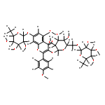
A novel metalloanthocyanin, nemophilin (1), was found in the blue petals of Nemophila menziesii. The major anthocyanin and flavone components were identified as 3-O-(6-O-p-coumaroyl-β-glucopyranosyl)-5-O-(6-O-malonyl-β-glucopyranosyl)petunidin (2, NM) and 7-O-β-glucopyranosyl-4′-O-(6-O-malonyl-β-glucopyranosyl)apigenin (3, MP), respectively. Reconstruction experiments using 2 and 3 with various combinations of metal ions were conducted, and the colors and stabilities of the obtained metalloanthocyanins were measured. NM and MP with magnesium ions yielded purplish-blue-colored metalloanthocyanin (Mg–Mg nemophilin, 1a), and NM and MP with both magnesium and iron ions generated a blue metalloanthocyanin (Fe–Mg nemophilin, 1b). Each pigment exhibited UV–vis and CD spectra typical of metalloanthocyanins; the spectra of the Mg–Mg nemophilin were very similar to those of commelinin from blue dayflower petals, and the spectra of the Fe–Mg nemophilin were similar to those of protocyanin in blue cornflower petals. Nemophilin is the first example of a petunidin glycoside as the anthocyanin component in metalloanthocyanin. In addition, both Mg–Mg nemophilin (1a) and Fe–Mg nemophilin (1b) may coexist in natural petals to develop the petal color.
Co-reporter:Mihoko Mori, Naoko Miki, Daisuke Ito, Tadao Kondo, Kumi Yoshida
Tetrahedron 2014 70(45) pp: 8657-8664
Publication Date(Web):
DOI:10.1016/j.tet.2014.09.046
Co-reporter:Kumi Yoshida, Takashi Negishi
Phytochemistry 2013 Volume 94() pp:60-67
Publication Date(Web):October 2013
DOI:10.1016/j.phytochem.2013.04.017
•Vacuolar iron transporter gene (CcVIT) was identified from blue cornflower petals.•Amino acid sequence of CcVIT gene has a high similarity to AtVIT1 and TgVit1.•In the mutant-line petal with purple phenotype, mutated CcVIT mRNA was expressed.•The A236E replacement of CcVIT protein affect Fe3+ defection to reduce protocyanin content.The blue petal color of the cornflower (Centaurea cyanus) is caused by protocyanin, a kind of metalloanthocyanin, which is a self-assembled supramolecular metal complex pigment. Protocyanin is composed of six molecules of anthocyanin, six molecules of flavone, one ferric ion, and one magnesium ion. The ferric ion is essential for blue color development. Here, we identify the vacuolar iron transporter gene (CcVIT) from the blue petals of C. cyanus and its function is identified and characterized. The CcVIT transcript was observed only in the petals. Its amino acid sequence is highly homologous to the Arabidopsis thaliana (AtVIT1) and Tulipa gesneriana (TgVit1) vacuolar iron transporters. Heterologous expression of the CcVIT gene in yeast indicated that the corresponding gene product transports ferrous ion into vacuoles. Analysis of purple mutant-line petals clarified that the anthocyanin and flavone components were the same as those found in plants with blue petals, but the amount of iron ions in the colored cells decreased, and consequently the amount of blue protocyanin was reduced. The CcVIT gene was expressed even in purple mutant petals, however, an amino acid substitution (A236E) occurred in that case. This change in the CcVIT gene sequence also resulted in loss of iron transport activity. The CcVIT protein thus plays a critical role in the blue coloration of cornflower petals.Vacuolar iron transporter (CcVIT) was critical for blue coloration of cornflower and amino acid replacement (A236E) reduced the activity to give purple color petals.
Co-reporter:Kumi Yoshida, Mihoko Mori and Tadao Kondo
Natural Product Reports 2009 vol. 26(Issue 7) pp:884-915
Publication Date(Web):06 May 2009
DOI:10.1039/B800165K
Covering: 1992 to 2007
Co-reporter:Mihoko Mori, Tadao Kondo, Kumi Yoshida
Phytochemistry 2008 Volume 69(Issue 18) pp:3151-3158
Publication Date(Web):December 2008
DOI:10.1016/j.phytochem.2008.03.015
A metalloanthocyanin, cyanosalvianin, was found in blue petals of Salvia uliginosa. Cyanosalvianin consisted of 3-O-(6-O-p-coumaroylglucopyranosyl)-5-O-(4-O-acetyl-6-O-malonylglucopyranosyl) delphinidin, 7,4′-di-O-glucopyranosylapigenin and magnesium ion. We reproduced the same blue color as the petals by mixing the three components together. An ESI-MS measurement gave a molecular weight of 9014 indicating the composition of cyanosalvianin to be six molecules of the anthocyanin component, six molecules of the flavone component and two magnesium ions. The special arrangement of the organic components in cyanosalvianin was analyzed by CD and 2D-NMR spectroscopy. It was clarified that cyanosalvianin has a similar structure to that of commelinin, a metalloanthocyanin isolated from blue dayflower, Commelina communis.A metalloanthocyanin, cyanosalvianin, was found in the blue petals of Salvia uliginosa. Cyanosalvianin consisted of six molecules of anthocyanin, six molecules of flavone and two atoms of Mg2+ and has a similar structure to that of commelinin.
Co-reporter:Kumi Yoshida, Daisuke Ito, Yosuke Shinkai, Tadao Kondo
Phytochemistry 2008 Volume 69(Issue 18) pp:3159-3165
Publication Date(Web):December 2008
DOI:10.1016/j.phytochem.2008.02.024
The sepal color of a chameleon hydrangea, Hydrangea macrophylla cv. Hovaria™ ‘Homigo’ changes in four stages, from colorless to blue, then to green, and finally to red, during maturation and the senescence periods. To clarify the chemical mechanism of the color change, we analyzed the components of the sepals at each stage. Blue-colored sepals contained 3-O-sambubiosyl- and 3-O-glucosyldelphinidin along with three co-pigments, 5-O-p-coumaroyl-, 5-O-caffeoyl- and 3-O-caffeoylquinic acids. The contents of glycosyldelphinidins decreased toward the green-colored stage, with a coincident increase in the number of chloroplasts. During the last red colored stage, the two species of 3-O-glycosyldelphinidin almost disappeared, and another two anthocyanins, 3-O-sambubiosyl- and 3-O-glucosylcyanidin, increased in amounts. Mixing of 3-O-glycosylcyanidins, co-pigments, and Al3+ in a buffered solution at pH 3.0–3.5 gave not a blue, but a red, colored solution that was the same as that of the sepal color of the 4th stage. Sepals of hydrangea grown in an highland area also turned red in autumn, and contained the same cyanidin glycosides. The red coloration of the hydrangea during senescence was due to a change in anthocyanin biosynthesis.The sepal color change of a chameleon hydrangea, Hydrangea macrophylla cv. Hovaria™ ‘Homigo’ was due to the change of anthocyanin component form delphinidin glycoside to cyanidin glycoside.
Co-reporter:Kumi Yoshida, Sayoko Kitahara, Daisuke Ito, Tadao Kondo
Phytochemistry 2006 Volume 67(Issue 10) pp:992-998
Publication Date(Web):May 2006
DOI:10.1016/j.phytochem.2006.03.013
The Himalayan blue poppy, Meconopsis grandis, has sky blue-colored petals, although the anthocyanidin nucleus of the petal pigment is cyanidin. The blue color development in this blue poppy involving ferric ions was therefore studied. We analyzed the vacuolar pH, and the organic and inorganic components of the colored cells. A direct measurement by a proton-selective microelectrode revealed that the vacuolar pH value was 4.8. The concentrations of the total anthocyanins in the colored cells were around 5 mM, and ca. three times more concentrated flavonols were detected. Fe was detected by atomic analysis of the colored cells, and the ratio of Fe to anthocyanins was ca. 0.8 eq. By mixing the anthocyanin, flavonol and metal ion components in a buffered aq. solution at pH 5.0, we were able to reproduce the same blue color; the visible absorption spectrum and CD were identical to those in the petals, with Fe3+, Mg2+ and flavonol being essential for the blue color. The blue pigment in Meconopsis should be a new type of metal complex pigment that is different from a stoichiometric supramolecular pigment such as commelinin or protocyanin.The underlying reasons for petal color development in the Himalayan blue poppy, Meconopsis grandis, was investigated and established.
Co-reporter:Mihoko Mori, Tadao Kondo, Kenjiro Toki, Kumi Yoshida
Phytochemistry 2006 Volume 67(Issue 6) pp:622-629
Publication Date(Web):March 2006
DOI:10.1016/j.phytochem.2005.12.024
The dicaffeoyl anthocyanin, phacelianin, was isolated from blue petals of Phacelia campanularia. Its structure was determined to be 3-O-(6-O-(4′-O-(6-O-(4′-O-β-d-glucopyranosyl-(E)-caffeoyl)-β-d-glucopyranosyl)-(E)-caffeoyl)-β-d-glucopyranosyl)-5-O-(6-O-malonyl-β-d-glucopyranosyl)delphinidin. The CD of the blue petals of the phacelia showed a strong negative Cotton effect and that of the suspension of the colored protoplasts was the same, indicating that the chromophores of phacelianin may stack intermolecularly in an anti-clockwise stacking manner in the blue-colored vacuoles. In a weakly acidic aqueous solution, phacelianin displayed the same blue color and negative Cotton effect in CD as those of the petals. However, blue-black colored precipitates gradually formed without metal ions. A very small amount of Al3+ or Fe3+ may be required to stabilize the blue solution. Phacelianin may take both an inter- and intramolecular stacking form and shows the blue petal color by molecular association and the co-existence of a small amount of metal ions. We also isolated a major anthocyanin from the blue petals of Evolvulus pilosus and revised the structure identical to phacelianin.The dicaffeoyl anthocyanin, phacelianin, was isolated from the blue petals of Phacelia campanularia and its structure was determined. Phacelianin may take both an inter- and intramolecular stacking form and shows the blue petal color by molecular association and the co-existence of a small amount of metal ions.
Co-reporter:Mihoko Mori, Kumi Yoshida, Yasuhito Ishigaki, Tsukasa Matsunaga, Osamu Nikaido, Kiyoshi Kameda, Tadao Kondo
Bioorganic & Medicinal Chemistry 2005 Volume 13(Issue 6) pp:2015-2020
Publication Date(Web):15 March 2005
DOI:10.1016/j.bmc.2005.01.011
The protective effects of polyacylated anthocyanin, heavenly blue anthocyanin (HBA), in blue flower petals of morning glory (Ipomoea tricolor cv. Heavenly Blue) against UV-B induced DNA damage were examined. We first clarified the concentration of HBA in epidermal vacuoles to be 12 mM, and then constructed a UV-B irradiating apparatus resembling flower petal tissue to assess the screening effect of HBA. Monochromatic (280 and 310 nm) or broad UV-B induced DNA lesions were reduced completely by the HBA filter to the same molecular numbers as those in living petal epidermis. However, diluted HBA solution and trisdeacyl HBA did not have the same reduction effect. HBA was more tolerant to solar radiation than trisdeacyl HBA. These data strongly suggest that polyacylated anthocyanins in flower petals can screen harmful UV-B efficiently. This action might be largely due to aromatic acyl residues.HBA, a polyacylated anthocyanin of blue morning glory completely prevents UV-B induced DNA damage at a physiological concentration (5 mM).
Co-reporter:Kumi Yoshida, Minako Osanai, Tadao Kondo
Phytochemistry 2003 Volume 63(Issue 6) pp:721-726
Publication Date(Web):July 2003
DOI:10.1016/S0031-9422(03)00273-5
The mechanism of dusky reddish-brown “kaki” color development of morning glory, Ipomoea nil cv. Danjuro, was studied. Three major known anthocyanins were isolated as glucosylated pelargonidin derivatives. Measurement of the vacuolar pH with proton-selective microelectrodes revealed the vacuolar pH of the colored cell of open flowers to be 6.8, while that of buds was 5.8. Mixing of the three anthocyanins according to the composition ratio in petals at pH 6.8 allowed the identical color to that of petals to be reproduced. The typical “kaki” color development was mostly caused by 5-OH free acylated anthocyanins, which have two λmax around 435 and 535 nm in the visible region.The mechanism of dusky reddish-brown “kaki” color development of morning glory, Ipomoea nil cv. Danjuro, was clarified by combination of vacuolar pH and color measurements of single cell and reproduction by mixing isolated components.
Co-reporter:Yuki Kimura, Kin-ichi Oyama, Tadao Kondo, Kumi Yoshida
Tetrahedron Letters (8 March 2017) Volume 58(Issue 10) pp:919-922
Publication Date(Web):8 March 2017
DOI:10.1016/j.tetlet.2017.01.065


![2-Propenoic acid, 3-[3,4-bis(methoxymethoxy)phenyl]-, (2E)-](http://img.cochemist.com/ccimg/850200/850177-05-8.png)
![2-Propenoic acid, 3-[3,4-bis(methoxymethoxy)phenyl]-, (2E)-](http://img.cochemist.com/ccimg/850200/850177-05-8_b.png)
![2-PROPENOIC ACID, 3-[4-(METHOXYMETHOXY)PHENYL]-, METHYL ESTER, (2E)-](http://img.cochemist.com/ccimg/84200/84184-55-4.png)
![2-PROPENOIC ACID, 3-[4-(METHOXYMETHOXY)PHENYL]-, METHYL ESTER, (2E)-](http://img.cochemist.com/ccimg/84200/84184-55-4_b.png)
![2-Propenoic acid, 3-[4-(methoxymethoxy)phenyl]-, (2E)-](http://img.cochemist.com/ccimg/61900/61844-66-4.png)
![2-Propenoic acid, 3-[4-(methoxymethoxy)phenyl]-, (2E)-](http://img.cochemist.com/ccimg/61900/61844-66-4_b.png)