Co-reporter:Xiufang Zhang, Lu Chen, Weiyun Chai, Xiao-Yuan Lian, Zhizhen Zhang
Phytochemistry 2017 Volume 144(Volume 144) pp:
Publication Date(Web):1 December 2017
DOI:10.1016/j.phytochem.2017.09.010
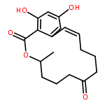
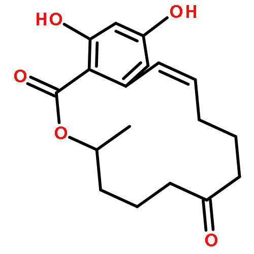
•Streptopertusacin A and bioactive bafilomycins were isolated from Streptomyces sp.•Streptopertusacin A is a unique indolizinium alkaloid existing as a zwitterion.•Bafilomycins showed potent activity against the proliferation of glioma cells.•Bafilomycins had activity against methicillin-resistant Staphylococcus aureus.Streptopertusacin A, a unique indolizinium alkaloid existing as a zwitterion, and six bafilomycins including two previously undescribed ones of 21,22-en-bafilomycin D and 21,22-en-9-hydroxybafilomycin D were isolated from a culture of the seaweed-derived Streptomyces sp. HZP-2216E. Structures of these isolated compounds were determined based on extensive NMR spectroscopic analyses, HRESIMS and MS-MS data. The stereochemical assignments were achieved by NOE information, chemical degradation, Marfey's method, and electronic circular dichroism (ECD) calculation. Streptopertusacin A is the first example of this type of indolizinium alkaloid from microorganisms and showed moderate activity against the growth of methicillin-resistant Staphylococcus aureus (MRSA). 21,22-en-bafilomycin D and 21,22-en-9-hydroxybafilomycin D had potent activities in inhibiting the proliferation of glioma cells and the growth of MRSA.A unique indolizinium alkaloid streptopertusacin A (1) existing as a zwitterion and two previously undescribed bafilomycin analogues of 21,22-en-bafilomycin D (2) and 21,22-en-9-hydroxybafilomycin D (3) were isolated from a culture of seaweed-derived Streptomyces sp. HZP-2216E. Bafilomycins 2 and 3 had potent activities in inhibiting the proliferation of glioma cells and the growth of methicillin-resistant Staphylococcus aureus.Download high-res image (519KB)Download full-size image
Co-reporter:Lu CHEN, Wen-Ling WANG, Teng-Fei SONG, Xin XIE, ... Zhi-Zhen ZHANG
Chinese Journal of Natural Medicines 2017 Volume 15, Issue 8(Volume 15, Issue 8) pp:
Publication Date(Web):1 August 2017
DOI:10.1016/S1875-5364(17)30085-7
Tripolinolate A (TLA) is recently identified as a new compound from a halophyte plant Tripolium vulgare and has been shown to have significant in vitro activity against the proliferation of colorectal cancer and glioma cells. This study was designed to further investigate the effects of TLA on the proliferation of human normal cells, and the apoptosis and cell cycle in colorectal cancer cells, and the growth of tumors in the colorectal cancer-bearing animals. The data obtained from this study demonstrated that: 1) TLA had much less cytotoxicity in the human normal cells than the colorectal cancer cells; 2) TLA remarkably induced apoptosis in the human colorectal cancer cells and blocked cell cycle at G2/M phase, and 3) TLA had significant anti-colorectal cancer activity in the tumor-bearing animals.
Co-reporter:Lu Chen, Ying Liang, Tengfei Song, Komal Anjum, Wenling Wang, Siran Yu, Haocai Huang, Xiao-Yuan Lian, Zhizhen Zhang
Bioorganic & Medicinal Chemistry Letters 2015 Volume 25(Issue 13) pp:2629-2633
Publication Date(Web):1 July 2015
DOI:10.1016/j.bmcl.2015.04.091
A new coniferol derivative, named as tripolinolate A (1), and 11 known compounds (2–12) were isolated from whole plants of Tripolium vulgare Nees. The structure of this new compound was determined as 4-(2S-methylbutyryl)-9-acetyl-coniferol based on its NMR and HRESIMS spectral analyses. A simple and efficient method was designed to prepare tripolinolate A and its 19 analogs including nine new chemical entities for bioactive assay. Tripolinolate A and its analog 4,9-diacetyl-coniferol were found to be the two most active compounds that significantly inhibited the proliferation of different cancer cell lines with IC50 values ranging from 0.36 to 12.9 μM and induced apoptosis in tumor cells. Structure–activity relationship analysis suggested that the molecular size of acyl moieties at C-4 and C-9 position might have an effect on the activity of this type of coniferol derivatives.
Co-reporter:Xuewei Ye, Siran Yu, Xiao-Yuan Lian, Zhizhen Zhang
Journal of Pharmaceutical and Biomedical Analysis 2014 Volume 88() pp:472-476
Publication Date(Web):25 January 2014
DOI:10.1016/j.jpba.2013.09.017
•A HPLC analytic method for the analysis of glycosides in Fatsia japonica was developed and validated.•The established HPLC method has been applied for the qualification of 11 glycosides in the different parts of F. japonica.•This HPLC method could be used for quantitive analysis of this herb and its products.Fatsia japonica Decne. & Planch. is a triterpenoid glycoside-rich herb with anti-inflammatory activity for the treatment of rheumatoid arthritis. A method for quantitative analysis of the complex triterpenoid glycosides in this medicinal plant has not been established so far. In this study, a high performance liquid chromatography (HPLC) method was developed for simultaneous qualification of 11 glycosides in F. japonica. The analysis was performed on an ODS-2 Hypersil column (250 mm × 4.6 mm, 5 μm) with a binary gradient mobile phase of water and acetonitrile. The established HPLC method was validated in terms of linearity, sensitivity, stability, precision, accuracy, and recovery. Results showed that this method had good linearity with R2 at 0.99992–0.99999 in the test range of 0.04–9.00 μg/μL. The limit of detection (LOD) and limit of quantification (LOQ) for the standard compounds were 0.013–0.020 μg/μL and 0.040–0.060 μg/μL. The relative standard deviations (RSDs%) of run variations were 0.83–1.40% for intra-day and 0.84–3.59% for inter-day. The analyzed compounds in the samples were stable for at least 36 h, and the spike recoveries of the detected glycosides were 99.67–103.11%. The developed HPLC method was successfully applied for the measurements of the contents of 11 triterpenoid glycoside in different parts of F. japonica. Taken together, the HPLC method newly developed in this study could be used for qualitative and quantitative analysis of the bioactive triterpenoid glycosides in F. japonica and its products.A HPLC analytic method for the qualification of the bioactive glycosides in Fatsia japonica was developed and validated.
Co-reporter:Xuewei Ye, Siran Yu, Ying Liang, Haocai Huang, Xiao-Yuan Lian, Zhizhen Zhang
Bioorganic & Medicinal Chemistry Letters 2014 Volume 24(Issue 22) pp:5157-5163
Publication Date(Web):15 November 2014
DOI:10.1016/j.bmcl.2014.09.087
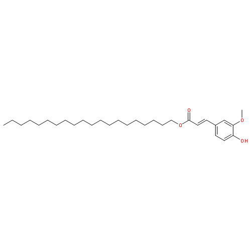
A total of 54 natural origin compounds were evaluated for their activity in inhibiting the proliferation of glioma cells. Results showed that four Aesculus polyhydroxylated triterpenoid saponins (3–6), six Gleditsia triterpenoid saponins (7–12), and five phenolic compounds (43–46, 51) had dose-dependent activity suppressing the proliferation of both C6 and U251 cells. Structure–activity relationship analysis suggested that the acetyl group at C-28 for the Aesculus saponins and the monoterpenic acid moiety for the Gleditsia saponins could be critical for the activity of these active compounds. Aesculioside H (4), gleditsioside A (7), and feuric acid 3,4-dihydroxyphenethyl ester (FADPE, 46) were the three most active compounds from the different types of the active compounds and induced apoptosis and necrosis in glioma cells.
Co-reporter:Xiao-Yuan Lian, Zhizhen Zhang
Journal of Pharmaceutical and Biomedical Analysis 2013 Volume 75() pp:41-46
Publication Date(Web):5 March 2013
DOI:10.1016/j.jpba.2012.11.007
A high performance liquid chromatography method was developed for simultaneous quantification of 29 ingredients occurring as a very complex mixture in the gleditsia fruits. The analysis was performed on an ODS-2 Hypersil column (250 mm × 4.6 mm, 5 μm) with a binary gradient mobile phase of water and acetonitrile both containing 0.1% acetic acid. The method was validated in terms of linearity, sensitivity, stability, precision, and accuracy. It was found that this method had linearity with R2 at 0.99889–0.99997 in the test range of 1.0–24.0 μg. The limit of detection (LOD) and limit of quantification (LOQ) for 16 tested reference saponins were 0.24–0.39 μg and 1.0–1.2 μg, respectively. The relative standard deviations (RSDs %) for intra-day and inter-day repeatability were not more than 3.11% and 4.02%, respectively. The analyzed samples were stable for at least 48 h. The spike recoveries for eight analyzed saponins were 99.68–102.17%. The established HPLC analytic method was successfully used to determine the concentrations of 29 compounds including 19 gleditsia saponins and ten unidentified ingredients in eight commercial gleditsia fruits from different sources. The results from this study suggested that this newly developed HPLC method could be used for qualitative and quantitative analysis of the saponins in the gleditsia fruits and the gleditsia extracts that used for animal study and other purpose.Highlights► A HPLC method was developed for analyzing the ingredients of gleditsia fruits. ► A total of 29 ingredients in gleditsia fruits were simultaneously determined. ► The established method could be used for quantitive analysis of gleditsia saponins.
Co-reporter:Siran Yu, Xuewei Ye, Lu Chen, Xiao-Yuan Lian, Zhizhen Zhang
Steroids (October 2014) Volume 88() pp:19-25
Publication Date(Web):1 October 2014
DOI:10.1016/j.steroids.2014.06.013
•Six rare polyoxygenated 24,28-epoxyergosterols were isolated from Anthopleura midori.•The structures of these epoxyergosterols were determined by NMR and HRESIMS analyses.•These epoxyergosterols showed activity inhibiting the proliferation of glioma cells.Eleven sterols (1–11) and one carotenoid (12) were isolated and identified from sea anemone Anthopleura midori. Compounds 1–6 are rare polyoxygenated ergosterols with a 24,28-epoxy moiety. The structures of these epoxyergosterols were determined by NMR and HRESIMS analyses as well as their chemical-physical properties. Epoxyergosterols 1 and 2 were found to be new natural products and 3–6 are new compounds. Bioactive assay showed that compounds 1, 2, 3, 5, 7, 8, 11, and 12 inhibited the proliferation of rat glioma C6 and human glioma U251 cells with IC50 in a range of 2.41–80.45 μM. Further investigation suggested that 1 and 3 induced apoptosis in glioma cells and 1 blocked U251 cells at the G0/G1 phase.Graphical abstractDownload full-size image
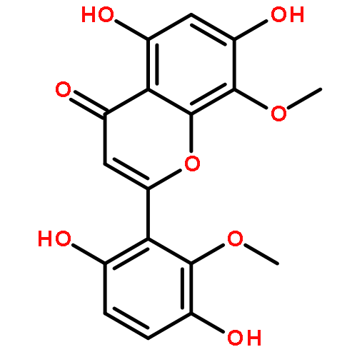
Co-reporter:Xuewei Ye, Komal Anjum, Tengfei Song, Wenling Wang, Ying Liang, Mengxuan Chen, Haocai Huang, Xiao-Yuan Lian, Zhizhen Zhang
Phytochemistry (March 2017) Volume 135() pp:151-159
Publication Date(Web):March 2017
DOI:10.1016/j.phytochem.2016.12.010


![Actinomycin D,3A-[(4R)-4-hydroxy-L-proline]- (9CI)](http://img.cochemist.com/ccimg/18900/18865-46-8.png)
![Actinomycin D,3A-[(4R)-4-hydroxy-L-proline]- (9CI)](http://img.cochemist.com/ccimg/18900/18865-46-8_b.png)


![(3S-trans)-hexahydro-3-isobutylpyrrolo[1,2-a]pyrazine-1,4-dione](http://img.cochemist.com/ccimg/2900/2873-36-1.png)
![(3S-trans)-hexahydro-3-isobutylpyrrolo[1,2-a]pyrazine-1,4-dione](http://img.cochemist.com/ccimg/2900/2873-36-1_b.png)


![N-[2-(1H-INDOL-3-YL)ETHYL]ACETAMIDE](http://img.cochemist.com/ccimg/1100/1016-47-3.png)
![N-[2-(1H-INDOL-3-YL)ETHYL]ACETAMIDE](http://img.cochemist.com/ccimg/1100/1016-47-3_b.png)
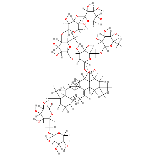
![(20S)-3-O-{beta-D-6-O-[(E)-but-2-enoyl]glucopyranosyl-(1->2)-beta-D-glucopyranosyl}-20-O-[beta-D-glucopyranosyl-(1->6)-beta-D-glucopyranosyl]protopanaxadiol](http://img.cochemist.com/ccimg/1346600/1346522-89-1.png)
![(20S)-3-O-{beta-D-6-O-[(E)-but-2-enoyl]glucopyranosyl-(1->2)-beta-D-glucopyranosyl}-20-O-[beta-D-glucopyranosyl-(1->6)-beta-D-glucopyranosyl]protopanaxadiol](http://img.cochemist.com/ccimg/1346600/1346522-89-1_b.png)


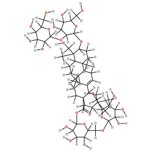
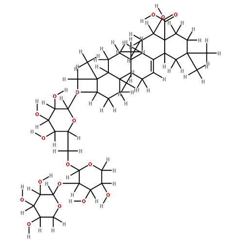
![â-D-Glucopyranosiduronic acid,(3â,4â,16R,21â,22R)-28-(acetyloxy)-16,- 22,23-trihydroxy-21-[[(2Z)-2-methyl-1-oxo- 2-butenyl]oxy]olean-12-en-3-yl O-â-D-glucopyranosyl-(1f2)-O-[â-Dglucopyranosyl-( 1f4)]-](http://img.cochemist.com/ccimg/220000/219944-46-4.png)
![â-D-Glucopyranosiduronic acid,(3â,4â,16R,21â,22R)-28-(acetyloxy)-16,- 22,23-trihydroxy-21-[[(2Z)-2-methyl-1-oxo- 2-butenyl]oxy]olean-12-en-3-yl O-â-D-glucopyranosyl-(1f2)-O-[â-Dglucopyranosyl-( 1f4)]-](http://img.cochemist.com/ccimg/220000/219944-46-4_b.png)


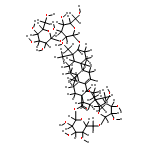


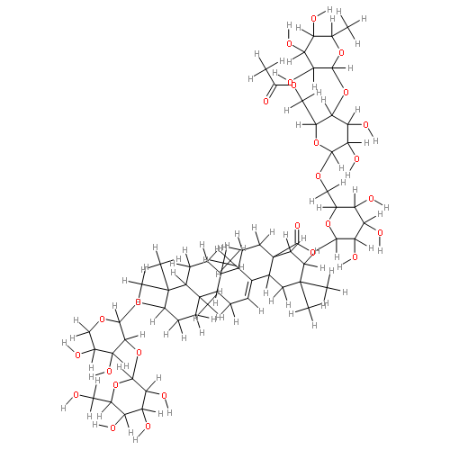
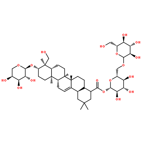
![Olean-12-en-28-oicacid, 3-[(2-O-b-D-glucopyranosyl-a-L-arabinopyranosyl)oxy]-23-hydroxy-,O-6-deoxy-a-L-mannopyranosyl-(1®4)-O-b-D-glucopyranosyl-(1®6)-b-D-glucopyranosyl ester, (3b,4a)-](http://img.cochemist.com/ccimg/60500/60454-69-5.png)
![Olean-12-en-28-oicacid, 3-[(2-O-b-D-glucopyranosyl-a-L-arabinopyranosyl)oxy]-23-hydroxy-,O-6-deoxy-a-L-mannopyranosyl-(1®4)-O-b-D-glucopyranosyl-(1®6)-b-D-glucopyranosyl ester, (3b,4a)-](http://img.cochemist.com/ccimg/60500/60454-69-5_b.png)


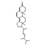
![(3S,5R,6R,8S,9S,10R,13R,14S,17R)-5,6-dihydroxy-10,13-dimethyl-17-((R)-6-methylheptan-2-yl)hexadecahydro-1H-cyclopenta[a]phenanthren-3-yl acetate](http://img.cochemist.com/ccimg/35100/35089-26-0.png)
![(3S,5R,6R,8S,9S,10R,13R,14S,17R)-5,6-dihydroxy-10,13-dimethyl-17-((R)-6-methylheptan-2-yl)hexadecahydro-1H-cyclopenta[a]phenanthren-3-yl acetate](http://img.cochemist.com/ccimg/35100/35089-26-0_b.png)


![2,2'-[(2,4-DIMETHYL-3-PENTANYL)IMINO]DIETHANOL](http://img.cochemist.com/ccimg/32900/32811-40-8.png)
![2,2'-[(2,4-DIMETHYL-3-PENTANYL)IMINO]DIETHANOL](http://img.cochemist.com/ccimg/32900/32811-40-8_b.png)


![5,7-dihydroxy-2-(4-hydroxyphenyl)-3-[(2s,3r,4s,5s,6r)-3,4,5-trihydroxy-6-(hydroxymethyl)oxan-2-yl]oxychromen-4-one](http://img.cochemist.com/ccimg/500/480-10-4.png)
![5,7-dihydroxy-2-(4-hydroxyphenyl)-3-[(2s,3r,4s,5s,6r)-3,4,5-trihydroxy-6-(hydroxymethyl)oxan-2-yl]oxychromen-4-one](http://img.cochemist.com/ccimg/500/480-10-4_b.png)


