Co-reporter:Heajin Lee, Ralf Landgraf, James N. Wilson
Bioorganic & Medicinal Chemistry 2017 Volume 25, Issue 21(Issue 21) pp:
Publication Date(Web):1 November 2017
DOI:10.1016/j.bmc.2017.09.034
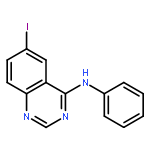
A fluorescent probe targeting the ERBB2 receptor tyrosine was designed, synthesized and evaluated as reporter of ERBB2 dynamics in overexpressing BT474, i.e. Her2(+), cells. Two cyanoquinazoline (CQ) probes modeled after type-I (CQ1) or active state and type-II (CQ2) or inactive state inhibitors were designed and synthesized with extended π systems that impart binding-induced, turn-on fluorescence. Solution spectroscopy revealed that CQ1 exhibited attractive photophysical properties and displayed turn-on emission in the presence of purified, soluble ERBB2 kinase domain, while CQ2 was found to be non-emissive, likely due to quenching via a photoinduced electron transfer mechanism. Live cell imaging with CQ1 revealed that this probe targeted an intracellular population of ERBB2, which increased following treatment with type-I inhibitors, gefinitib and canertinib, but showed no response to type-II inhibitors. CQ1 thus provides a novel means of imaging the dynamic response of ERBB2(+) cells to kinase inhibitors.Download high-res image (134KB)Download full-size image
Co-reporter:Anthony N. Cauley;James N. Wilson
Biomaterials Science (2013-Present) 2017 vol. 5(Issue 10) pp:2114-2121
Publication Date(Web):2017/09/26
DOI:10.1039/C7BM00518K
We report a library of functionalized lignins and demonstrate their utility as nanocontainers for organic dyes in biologically relevant applications. Kraft lignin was modified via SN2 reaction at the phenolic –OH group utilizing a mild base, potassium carbonate, and various alkyl halides, several bearing additional functionalities, with dimethylsulfoxide as solvent. The resulting phenoxy ethers were characterized by 1H-NMR and IR spectroscopy, as well as DLS and SEM to evaluate their morphology and supramolecular organization. Lignin modified with long-chain hydrocarbon tails was found to effectively encapsulate DiD, a cyanine dye, decrease aggregation, enhance optical transitions and exert a photoprotective effect. The dye–lignin assemblies were also examined as imaging agents, via confocal microscopy, and found to accumulate intracellularly with no leaching of the dye to hydrophobic subcellular components observed. Lignin functionalized with short chain carboxylic acids interacts with ligands directed at the norepinephrine transporter (NET), suggesting applications in sequestration of neuroactive compounds.
Co-reporter:Sicheng Tang, Yang Zhang, Ek Raj Thapaliya, Adrienne S. Brown, James N. WilsonFrançisco M. Raymo
ACS Sensors - New in 2016 2017 Volume 2(Issue 1) pp:
Publication Date(Web):December 21, 2016
DOI:10.1021/acssensors.6b00592
Halochromic coumarin–oxazine prefluorophores and targeting folate ligands can be connected covalently to the side chains of amphiphilic polymers. The resulting macromolecular constructs assemble into nanoparticles in aqueous environments. The prefluorophores do not produce any detectable fluorescence at neutral pH, but are converted into fluorophores with intense visible emission at acidic pH. Protonation opens the oxazine heterocycle to shift bathochromically the coumarin absorption and activate fluorescence with a brightness per nanoparticle approaching 5 × 10⁵ M–1 cm–1. This value translates into a 170-fold enhancement relative to the isolated fluorophores dissolved in organic solvent. The folate ligands direct these multicomponent constructs into acidic intracellular compartments of folate-positive cells, where the prefluorophores switch to the corresponding fluorophores and produce fluorescence. The pH-induced activation of the signaling units ensures negligible background fluorescence from the extracellular matrix, which instead limits considerably the contrast accessible with model systems incorporating conventional nonactivatable fluorophores. Furthermore, no intracellular fluorescence can be detected when the very same measurements are performed with folate-negative cells. Nonetheless, control experiments demonstrate that the covalent connection of the prefluorophores to the polymer backbone of the amphiphilic constructs is essential to ensure selectivity. Model systems with prefluorophores noncovalently encapsulated cannot discriminate folate-positive from -negative cells. Thus, our structural design for the covalent integration of activatable signaling units and targeting ligands within the same nanostructured assembly together with the photophysical properties engineered into the emissive components offer the opportunity to highlight cancer cells selectively with high brightness and optimal contrast.Keywords: activatable fluorophores; amphiphilic polymers; cancer detection; folate targeting; halochromism; intracellular pH sensors; molecular switches; supramolecular nanocarriers;
Co-reporter:Heajin Lee, Wenjun Liu, Adrienne S. Brown, Ralf Landgraf, and James N. Wilson
Analytical Chemistry 2016 Volume 88(Issue 23) pp:
Publication Date(Web):November 18, 2016
DOI:10.1021/acs.analchem.6b03836
The human epidermal growth factor receptor, EGFR/ERBB/HER, family of receptor tyrosine kinases is central to many signaling pathways and a validated chemotherapy target in multiple cancers. While EGFR/ERBB-targeted therapies, including monoclonal antibodies, e.g., trastuzumab, and small molecule kinase inhibitors, such as lapatinib, have been developed, rapid identification and classification of cancer cells is key to identifying the best treatment regime. We report ERBB2 (also HER2) targeting kinase probes that exhibit a “turn-on” emission response upon binding. These live cell compatible probes differentiate ERBB2(+) cells from low-level, ERBB2(−) cells by targeting the intracellular ATP-binding pocket of ERBB2 with therapeutic inhibitor-like specificity. Beyond kinase expression levels, probe signal is linked to the phosphotyrosine-correlated activation state of the ERBB2 population. Additionally, the rapid signaling capability of the probes can report changes in activation state in live cells providing a unique type of complementary information to immunohistochemical assays of receptor kinase populations.
Co-reporter:James N. Wilson, Wenjun Liu, Adrienne S. Brown and Ralf Landgraf
Organic & Biomolecular Chemistry 2015 vol. 13(Issue 17) pp:5006-5011
Publication Date(Web):20 Mar 2015
DOI:10.1039/C5OB00239G
We report the photophysical properties, binding-induced turn-on emission, and fluorescence imaging of the cellular uptake and distribution of lapatinib, an EGFR/ERBB inhibitor. Lapatinib, a type II, i.e. inactive state, inhibitor that targets the ATP binding pocket of the EGFR family of receptor tyrosine kinases. DFT calculations predict that the 6-furanylquinazoline core of lapatinib should exhibit an excited state with charge transfer character and an S0 to S1 transition energy of 3.4 eV. Absorption confirms an optical transition in the near UV to violet, while fluorescence spectroscopy shows that photoemission is highly sensitive to solvent polarity. The hydrophobicity of lapatinib leads to fluorescent aggregates in solution, however, binding to the lipid-carrier protein, BSA or to the kinase domain of ERBB2, produces spectroscopically distinct photoemission. Confocal fluorescence microscopy imaging of lapatinib uptake in ERBB2-overexpressing MCF7 and BT474 cells reveals pools of intracellular inhibitor with emission profiles consistent with aggregated lapatinib.
Co-reporter:Demar R. G. Pitter, Adrienne S. Brown, James D. Baker and James N. Wilson
Organic & Biomolecular Chemistry 2015 vol. 13(Issue 36) pp:9477-9484
Publication Date(Web):04 Aug 2015
DOI:10.1039/C5OB01428J
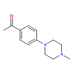
Several new DNA-targeting probes that exhibit binding-induced ‘turn on’ fluorescence are presented. Two of the dyes, orange emissive 1, (E)-4-(4(-4-methylpiperazin-1-yl)phenyl)6-(4-(4-methylpi-perazin-1-yl)styryl)pyrimidin-2-ol), and red emissive 2, (E)-4-(4(-4-methyl-piperazin-1-yl)-phenyl)6-(4-(4-methylpiperazin-1-yl)styryl)-1,3-propanedionato-κO,κO′]difluoroborane), are brightly fluorescent when bound to DNA, but are virtually non-fluorescent in aqueous solutions. Confocal fluorescence microscopy of live BT474, MCF7 and HEK293 cells demonstrates that both probes are cell permeable and rapidly accumulated intracellularly into cell nuclei and the cytosol. Taking advantage of their environmental sensitivity, these two pools of fluorophores are readily resolved into separate channels, and thus, a single dye allows two-color imaging of the nuclear and cytosolic compartments.
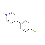
Co-reporter:James N. Wilson, Lucy Kate Ladefoged, W. Michael Babinchak, and Birgit Schiøtt
ACS Chemical Neuroscience 2014 Volume 5(Issue 4) pp:296
Publication Date(Web):January 24, 2014
DOI:10.1021/cn400230x
The binding-induced fluorescence of 4-(4-(dimethylamino)-phenyl)-1-methylpyridinium (APP+) and two new serotonin transporter (SERT)-binding fluorescent analogues, 1-butyl-4-[4-(1-dimethylamino)phenyl]-pyridinium bromide (BPP+) and 1-methyl-4-[4-(1-piperidinyl)phenyl]-pyridinium (PPP+), has been investigated. Optical spectroscopy reveals that these probes are highly sensitive to their chemical microenvironment, responding to variations in polarity with changes in transition energies and responding to changes in viscosity or rotational freedom with emission enhancements. Molecular docking calculations reveal that the probes are able to access the nonpolar and conformationally restrictive binding pocket of SERT. As a result, the probes exhibit previously not identified binding-induced turn-on emission that is spectroscopically distinct from dyes that have accumulated intracellularly. Thus, binding and transport dynamics of SERT ligands can be resolved both spatially and spectroscopically.Keywords: fluorescence; induced fit docking; LeuT; quantum polarized ligand docking; Serotonin; SERT
Co-reporter:Jyothi Dhuguru, Wenjun Liu, Walter G. Gonzalez, W. Michael Babinchak, Jaroslava Miksovska, Ralf Landgraf, and James N. Wilson
The Journal of Organic Chemistry 2014 Volume 79(Issue 11) pp:4940-4947
Publication Date(Web):May 2, 2014
DOI:10.1021/jo500520x
Fluorescent N-phenyl-4-aminoquinazoline probes targeting the ATP-binding pocket of the ERBB family of receptor tyrosine kinases are reported. Extension of the aromatic quinazoline core with fluorophore “arms” through substitution at the 6- position of the quinazoline core with phenyl, styryl, and phenylbutadienyl moieties was predicted by means of TD-DFT calculations to produce probes with tunable photoexcitation energies and excited states possessing charge-transfer character. Optical spectroscopy identified several synthesized probes that are nonemissive in aqueous solutions and exhibit emission enhancements in solvents of low polarity, suggesting good performance as turn-on fluorophores. Ligand-induced ERBB2 phosphorylation assays demonstrate that despite chemical modification to the quinazoline core these probes still function as ERBB2 inhibitors in MCF7 cells. Two probes were found to exhibit ERBB2-induced fluorescence, demonstrating the utility of these probes as turn-on, fluoroescent kinase inhibitors.
Co-reporter:Demar R. G. Pitter, Jens Wigenius, Adrienne S. Brown, James D. Baker, Fredrik Westerlund, and James N. Wilson
Organic Letters 2013 Volume 15(Issue 6) pp:1330-1333
Publication Date(Web):March 5, 2013
DOI:10.1021/ol400268t
DNA-binding, green and yellow fluorescent probes with excellent brightness and high on/off ratios are reported. The probes are membrane permeable, live-cell compatible, and optimally matched to 405 nm and 514 nm laser lines, making them attractive alternatives to UV-excited and blue emissive Hoechst 33342 and DAPI nuclear stains. Their electronic structure was investigated by optical spectroscopy supported by TD-DFT calculations. DNA binding is accompanied by 27- to 75-fold emission enhancements, and linear dichroism demonstrates that one dye is a groove binder while the other intercalates into DNA.
Co-reporter:James N. Wilson, Jens Wigenius, Demar R. G. Pitter, Yanhua Qiu, Maria Abrahamsson, and Fredrik Westerlund
The Journal of Physical Chemistry B 2013 Volume 117(Issue 40) pp:12000-12006
Publication Date(Web):September 17, 2013
DOI:10.1021/jp406993m
The photophysical properties of two recently reported live cell compatible, DNA-binding dyes, 4,6-bis(4-(4-methylpiperazin-1-yl)phenyl)pyrimidin-2-ol, 1, and [1,3-bis[4-(4-methylpiperazin-1-yl)phenyl]-1,3-propandioato-κO, κO′]difluoroboron, 2, are characterized. Both dyes are quenched in aqueous solutions, while binding to sequences containing only AT pairs enhances the emission. Binding of the dyes to sequences containing only GC pairs does not produce a significant emission enhancement, and for sequences containing both AT and GC base pairs, emission is dependent on the length of the AT pair tracts. Through emission lifetime measurements and analysis of the dye redox potentials, photoinduced electron transfer with GC pairs is implicated as a quenching mechanism. Binding of the dyes to AT-rich regions is accompanied by bathochromic shifts of 26 and 30 nm, respectively. Excitation at longer wavelengths thus increases the ON/OFF ratio of the bound probes significantly and provides improved contrast ratios in solution as well as in fluorescence microscopy of living cells.
Co-reporter:James N. Wilson, Adrienne S. Brown, W. Michael Babinchak, Clark D. Ridge and Jamie D. Walls
Organic & Biomolecular Chemistry 2012 vol. 10(Issue 43) pp:8710-8719
Publication Date(Web):14 Sep 2012
DOI:10.1039/C2OB26633D
We report the synthesis, binding kinetics, optical spectroscopy and predicted binding modes of a series of sterically demanding, fluorescent norepinephrine transporter (NET) ligands. A series of bulky stilbazolium dyes, including six newly synthesized compounds, were evaluated to determine the effect of extending the molecular probes’ ‘heads’ or ‘tails’. Taking advantage of the dyes’ characteristic ‘turn-on’ emission, the kinetic binding parameters, kon and koff were determined revealing that extension of the molecules’ tails is well tolerated while expansion of the head is not. Additionally, a ‘headfirst’ orientation appears to be preferred over a ‘tail-first’ binding pose. Further details of the possible binding modes were obtained from the emission spectra of the bound probes. A small range of interplanar twist angles, approximately 35° to 60°, is predicted to produce the observed emission. Docking experiments and molecular modelling support the kinetic and spectroscopic data providing structural insights into substrate binding.
Co-reporter:Erika L. Smith, Adrienne S. Brown, Edward Adjaye-Mensah and James N. Wilson
Organic & Biomolecular Chemistry 2012 vol. 10(Issue 8) pp:1493-1496
Publication Date(Web):30 Nov 2011
DOI:10.1039/C2OB06796J
A series of stilbazolium dimers were synthesized and investigated as sterically demanding ligands targeting the norepinephrine transporter (NET). The environmentally sensitive fluorescence of the dyes enables their use as self-reporting ligands; binding to and displacement from NET can be monitored by fluorescence microscopy.
Co-reporter:Renaud Sicard, Jyothi Dhuguru, Wenjun Liu, Nirav Patel, Ralf Landgraf, James N. Wilson
Bioorganic & Medicinal Chemistry Letters 2012 Volume 22(Issue 17) pp:5532-5535
Publication Date(Web):1 September 2012
DOI:10.1016/j.bmcl.2012.07.034
ERBB receptor kinases play a crucial role in normal development and cancer malignancies. A broad range of modifications creates receptor subpopulations with distinct functional properties in live cells. Their apparent activation state, typically assayed by tyrosine phosphorylation of substrates, reflects a complex equilibrium of competing reactions. With the aim of developing optical tools to investigate ERBB populations and their state of activation, we have synthesized a fluorescent ‘turn-on’ probe, DMAQ, targeting the ERBB ATP binding pocket. Upon binding, probe emission increases due to the hydrophobic environment and restricted geometry of the ERBB2 kinase domain, facilitating the analysis of receptor states at low occupancy and without the removal of unbound probes. Cellular ERBB2 autophosphorylation is inhibited with saturation kinetics that correlate with the increase in probe fluorescence. Thus, DMAQ is an example of a new generation of ‘turn-on’ probes with potential applications in querying receptor kinase populations both in vitro and in live cells.
Co-reporter:Edward Adjaye-Mensah, Walter G. Gonzalez, David R. Bussé, Burjor Captain, Jaroslava Miksovska, and James N. Wilson
The Journal of Physical Chemistry A 2012 Volume 116(Issue 34) pp:8671-8677
Publication Date(Web):August 2, 2012
DOI:10.1021/jp3036956
The photophysics of 1-ethyl-4,6-bis(4-methoxyphenyl)-2(1H)-pyrimidone (1) and 1-ethyl-4,6-bis(4-(dimethylamino)phenyl)-2(1H)-pyrimidone (2) were investigated to determine the mechanisms of emission switching in response to protonation. UV–vis and steady state emission spectroscopy of the protonated and unprotonated forms across a range of solvents reveal the polarity dependence of the vertical excitation energies. Emission lifetimes and quantum yields show the solvent dependency of the excited states. Emission enhancements were observed in polyethylene glycol solutions and in the solid state (both thin film and single crystal), demonstrating the role of intramolecular rotation in thermal relaxation of the excited states. TD-DFT calculations provide insights into the excited state geometries and the role of intramolecular charge transfer. The collected data show that emission of diphenylpyrimidones can be modulated by four factors, including the identity of the electron-donating auxochrome, protonation state, solvent polarity, and viscosity.
Co-reporter:Edward Adjaye-Mensah, Walter G. Gonzalez, Jaroslava Miksovska, and James N. Wilson
The Journal of Physical Chemistry A 2012 Volume 116(Issue 51) pp:12470-12475
Publication Date(Web):December 6, 2012
DOI:10.1021/jp3106869
The photophysical properties of 4-[2-(6-hydroxy-2-naphthalenyl)-ethenyl]-1-methyl-pyridinium (HNEP+) and its deprotonated form (NEP), a benzofused derivative of Brooker’s merocyanine (BM), were investigated through a combined spectroscopic and computational approach. Despite their structural similarities and similar pKa values, HNEP+/NEP and BMH+/BM differ in the extent of charge delocalization in the ground and excited states. NEP exhibits the spectral characteristics of a charge transfer species in solvents in which BM exists in a charge-delocalized quinoid; however, quantum chemical calculations show that the CT absorption of NEP is not necessarily a consequence of the zwitterionic character. HNEP+ displays larger Stokes shifts than BMH+, and NEP demonstrates enhanced solvatochromism relative to BM as a consequence of benzofusion.
Co-reporter:Adrienne S. Brown, Lisa-Marie Bernal, Teresa L. Micotto, Erika L. Smith and James N. Wilson
Organic & Biomolecular Chemistry 2011 vol. 9(Issue 7) pp:2142-2148
Publication Date(Web):15 Dec 2010
DOI:10.1039/C0OB00849D
A set of spectrally diverse stilbazolium dyes was identified in an uptake assay using cultured brainstem and cerebellum cells isolated from e19 chicks. Pretreatment of cells with indatraline, a monoamine reuptake inhibitor, allowed identification of dyes that may interact with monoamine transporters. Two structurally related, yet spectrally segregated, probes, (E)-1-methyl-4-[2-(2-naphthalenyl)ethenyl]-pyridinium iodide (NEP+, 3A) and (E)-4-[2-(6-hydroxy-2-naphthalenyl)ethenyl]-1-methyl-pyridinium iodide (HNEP+, 4A), were selected and further investigated using HEK-293 cells selectively expressing dopamine, norepinephrine or serotonin transporters. HNEP+ was selectively accumulated viacatecholamine transporters, with the norepinephrine transporter (NET) giving the highest response; NEP+ was not transported, though possible binding was observed. The alternate modes of interaction enable the use of NEP+ and HNEP+ to image distinct cell populations in live brain tissue explants. The preference for HNEP+ accumulation viaNET was confirmed by imaging uptake in the absence and presence of desipramine, a norepinephrine reuptake inhibitor.
Co-reporter:Sharanappa M. Bagale, Adrienne S. Brown, Melissa M. Carballosa Gonzalez, Alberto Vitores, Teresa L. Micotto, N.S. Saleesh Kumar, Ian D. Hentall, James N. Wilson
Bioorganic & Medicinal Chemistry Letters 2011 Volume 21(Issue 24) pp:7387-7391
Publication Date(Web):15 December 2011
DOI:10.1016/j.bmcl.2011.10.007
Serotonin is a monoamine serving as a chemical messenger in diverse brain regions, as well as in blood and various other organs. We synthesized six ethylamine functionalized fluorophores as fluorescent probes for serotonin. The one with best spectral properties and aqueous solubility, 6-amino-2-(2-aminoethyl)-1H-benzo[de]isoquinoline-1,3(2H)-dione, was studied in detail both in vivo and in vitro. It was shown to act as a ligand for serotonin transporter (SERT) without acute cerebral or cardiovascular toxicity or adverse effects. Fluorescent serotonin analogs can be used for direct visualization of SERT distribution and activity in live tissue.
Co-reporter:Maneesh D. Gujrati, N. S. Saleesh Kumar, Adrienne S. Brown, Burjor Captain, and James N. Wilson
Langmuir 2011 Volume 27(Issue 11) pp:6554-6558
Publication Date(Web):May 6, 2011
DOI:10.1021/la2012809
Charge-transfer (CT) complexes composed of a π-electron-poor naphthalene diimide (NDI) derivative combined with a series of π-electron-rich donors were investigated. Solutions of the CT complexes are nonemissive; however, solid-state complexes and aqueous suspensions display emission that is dependent on the energy of the HOMO of the electron donor. Crystallographic analysis of a pyrene–NDI complex reveals columnar packing and a high degree of frontier molecular orbital (FMO) overlap that likely contributes to the observed optical properties. The fluorescent CT particles are utilized as imaging agents; additional luminescent CT complexes may be realized by considering FMO energies and topologies.
Co-reporter:N. S. Saleesh Kumar, Maneesh D. Gujrati and James N. Wilson
Chemical Communications 2010 vol. 46(Issue 30) pp:5464-5466
Publication Date(Web):02 Jul 2010
DOI:10.1039/C0CC00249F
Nine combinations of π-electron donors and acceptors were examined by UV-vis, fluorescence and 1H-NMR spectroscopy to identify π-stacked charge transfer complexes in macromolecular and supramolecular constructs. The high association constant of pyrene and naphthalene diimide suggests a preferentially π-stacking pair rationalized by frontier orbital congruence.
Co-reporter:Teresa L. Micotto, Adrienne S. Brown and James N. Wilson
Chemical Communications 2009 (Issue 48) pp:7548-7550
Publication Date(Web):09 Nov 2009
DOI:10.1039/B917578D
Fluorescent probes based on a 6-hydroxycarbostyril core accumulate inside neurons and astroglia in the absence of a serotonin uptake inhibitor.
Co-reporter:Erika L. Smith, Adrienne S. Brown, Edward Adjaye-Mensah and James N. Wilson
Organic & Biomolecular Chemistry 2012 - vol. 10(Issue 8) pp:NaN1496-1496
Publication Date(Web):2011/11/30
DOI:10.1039/C2OB06796J
A series of stilbazolium dimers were synthesized and investigated as sterically demanding ligands targeting the norepinephrine transporter (NET). The environmentally sensitive fluorescence of the dyes enables their use as self-reporting ligands; binding to and displacement from NET can be monitored by fluorescence microscopy.
Co-reporter:James N. Wilson, Adrienne S. Brown, W. Michael Babinchak, Clark D. Ridge and Jamie D. Walls
Organic & Biomolecular Chemistry 2012 - vol. 10(Issue 43) pp:NaN8719-8719
Publication Date(Web):2012/09/14
DOI:10.1039/C2OB26633D
We report the synthesis, binding kinetics, optical spectroscopy and predicted binding modes of a series of sterically demanding, fluorescent norepinephrine transporter (NET) ligands. A series of bulky stilbazolium dyes, including six newly synthesized compounds, were evaluated to determine the effect of extending the molecular probes’ ‘heads’ or ‘tails’. Taking advantage of the dyes’ characteristic ‘turn-on’ emission, the kinetic binding parameters, kon and koff were determined revealing that extension of the molecules’ tails is well tolerated while expansion of the head is not. Additionally, a ‘headfirst’ orientation appears to be preferred over a ‘tail-first’ binding pose. Further details of the possible binding modes were obtained from the emission spectra of the bound probes. A small range of interplanar twist angles, approximately 35° to 60°, is predicted to produce the observed emission. Docking experiments and molecular modelling support the kinetic and spectroscopic data providing structural insights into substrate binding.
Co-reporter:Teresa L. Micotto, Adrienne S. Brown and James N. Wilson
Chemical Communications 2009(Issue 48) pp:
Publication Date(Web):
DOI:10.1039/B917578D
Co-reporter:Demar R. G. Pitter, Adrienne S. Brown, James D. Baker and James N. Wilson
Organic & Biomolecular Chemistry 2015 - vol. 13(Issue 36) pp:NaN9484-9484
Publication Date(Web):2015/08/04
DOI:10.1039/C5OB01428J
Several new DNA-targeting probes that exhibit binding-induced ‘turn on’ fluorescence are presented. Two of the dyes, orange emissive 1, (E)-4-(4(-4-methylpiperazin-1-yl)phenyl)6-(4-(4-methylpi-perazin-1-yl)styryl)pyrimidin-2-ol), and red emissive 2, (E)-4-(4(-4-methyl-piperazin-1-yl)-phenyl)6-(4-(4-methylpiperazin-1-yl)styryl)-1,3-propanedionato-κO,κO′]difluoroborane), are brightly fluorescent when bound to DNA, but are virtually non-fluorescent in aqueous solutions. Confocal fluorescence microscopy of live BT474, MCF7 and HEK293 cells demonstrates that both probes are cell permeable and rapidly accumulated intracellularly into cell nuclei and the cytosol. Taking advantage of their environmental sensitivity, these two pools of fluorophores are readily resolved into separate channels, and thus, a single dye allows two-color imaging of the nuclear and cytosolic compartments.
Co-reporter:N. S. Saleesh Kumar, Maneesh D. Gujrati and James N. Wilson
Chemical Communications 2010 - vol. 46(Issue 30) pp:NaN5466-5466
Publication Date(Web):2010/07/02
DOI:10.1039/C0CC00249F
Nine combinations of π-electron donors and acceptors were examined by UV-vis, fluorescence and 1H-NMR spectroscopy to identify π-stacked charge transfer complexes in macromolecular and supramolecular constructs. The high association constant of pyrene and naphthalene diimide suggests a preferentially π-stacking pair rationalized by frontier orbital congruence.
Co-reporter:Adrienne S. Brown, Lisa-Marie Bernal, Teresa L. Micotto, Erika L. Smith and James N. Wilson
Organic & Biomolecular Chemistry 2011 - vol. 9(Issue 7) pp:NaN2148-2148
Publication Date(Web):2010/12/15
DOI:10.1039/C0OB00849D
A set of spectrally diverse stilbazolium dyes was identified in an uptake assay using cultured brainstem and cerebellum cells isolated from e19 chicks. Pretreatment of cells with indatraline, a monoamine reuptake inhibitor, allowed identification of dyes that may interact with monoamine transporters. Two structurally related, yet spectrally segregated, probes, (E)-1-methyl-4-[2-(2-naphthalenyl)ethenyl]-pyridinium iodide (NEP+, 3A) and (E)-4-[2-(6-hydroxy-2-naphthalenyl)ethenyl]-1-methyl-pyridinium iodide (HNEP+, 4A), were selected and further investigated using HEK-293 cells selectively expressing dopamine, norepinephrine or serotonin transporters. HNEP+ was selectively accumulated viacatecholamine transporters, with the norepinephrine transporter (NET) giving the highest response; NEP+ was not transported, though possible binding was observed. The alternate modes of interaction enable the use of NEP+ and HNEP+ to image distinct cell populations in live brain tissue explants. The preference for HNEP+ accumulation viaNET was confirmed by imaging uptake in the absence and presence of desipramine, a norepinephrine reuptake inhibitor.
Co-reporter:James N. Wilson, Wenjun Liu, Adrienne S. Brown and Ralf Landgraf
Organic & Biomolecular Chemistry 2015 - vol. 13(Issue 17) pp:NaN5011-5011
Publication Date(Web):2015/03/20
DOI:10.1039/C5OB00239G
We report the photophysical properties, binding-induced turn-on emission, and fluorescence imaging of the cellular uptake and distribution of lapatinib, an EGFR/ERBB inhibitor. Lapatinib, a type II, i.e. inactive state, inhibitor that targets the ATP binding pocket of the EGFR family of receptor tyrosine kinases. DFT calculations predict that the 6-furanylquinazoline core of lapatinib should exhibit an excited state with charge transfer character and an S0 to S1 transition energy of 3.4 eV. Absorption confirms an optical transition in the near UV to violet, while fluorescence spectroscopy shows that photoemission is highly sensitive to solvent polarity. The hydrophobicity of lapatinib leads to fluorescent aggregates in solution, however, binding to the lipid-carrier protein, BSA or to the kinase domain of ERBB2, produces spectroscopically distinct photoemission. Confocal fluorescence microscopy imaging of lapatinib uptake in ERBB2-overexpressing MCF7 and BT474 cells reveals pools of intracellular inhibitor with emission profiles consistent with aggregated lapatinib.