Co-reporter:Ruslan G. Tuguntaev, Shizhu Chen, Ahmed Shaker Eltahan, Anbu Mozhi, Shubin Jin, Jinchao Zhang, Chan Li, Paul C. Wang, and Xing-Jie Liang
ACS Applied Materials & Interfaces May 24, 2017 Volume 9(Issue 20) pp:16900-16900
Publication Date(Web):May 2, 2017
DOI:10.1021/acsami.7b03877
Vitamin E derivatives possess many essential features for drug-delivery applications, such as biocompatibility, stability, improvement of water solubility of hydrophobic compounds, anticancer activity, and the ability to overcome multidrug resistance (MDR). Herein, vitamin E derivatives are used to overcome MDR through a combined P-glycoprotein (P-gp) inhibition and mitochondrial impairment strategy. A novel nanomicellar drug-delivery system as a carrier for doxorubicin (DOX) was developed, in which d-α-tocopheryl polyethylene glycol 1000 succinate was used as a P-gp inhibitor, α-tocopheryl succinate was introduced as a mitochondrial disrupting agent, and d-α-tocopheryl polyethylene glycol 2000 succinate was used as the main building block of micelles. The optimal ratio between the components of the nanocarrier was determined. The resultant DOX-loaded mixed micelles exhibited a suitable size of 52.08 nm, high drug-loading encapsulation efficiency (>98%), high stability, and pH-dependent drug release. In vitro experiments demonstrated a significantly increased cytotoxic activity of DOX-loaded mixed micelles against resistant MCF-7/Adr cells (45-fold higher than DOX after 48 h of treatment). In vivo studies revealed superior antitumor efficiency with less cardio- and hepatotoxicities of DOX-loaded micelles compared with that of free DOX. These results highlight that the developed DOX-loaded mixed micelles have a promising potential to overcome MDR in chemotherapy for clinical usage.Keywords: drug delivery; mitochondrial impairment; multidrug resistance; P-gp inhibition; vitamin E derivatives;
Co-reporter:Tingbin Zhang, Weisheng Guo, Chunqiu Zhang, Jing Yu, Jing Xu, Shuyi Li, Jian-Hua Tian, Paul C. Wang, Jin-Feng Xing, and Xing-Jie Liang
ACS Applied Materials & Interfaces May 17, 2017 Volume 9(Issue 19) pp:16006-16006
Publication Date(Web):April 27, 2017
DOI:10.1021/acsami.7b03402
Viruses have evolved to be outstandingly efficient at gene delivery, but their use as vectors is limited by safety risks. Inspired by the structure of viruses, we constructed a virus-mimicking vector (denoted as TR4@siRNA@Tf NCs) with virus-like architecture and infection properties. Composed of a hydrophilic peptide, an aggregation-induced emission (AIE) luminogen, and a lipophilic tail, TR4 imitates the viral capsid and endows the vector with AIE properties as well as efficient siRNA compaction. The outer glycoprotein transferrin (Tf) mimics the viral envelope protein and endows the vector with reduced cytotoxicity as well as enhanced targeting capability. Because of the strong interaction between Tf and transferrin receptors on the cell surface, the Tf coating can accelerate the intracellular release of siRNA into the cytosol. Tf and TR4 are eventually cycled back to the cell membrane. Our results confirmed that the constructed siRNA@TR4@Tf NCs show a high siRNA silencing efficiency of 85% with significantly reduced cytotoxicity. These NCs have comparable transfection ability to natural viruses while avoiding the toxicity issues associated with typical nonviral vectors. Therefore, this proposed virus-like siRNA vector, which integrates the advantages of both viral and nonviral vectors, should find many potential applications in gene therapy.Keywords: active targeting; aggregation-induced emission; gene delivery; transferrin; virus-like vectors;
Co-reporter:Shizhu Chen;Juan Liu;Yuanyuan Zhao;Xianlei Li;Ningqiang Gong;Shuaidong Huo
Bioconjugate Chemistry January 18, 2017 Volume 28(Issue 1) pp:239-243
Publication Date(Web):October 12, 2016
DOI:10.1021/acs.bioconjchem.6b00488
Ultrasmall nanoparticles provide us with essential alternatives for designing more efficient nanocarriers for drug delivery. However, the fast clearance of ultrasmall nanoparticles limits their application to some extent. One of the most frequently used compound to slow the clearance of nanocarriers and nanodrugs is PEG, which is also approved by FDA. Nonetheless, few reports explored the effect of the PEGylation of ultrasmall nanoparticles on their behavior in vivo. Herein, we investigated the impact of different PEG grafting level of 2 nm core sized gold nanoparticles on their biological behavior in tumor-bearing mice. The results indicate that partial (∼50%) surface PEGylation could prolong the blood circulation and increase the tumor accumulation of ultrasmall nanoparticles to a maximum extent, which guide us to build more profitable small-sized nanocarriers for drug delivery.
Co-reporter:Chunqiu Zhang;Tingbin Zhang;Shubin Jin;Xiangdong Xue;Xiaolong Yang;Ningqiang Gong;Jinchao Zhang;Paul C. Wang;Jian-Hua Tian;Jinfeng Xing
ACS Applied Materials & Interfaces February 8, 2017 Volume 9(Issue 5) pp:4425-4432
Publication Date(Web):January 11, 2017
DOI:10.1021/acsami.6b11536
High-efficiency gene transfer and suitably low cytotoxicity are the main goals of gene transfection systems based on nonviral vectors. In addition, it is desirable to track the gene transfer process in order to observe and explain the mechanism. Herein, inspired by viral structures that are optimized for gene delivery, we designed a small-molecule gene vector (TR4) with aggregation-induced emission properties by capping a peptide containing four arginine residues with tetraphenylethene (TPE) and a lipophilic tail. This novel vector can self-assemble with plasmid DNA to form nanofibers in solution with low cytotoxicity, high stability, and high transfection efficiency. pDNA@TR4 complexes were able to transfect a variety of different cell lines, including stem cells. The self-assembly process induces bright fluorescence from TPE, which makes the nanofibers visible by confocal laser scanning microscopy (CLSM). This allows us for the tracking of the gene delivery process.Keywords: gene delivery; nanofibers; peptide; self-indicating; transfection;
Co-reporter:Xiaowei Ma, Raimo Hartmann, Dorleta Jimenez de Aberasturi, Fang Yang, Stefaan J. H. Soenen, Bella B. Manshian, Jonas Franz, Daniel Valdeperez, Beatriz Pelaz, Neus Feliu, Norbert Hampp, Christoph Riethmüller, Henning Vieker, Natalie Frese, Armin Gölzhäuser, Michael Simonich, Robert L. Tanguay, Xing-Jie Liang, and Wolfgang J. Parak
ACS Nano August 22, 2017 Volume 11(Issue 8) pp:7807-7807
Publication Date(Web):June 22, 2017
DOI:10.1021/acsnano.7b01760
Exposure of cells to colloidal nanoparticles (NPs) can have concentration-dependent harmful effects. Mostly, such effects are monitored with biochemical assays or probes from molecular biology, i.e., viability assays, gene expression profiles, etc., neglecting that the presence of NPs can also drastically affect cellular morphology. In the case of polymer-coated Au NPs, we demonstrate that upon NP internalization, cells undergo lysosomal swelling, alterations in mitochondrial morphology, disturbances in actin and tubulin cytoskeleton and associated signaling, and reduction of focal adhesion contact area and number of filopodia. Appropriate imaging and data treatment techniques allow for quantitative analyses of these concentration-dependent changes. Abnormalities in morphology occur at similar (or even lower) NP concentrations as the onset of reduced cellular viability. Cellular morphology is thus an important quantitative indicator to verify harmful effects of NPs to cells, without requiring biochemical assays, but relying on appropriate staining and imaging techniques.Keywords: Au NPs; cellular morphology; cellular response; cytotoxicity; nanoparticles; viability;
Co-reporter:Yuhua Weng, Juan Liu, Shubin Jin, Weisheng Guo, Xingjie Liang, Zhongbo Hu
Acta Pharmaceutica Sinica B 2017 Volume 7, Issue 3(Issue 3) pp:
Publication Date(Web):1 May 2017
DOI:10.1016/j.apsb.2016.09.001
Ocular diseases include various anterior and posterior segment diseases. Due to the unique anatomy and physiology of the eye, efficient ocular drug delivery is a great challenge to researchers and pharmacologists. Although there are conventional noninvasive and invasive treatments, such as eye drops, injections and implants, the current treatments either suffer from low bioavailability or severe adverse ocular effects. Alternatively, the emerging nanoscience and nanotechnology are playing an important role in the development of novel strategies for ocular disease therapy. Various active molecules have been designed to associate with nanocarriers to overcome ocular barriers and intimately interact with specific ocular tissues. In this review, we highlight the recent attempts of nanotechnology-based systems for imaging and treating ocular diseases, such as corneal d iseases, glaucoma, retina diseases, and choroid diseases. Although additional work remains, the progress described herein may pave the way to new, highly effective and important ocular nanomedicines.In this review, we highlights recent advances in development of nanotechnology-based systems, which could deliver both ocular drugs and gene to the eye via cornal absorption, periocular injection, and intravitreal injection, for ocular disease therapy and diagnosis. Both of nanosystems application and challenge in ophthalmology have been discussed and prospected.Download high-res image (198KB)Download full-size image
Co-reporter:Fei Duan, Xiaochen Feng, Yan Jin, Dawei Liu, Xinjian Yang, Guoqiang Zhou, Dandan Liu, Zhenhua Li, Xing-Jie Liang, Jinchao Zhang
Biomaterials 2017 Volume 144(Volume 144) pp:
Publication Date(Web):1 November 2017
DOI:10.1016/j.biomaterials.2017.08.024
The development of effective therapies to control methicillin-resistant Staphylococcus aureus (MRSA) infections is challenging because antibiotics can be degraded by the production of certain enzymes, for example, β-lactamases. Additionally, the antibiotics themselves fail to penetrate the full depth of biofilms formed from extracellular polymers. Nanoparticle-based carriers can deliver antibiotics with better biofilm penetration, thus combating bacterial resistance. In this study, we describe a general approach for the construction of β-lactam antibiotics and β-lactamase inhibitors co-delivery of nanoantibiotics based on metal–carbenicillin framework-coated mesoporous silica nanoparticles (MSN) to overcome MRSA. Carbenicillin, a β-lactam antibiotic, was used as an organic ligand that coordinates with Fe3+ to form a metal–carbenicillin framework to block the pores of the MSN. Furthermore, these β-lactamase inhibitor-loaded nanoantibiotics were stable under physiological conditions and could synchronously release antibiotic molecules and inhibitors at the bacterial infection site to achieve a better elimination of antibiotic resistant bacterial strains and biofilms. We confirmed that these β-lactamase inhibitor-loaded nanoantibiotics had better penetration depth into biofilms and an obvious effect on the inhibition of MRSA both in vitro and in vivo.
Co-reporter:Yan Wen;Wei Zhang;Ningqiang Gong;Yi-Feng Wang;Hong-Bo Guo;Weisheng Guo;Paul C. Wang
Nanoscale (2009-Present) 2017 vol. 9(Issue 38) pp:14347-14356
Publication Date(Web):2017/10/05
DOI:10.1039/C7NR03129G
Carrier-free nanodrugs formulated from the supramolecular self-assembly of pure drug molecules have emerged as an innovative and promising strategy for tumor therapy. We report herein a new and simple method to directly assemble a small hydrophobic anticancer drug, 10-hydroxycamptothecin (HCPT), with a photosensitizer chlorin e6 (Ce6) to form stable, discrete nanorods (NRs), which not only circumvent the extreme hydrophobicity of HCPT but also incorporate two different modalities into one delivery system for combination therapy. Different ratios of HCPT to Ce6 were evaluated to afford the optimal nanoformulation. The as-prepared HCPT/Ce6 NRs were fully characterized, indicating a relatively uniform size of about 360 nm in length and 135 nm in width, and a surface charge of about −33 mV. Efficient internalization of the NRs by cancer cells was observed by using a confocal microscope and the generation of singlet oxygen species arising from the NRs under 655 nm laser irradiation was detected by DCFH-DA. As a result, very potent in vitro efficacy against several kinds of cancer cell lines was achieved through chemo-photodynamic dual therapy. The in vivo tumor suppression effect of HCPT/Ce6 NRs was verified on a subcutaneous xenograft mouse model, achieving almost complete inhibition of the tumor growth, which may benefit from the superiority of nanomedicine and combination therapy. The rationale of this facile and green strategy for carrier-free nanodrug formulation via the self-assembly approach might provide new opportunities for the development of combinatorial therapeutics for tumors.
Co-reporter:Shuyi Li, Chan Li, Shubin Jin, Juan Liu, Xiangdong Xue, Ahmed Shaker Eltahan, Jiadong Sun, Jingjie Tan, Jinchen Dong, Xing-Jie Liang
Biomaterials 2017 Volume 144(Volume 144) pp:
Publication Date(Web):1 November 2017
DOI:10.1016/j.biomaterials.2017.08.021
Platinum-based DNA-adducting agents are used extensively in the clinic for cancer chemotherapy. However, the anti-tumor efficacy of these drugs is severely limited by cisplatin resistance, and this can lead to the failure of chemotherapy. One of cisplatin resistance mechanisms is associated with overexpression of glutathione S-transferases (GSTs), which would accelerate the deactivation of cisplatin and decrease its antitumor efficiency. Nanoscale micelles encapsulating ethacraplatin, a conjugate of cisplatin and ethacrynic acid (an effective GSTs inhibitor), can enhance the accumulation of active cisplatin in cancer cells by inhibiting the activity of GSTs and circumventing deactivation of cisplatin. In vitro and in vivo results provide strong evidence that GSTs inhibitor-modified cisplatin prodrug combined with nanoparticle encapsulation favor high effective platinum accumulation, significantly enhanced antitumor efficacy against cisplatin-resistant cancer and decreased system toxicity. It is believed that these ethacraplatin-loaded micelles have the ability of overcoming resistance of cancers toward cisplatin and will improve the prospects for chemotherapy of cisplatin-resistant cancers in the near future.Download high-res image (313KB)Download full-size image
Co-reporter:Xiangdong Xue;Shubin Jin;Zhipeng Li;Chunqiu Zhang;Weisheng Guo;Liming Hu;Paul C. Wang;Jinchao Zhang
Advanced Science 2017 Volume 4(Issue 12) pp:
Publication Date(Web):2017/12/01
DOI:10.1002/advs.201700229
AbstractOrganic dyes generally suffer from small Stokes shift that usually leads to self-quenching and -gaining errors during the fluorescent imaging process. Here, a through-bond energy transfer (TBET) cassette is developed with large Stokes shift to pursue precise cell imaging. The TBET is constructed by covalently conjugated tetraphenylethene (acts as donor) and rhodamine (acceptor) through an acetylene bond. The constructed TBET cassette distinctly behaves as dual-Stokes shifts, including a large pseudo-Stokes shift caused by energy transfer, from donor's absorption to acceptor's emission (up to 260 nm) and a smaller Stokes shift of acceptor molecules itself. Due to the intrinsic dual-Stokes shifts, TBET cassette exhibits specific “dual distinct absorbances, single shared emission” properties, which can be excitated under two different laser channels. By colocalization of the imaging readouts of these two channels, the precisely “double checked” fluorescent imaging is achieved in living cells.
Co-reporter:Yuanyuan Zhao;Xu Zhang;Zhipeng Li;Shuaidong Huo;Ke Zhang;Juntao Gao;Hao Wang
Advanced Materials 2017 Volume 29(Issue 32) pp:
Publication Date(Web):2017/08/01
DOI:10.1002/adma.201601128
Simultaneous precise localization and activity evaluation of a biomolecule in a single living cell is through an enzyme-specific signal-amplification process, which involves the localized, site-specific self-assembly, and activation of a presignaling molecule. The inactive presignaling tetraphenylethylene (TPE)-peptide derivative, TPE-YpYY, is nondetectable and highly biocompatible and these small molecules rapidly diffuse into living cells. Upon safely arriving at an active site, and accessing the catalytic pocket of an enzyme, TPE-YpYY immediately and quantitatively accumulates in situ in response to enzymatic activity, forms an enzyme anchor TPE-YYY nanoassembly, displays aggregation-induced emission behavior, and finally lights up the active enzyme, indicating its activity, and allowing its status in living cells to be tracked. This simple and direct self-portrait method can be used to monitor dynamic self-assembly processes in individual living cells and may provide new insights that reveal undiscovered biological processes and that aid in developing biomedical hybrid devices. In the future, this strategy of molecular design can be further expanded to the noninvasive investigation of other bioactive molecules, thus facilitating quantitative imaging.
Co-reporter:Zhipeng Li, Yi-Feng Wang, Xu Zhang, Chengchu Zeng, Liming Hu, Xing-Jie Liang
Sensors and Actuators B: Chemical 2017 Volume 242() pp:189-194
Publication Date(Web):April 2017
DOI:10.1016/j.snb.2016.11.011
•A series of “turn-on” response mechanism compounds (F1-F4) to TYR have been designed and synthesized.•Based on tyrosinase-triggered oxidative reaction and urea hydrolysis reaction, F2 have excellent sensitivity and selectivity toward tyrosinase in vitro.•F2 was successfully used to imaging living melanoma cells as a result of the conversion of F2 to F0 by TYR catalysis.Melanoma, with poor prognosis and highly metastatic spread, is the most deadly skin cancer, but the monitoring and diagnosis of melanoma is still a challenging. The rate-limiting enzyme tyrosinase is crucial for controlling melanin production, and is a well-known biomarker closely associated with the level of malignancy. However, effective probes for detecting tyrosinase in living cells are currently lacking. This paper describes the design, synthesis and characterization of F2, a high sensitive type of “turn-on” fluorescent probe for imaging living melanoma cells. F2 could be activated by tyrosinase-catalyzed oxidation followed by hydrolysis of a urea linkage. The results demonstrate that F2 displays cyan fluorescence when activated by tyrosinase, and has sufficient sensitivity and selectivity to detect tyrosinase in aqueous solution and in living cells. F2 has potential for the noninvasive real-time diagnosis and tracking of melanoma.Download high-res image (216KB)Download full-size image
Co-reporter:Anbu Mozhi;Israr Ahmad;Chukwunweike Ikechukwu Okeke;Chan Li
RSC Advances (2011-Present) 2017 vol. 7(Issue 21) pp:12886-12896
Publication Date(Web):2017/02/21
DOI:10.1039/C6RA27054A
Mitochondria plays a vital role in a wide range of biological processes in human health and diseases. They are considered to be important organelles responsible for cellular apoptosis or programmed cell death. Therefore, the targeting of chemotherapy towards mitochondria would be highly desirable. Herein, we developed a pH-sensitive polymer that is designed for the subcellular co-delivery of anticancer drugs and therapeutic peptides to tumor cells. The amphiphilic copolymer poly(β-amino esters)-poly(ethylene glycol) was synthesized and conjugated with the dual-targeting proapoptotic peptide CGKRKD(KLAKLAK)2. The conjugate can self-assemble into a core–shell micellar structure at the physiological pH of 7.4. The anticancer drug docetaxel (DTX) was encapsulated inside the core of the micelles. The CGKRK peptide is specifically targeted to angiogenic blood vessels in tumors and tumor cells, whereby the micelles are efficiently internalized into tumor cells via an energy-dependent, lipid draft/caveolae-mediated endocytosis pathway. Once inside the acidic endosomal compartment, the stimuli-responsive micellar carriers disassemble and release both pharmacological agents. CGKRK efficiently transports D(KLAKLAK)2 towards mitochondria to trigger mitochondria-dependent apoptosis. DTX affects microtubulin for arresting the cancer cell cycle. Thus, the combination of DTX and the therapeutic peptide displayed a synergistic antitumor effect in an MCF-7 cell line.
Co-reporter:Juan Liu, Xiaowei Ma, Chunni Lei, Xiangdong Xue, Tuo Wei, Jing Zhao, Shuyi Li and Xing-Jie Liang
Journal of Materials Chemistry A 2016 vol. 4(Issue 22) pp:3854-3858
Publication Date(Web):06 May 2016
DOI:10.1039/C6TB00761A
We developed a novel self-assembled DNA nanostructure for anticancer drug delivery. The resulting nanostructure was able to specifically target cancer cells and release the loaded drug at pH 5.0. More importantly, the drug-loaded DNA nanostructure effectively circumvented doxorubicin resistance of human lung adenocarcinoma epithelial cancer cells.
Co-reporter:Chunqiu Zhang, Shubin Jin, Xiangdong Xue, Tingbin Zhang, Yonggang Jiang, Paul C. Wang and Xing-Jie Liang
Journal of Materials Chemistry A 2016 vol. 4(Issue 21) pp:3842-3842
Publication Date(Web):10 May 2016
DOI:10.1039/C6TB90070D
Correction for ‘Tunable self-assembly of Irinotecan-fatty acid prodrugs with increased cytotoxicity to cancer cells’ by Chunqiu Zhang et al., J. Mater. Chem. B, 2016, DOI: 10.1039/c6tb00612d.
Co-reporter:Caiyan Zhao, Hongzhang Deng, Jing Xu, Shuyi Li, Lin Zhong, Leihou Shao, Yan Wu and Xing-jie Liang
Nanoscale 2016 vol. 8(Issue 20) pp:10832-10842
Publication Date(Web):21 Apr 2016
DOI:10.1039/C6NR02174C
PEGylated lipids confer longer systemic circulation and tumor accumulation via the enhanced permeability and retention (EPR) effect. However, PEGylation inhibits cellular uptake and subsequent endosomal escape. In order to balance the contradiction between the advantages of long circulation and the disadvantages of poor uptake of PEGylated lipids, we prepared a “sheddable” PEG-lipid micelle system based on the conjugation of PEG and phosphatidyl ethanolamine (DSPE) with a pH sensitive benzoic imine bond. In a physiological environment, the PEG-protected micelles were not readily taken up by the reticuloendothelial system (RES) and could be successfully delivered to tumor tissue by the EPR effect. In a tumor acidic microenvironment, the PEG chains detached from the surfaces of the micelles while the degree of linker cleavage could not cause a significant particle size change, which facilitated the carrier binding to tumor cells and improved the cellular uptake. Subsequently, the “sheddable” PEG-lipid micelles easily internalized into cells and the increased acidity in the lysosomes further promoted drug release. Thus, this “sheddable” PEG-lipid nanocarrier could be a good candidate for effective intracellular drug delivery in cancer chemotherapy.
Co-reporter:Jing Zhao, Juan Liu, Tuo Wei, Xiaowei Ma, Qiang Cheng, Shuaidong Huo, Chunqiu Zhang, Yanan Zhang, Xianglin Duan and Xing-Jie Liang
Nanoscale 2016 vol. 8(Issue 9) pp:5126-5138
Publication Date(Web):03 Feb 2016
DOI:10.1039/C5NR08966B
Prostate cancer is highly prevalent and has become the second leading cause of cancer-related death in men. Its treatment remains a challenge in the clinic, particularly in patients who have advanced to “castration-resistant prostate cancer” (CRPC). Thus, more effective therapeutic strategies are required. Quercetin (QCT) is a natural flavonoid compound that has attracted increasing interest due to its anticancer activity. However, the clinical application of quercetin is largely hampered by its poor water solubility and low bioavailability. The objective of this study was to evaluate the therapeutic potential of novel QCT-loaded nanomicelles (M-QCTs) assembled from DSPE-PEG2000 for prostate cancer treatment. Our results indicated that QCT was efficiently encapsulated into micelles up to 1 mg mL−1, which corresponds to a 450-fold increase of its water solubility. In vitro studies showed that the half-maximal inhibitory concentration (IC50) value (20.2 μM) of M-QCTs was much lower than free QCT (>200 μM). Thus, M-QCTs were considerably more effective than free QCT in proliferation inhibition and apoptosis induction of human androgen-independent PC-3 cells. Furthermore, M-QCTs showed superior antitumor efficacy and the tumor proliferation rate reduced by 52.03% compared to the control group in the PC-3 xenograft mouse model, possibly due to increased accumulation of M-QCTs at the tumor site by the enhanced permeability and retention (EPR) effect. Collectively, our studies demonstrated that M-QCTs significantly increase drug accumulation at the tumor site and exhibit superior anticancer activity in prostate cancer. Thus, our nanomicelle-based drug delivery system constitutes a promising and effective therapeutic strategy for clinical treatment.
Co-reporter:Xiangdong Xue, Jing Xu, Paul C. Wang and Xing-Jie Liang
Journal of Materials Chemistry A 2016 vol. 4(Issue 14) pp:2719-2730
Publication Date(Web):27 Jan 2016
DOI:10.1039/C5TC03651H
Nanopharmaceuticals possess a myriad of advantages for disease treatment, not only in delivering therapeutic agents, but also in deciphering their innate intracellular or subcellular behaviours, providing detailed diagnostic and prognostic information, quantifying treatment efficacy and designing better therapeutics. To evaluate the subcellular behaviour of nanopharmaceuticals, colourful fluorescence is the most potential technique, because it is capable of painting the subcellular detail in three dimensions with high resolution. Furthermore, the fluorescence is switchable, and thus the subcellular details can be lightened specifically without the undesirable background. However, most nanopharmaceuticals lack a fluorescent report group, and its introduction requires extra steps. Moreover, the introduced fluorescent groups can suffer from concentration quenching or aggregation-caused quenching (ACQ) when they are embedded in nanopharmaceuticals at a high concentration. The unique aggregation-induced emission (AIE) effect provides a straightforward solution. The aromatic cores of AIE molecules are always hydrophobic and do not undergo the ACQ effect even at high concentrations. Hence, AIE molecules can be directly introduced as building blocks to provide the driving force for the self-assembly of nanopharmaceuticals and can allow us to develop label-free, ACQ-free and luminescent nanopharmaceuticals that can simultaneously implement drug delivery and subcellular behaviour evaluation. This review presents different types of AIE molecules-based nanopharmaceuticals and their biological properties and applications for imaging subcellular behaviours, including the drug releasing process, metabolism of nanopharmaceuticals, subcellular distributions of drug and carriers, and therapeutic effect. With detailed acquaintance of these subcellular behaviours, we anticipate that the research we discuss in this review can inspire other scientists to develop next generation nanopharmaceuticals that can be guided by fluorescence imaging and thus can realize concisely controllable drug delivery.
Co-reporter:Chunqiu Zhang, Shubin Jin, Xiangdong Xue, Tingbin Zhang, Yonggang Jiang, Paul C. Wang and Xing-Jie Liang
Journal of Materials Chemistry A 2016 vol. 4(Issue 19) pp:3286-3291
Publication Date(Web):14 Apr 2016
DOI:10.1039/C6TB00612D
The development of a clinical chemotherapeutic is not an easy task. One challenge is how to deliver the agent to cancer cells. Nano-formulation of prodrugs, which combines the strengths of nanotechnology and prodrugs, possesses many advantages for chemotherapeutic drug delivery, including high drug loading efficiency, improved drug availability and enhanced accumulation in cancer cells. Here, we have constructed a small library of Irinotecan-derived prodrugs, in which the 20-hydroxyl group was derived with fatty-acid moieties through esterification. This conjugation fine-tuned the polarity of the Irinotecan molecule, thus enhancing the lipophilicity of the prodrugs and inducing their self-assembly into nanoparticles with different morphologies. These nano-formulated prodrugs accumulated at higher levels in cancer cells and were much more cytotoxic than free drugs. The rational design of prodrug-based nano-formulations opens a new avenue for the engineering of more efficient drug-delivery systems.
Co-reporter:Wei Zhang, Jianliang Shen, Hua Su, Ge Mu, Jing-Hua Sun, Cai-Ping Tan, Xing-Jie Liang, Liang-Nian Ji, and Zong-Wan Mao
ACS Applied Materials & Interfaces 2016 Volume 8(Issue 21) pp:13332-13340
Publication Date(Web):May 10, 2016
DOI:10.1021/acsami.6b03881
Combination therapy shows great promise in circumventing cisplatin resistance. We report herein the development of a novel nanoscale drug delivery system (nDDS) based nanotherapeutic that combines chemotherapy and photodynamic therapy (PDT) into one single platform to achieve synergistic anticancer capacity to conquer cisplatin resistance. Mesoporous silica nanoparticle (MSNs) was used as the drug delivery vector to conjugate cisplatin prodrug and to load photosensitizer chlorin e6 (Ce6) to afford the dual drug loaded delivery system MSNs/Ce6/Pt. The hybrid nanoparticles have an average diameter of about 100 nm and slightly positive surface charge of about 18.2 mV. The MSNs/Ce6/Pt nanoparticles can be efficiently internalized by cells through endocytosis, thereby achieving much higher cellular Pt uptake than cisplatin in cisplatin-resistant A549R lung cancer cells. After 660 nm light irradiation (10 mW/cm2), the cellular reactive oxygen species (ROS) level in MSNs/Ce6/Pt treated cells was elevated dramatically. As a result of these properties, MSNs/Ce6/Pt exhibited very potent anticancer activity against A549R cells, giving a half-maximal inhibitory concentration (IC50) value for the combination therapy of 0.53 μM, much lower than that of cisplatin (25.1 μM). This study suggests the great potential of nDDS-based nanotherapeutic for combined chemo-photodynamic therapy to circumvent cisplatin resistance.
Co-reporter:Juan Liu; Xiaowei Ma; Shubin Jin; Xiangdong Xue; Chunqiu Zhang; Tuo Wei; Weisheng Guo
Molecular Pharmaceutics 2016 Volume 13(Issue 5) pp:1723-1730
Publication Date(Web):April 12, 2016
DOI:10.1021/acs.molpharmaceut.6b00311
Multidrug resistance (MDR) of cancer is a challenge to effective chemotherapeutic interventions. The stimulus-responsive drug delivery system (DDS) based on nanotechnology provides a promising approach to overcome MDR. Through the development of a doxorubicin delivery system based on zinc oxide nanomaterials, we have demonstrated that MDR in breast cancer cell line can be significantly circumvented by a combination of efficient cellular uptake and a pH-triggered rapid drug release due to degradation of nanocarriers in acidic environment. Doxorubicin and zinc oxide nanoparticles, compared with free doxorubicin, effectively enhanced the intracellular drug concentration by simultaneously increasing cell uptake and decreasing cell efflux in MDR cancer cells. The acidic environment-triggered release of drug can be tracked real-time by the doxorubicin fluorescence recovery from its quenched state. Therefore, with the combination of therapeutic potential and the capacity to track release of drug in cancer cells, our system holds great potential in nanomedicine by serving dual roles of overcoming drug resistance and tracking intracellular drug release from the DDS.
Co-reporter:Shengliang Li, Xiaoyu Wang, Rong Hu, Hui Chen, Meng Li, Jianwu Wang, Yunxia Wang, Libing Liu, Fengting Lv, Xing-Jie Liang, and Shu Wang
Chemistry of Materials 2016 Volume 28(Issue 23) pp:
Publication Date(Web):November 2, 2016
DOI:10.1021/acs.chemmater.6b03738
Photothermal therapy (PTT) holds great promise for noninvasive cancer treatment. To fulfill this goal, highly effective and low-risk photothermal agents have been intensively explored. Here, we present a new PTT material based on conjugated polymer dots (Pdots) that exhibit strong near-infrared (NIR) absorption and high photostability. The Pdots result in a thermal response upon illumination with a NIR laser, leading to a high photothermal conversion efficiency of 65%. Thus, the photothermal ablation of cancer cells using the Pdots both in vitro and in vivo can be achieved, highlighting the potential of Pdots as a nanoplatform for clinical therapy. They also open up a new avenue to develop new photothermal therapeutic materials.
Co-reporter:Shizhu Chen, Keni Yang, Ruslan G. Tuguntaev, Anbu Mozhi, Jinchao Zhang, Paul C. Wang, Xing-Jie Liang
Nanomedicine: Nanotechnology, Biology and Medicine 2016 Volume 12(Issue 2) pp:269-286
Publication Date(Web):February 2016
DOI:10.1016/j.nano.2015.10.020
Multidrug resistance is one of the biggest obstacles in the treatment of cancer. Recent research studies highlight that tumor microenvironment plays a predominant role in tumor cell proliferation, metastasis, and drug resistance. Hence, targeting the tumor microenvironment provides a novel strategy for the evolution of cancer nanomedicine. The blooming knowledge about the tumor microenvironment merging with the design of PEG-based amphiphilic nanoparticles can provide an effective and promising platform to address the multidrug resistant tumor cells. This review describes the characteristic features of tumor microenvironment and their targeting mechanisms with the aid of PEG-based amphiphilic nanoparticles for the development of newer drug delivery systems to overcome multidrug resistance in cancer cells.From the Clinical EditorCancer is a leading cause of death worldwide. Many cancers develop multidrug resistance towards chemotherapeutic agents with time and strategies are urgently needed to combat against this. In this review article, the authors discuss the current capabilities of using nanomedicine to target the tumor microenvironments, which would provide new insight to the development of novel delivery systems for the future.Multidrug resistance in tumor involves multiple mechanisms, which mainly includes lowered extracellular pH, hypoxic region, and irregular vasculature in physiological level, and the alteration of apoptotic machineries, over-expression of efflux transporters, and enhanced repair mechanism of drug induced DNA damage in cellular level. With the increasing role of tumor microenvironment in multidrug resistance, cell proliferation and metastasis, this review will focus on the characteristics of tumor microenvironment and their targeting mechanisms with PEG-based amphiphilic nanoparticles to overcome chemoresistance.
Co-reporter:Juan Liu, Tuo Wei, Jing Zhao, Yuanyu Huang, Hua Deng, Anil Kumar, Chenxuan Wang, Zicai Liang, Xiaowei Ma, Xing-Jie Liang
Biomaterials 2016 91() pp: 44-56
Publication Date(Web):June 2016
DOI:10.1016/j.biomaterials.2016.03.013
By its unique advantages over traditional medicine, nanomedicine has offered new strategies for cancer treatment. In particular, the development of drug delivery strategies has focused on nanoscale particles to improve bioavailability. However, many of these nanoparticles are unable to overcome tumor resistance to chemotherapeutic agents. Recently, new opportunities for drug delivery have been provided by oligonucleotides that can self-assemble into three-dimensional nanostructures. In this work, we have designed and developed functional DNA nanostructures to deliver the chemotherapy drug doxorubicin (Dox) to resistant cancer cells. These nanostructures have two components. The first component is a DNA aptamer, which forms a dimeric G-quadruplex nanostructure to target cancer cells by binding with nucleolin. The second component is double-stranded DNA (dsDNA), which is rich in -GC- base pairs that can be applied for Dox delivery. We demonstrated that Dox was able to efficiently intercalate into dsDNA and this intercalation did not affect the aptamer's three-dimensional structure. In addition, the Aptamer-dsDNA (ApS) nanoparticle showed good stability and protected the dsDNA from degradation in bovine serum. More importantly, the ApS&Dox nanoparticle efficiently reversed the resistance of human breast cancer cells to Dox. The mechanism circumventing doxorubicin resistance by ApS&Dox nanoparticles may be predominantly by cell cycle arrest in S phase, effectively increased cell uptake and decreased cell efflux of doxorubicin. Furthermore, the ApS&Dox nanoparticles could effectively inhibit tumor growth, while less cardiotoxicity was observed. Overall, this functional DNA nanostructure provides new insights into the design of nanocarriers to overcome multidrug resistance through targeted drug delivery.
Co-reporter:Xiaoxi Zhuang, Xiaowei Ma, Xiangdong Xue, Qiao Jiang, Linlin Song, Luru Dai, Chunqiu Zhang, Shubin Jin, Keni Yang, Baoquan Ding, Paul C. Wang, and Xing-Jie Liang
ACS Nano 2016 Volume 10(Issue 3) pp:3486
Publication Date(Web):March 7, 2016
DOI:10.1021/acsnano.5b07671
Photodynamic therapy (PDT) offers an alternative for cancer treatment by using ultraviolet or visible light in the presence of a photosensitizer and molecular oxygen, which can produce highly reactive oxygen species that ultimately leading to the ablation of tumor cells by multifactorial mechanisms. However, this technique is limited by the penetration depth of incident light, the hypoxic environment of solid tumors, and the vulnerability of photobleaching reduces the efficiency of many imaging agents. In this work, we reported a cellular level dual-functional imaging and PDT nanosystem BMEPC-loaded DNA origami for photodynamic therapy with high efficiency and stable photoreactive property. The carbazole derivative BMEPC is a one- and two-photon imaging agent and photosensitizer with large two-photon absorption cross section, which can be fully excited by near-infrared light, and is also capable of destroying targets under anaerobic condition by generating reactive intermediates of Type I photodynamic reactions. However, the application of BMEPC was restricted by its poor solubility in aqueous environment and its aggregation caused quenching. We observed BMEPC-loaded DNA origami effectively reduced the photobleaching of BMEPC within cells. Upon binding to DNA origami, the intramolecular rotation of BMEPC became proper restricted, which intensify fluorescence emission and radicals production when being excited. After the BMEPC-loaded DNA origami are taken up by tumor cells, upon irradiation, BMEPC could generate free radicals and be released due to DNA photocleavage as well as the following partially degradation. Apoptosis was then induced by the generation of free radicals. This functional nanosystem provides an insight into the design of photosensitizer-loaded DNA origami for effective intracellular imaging and photodynamic therapy.Keywords: carbazole derivative photosensitizer; DNA origami; dual-functionality; intracellular imaging; photodynamic therapy
Co-reporter:Shuaidong Huo, Ying Jiang, Akash Gupta, Ziwen Jiang, Ryan F. Landis, Singyuk Hou, Xing-Jie Liang, and Vincent M. Rotello
ACS Nano 2016 Volume 10(Issue 9) pp:8732
Publication Date(Web):September 13, 2016
DOI:10.1021/acsnano.6b04207
Zwitterionic nanoparticles are generally considered nontoxic and noninteracting. Here, we report effective and selective antimicrobial activity of zwitterionic gold nanoparticles (AuNP) through modulation NP size and surface charge orientation. Using a set of 2, 4, and 6 nm core AuNPs, increasing particle size increased antimicrobial efficiency through bacterial membrane disruption. Further improvement was observed through control of the ligand structure, generating antimicrobial particles with low hemolytic activity and demonstrating the importance of size and surface structure in dictating the bioactivity properties of nanomaterials.Keywords: antimicrobial activity; charge orientation; size; zwitterionic gold nanoparticles
Co-reporter:Guoxian Lv, Weisheng Guo, Wei Zhang, Tingbin Zhang, Shuyi Li, Shizhu Chen, Ahmed Shaker Eltahan, Dongliang Wang, Yuqing Wang, Jinchao Zhang, Paul C. Wang, Jin Chang, and Xing-Jie Liang
ACS Nano 2016 Volume 10(Issue 10) pp:9637
Publication Date(Web):September 13, 2016
DOI:10.1021/acsnano.6b05419
Many theranostic nanomedicines (NMs) have been fabricated by packaging imaging and therapeutic moieties together. However, concerns about their potential architecture instability and pharmacokinetic complexity remain major obstacles to their clinical translation. Herein, we demonstrated the use of CuInS/ZnS quantum dots (ZCIS QDs) as “all-in-one” theranostic nanomedicines that possess intrinsic imaging and therapeutic capabilities within a well-defined nanostructure. ZCIS QDs were exploited for multispectral optical tomography (MSOT) imaging and synergistic PTT/PDT therapy. Due to the intrinsic fluorescence/MSOT imaging ability of the ZCIS QDs, their size-dependent distribution profiles were successfully visualized at tumor sites in vivo. Our results showed that the smaller nanomedicines (ZCIS NMs-25) have longer tumor retention times, higher tumor uptake, and deeper tumor penetration than the larger nanomedicines (ZCIS NMs-80). The ability of ZCIS QDs to mediate photoinduced tumor ablation was also explored. Our results verified that under a single 660 nm laser irradiation, the ZCIS NMs had simultaneous inherent photothermal and photodynamic effects, resulting in high therapy efficacy against tumors. In summary, the ZCIS QDs as “all-in-one” versatile nanomedicines allow high therapeutic efficacy as well as noninvasively monitoring tumor site localization profiles by imaging techniques and thus hold great potential as precision theranostic nanomedicines.Keywords: CuInS/ZnS quantum dots; multispectral optical tomography; photoacoustic imaging; photodynamic therapy; photothermal therapy
Co-reporter:Jinfeng Zhang, Shengliang Li, Fei-Fei An, Juan Liu, Shubin Jin, Jin-Chao Zhang, Paul C. Wang, Xiaohong Zhang, Chun-Sing Lee and Xing-Jie Liang
Nanoscale 2015 vol. 7(Issue 32) pp:13503-13510
Publication Date(Web):07 Jul 2015
DOI:10.1039/C5NR03259H
The use of different nanocarriers for delivering hydrophobic pharmaceutical agents to tumor sites has garnered major attention. Despite the merits of these nanocarriers, further studies are needed to improve their drug loading capacities (which are typically <10%) and reduce their potential systemic toxicity. Therefore, the development of alternative self-carried nanodrug delivery strategies without using inert carriers is highly desirable. In this study, we developed a self-carried curcumin (Cur) nanodrug for highly effective cancer therapy in vitro and in vivo with real-time monitoring of drug release. With a biocompatible C18PMH-PEG functionalization, the Cur nanoparticles (NPs) showed excellent dispersibility and outstanding stability in physiological environments with drug loading capacities >78 wt%. Both confocal microscopy and flow cytometry confirmed the cellular fluorescence “OFF–ON” activation and real-time monitoring of the Cur molecule release. In vitro and in vivo experiments clearly show that the therapeutic efficacy of the PEGylated Cur NPs is considerably better than that of free Cur. This self-carried strategy with real-time monitoring of drug release may open a new way for simultaneous cancer therapy and monitoring.
Co-reporter:Yi-Feng Wang, Jing Che, Yong-Chao Zheng, Yuan-Yuan Zhao, Fei Chen, Shu-Bin Jin, Ning-Qiang Gong, Jing Xu, Zhong-Bo Hu and Xing-Jie Liang
Journal of Materials Chemistry A 2015 vol. 3(Issue 45) pp:8775-8781
Publication Date(Web):06 Oct 2015
DOI:10.1039/C5TB01761K
Microenvironment in biology is diverse and complex which has been a great challenge for in vivo imaging materials, and so materials with environmental tolerance and photostability need to be explored. For aggregation-induced emission (AIE) molecules, the fluorescence is closely related to the restricted structure which is directly affected by the microenvironment. Inorganic silica nanoparticles can provide a rigid microenvironment which can stabilize AIE molecules to obtain fluorescent materials with environmental tolerance. Here, stable fluorescent SiO2 nanoparticles (CWQ-11@SiO2 NPs) have been prepared by doping with typical AIE molecules named CWQ-11. CWQ-11@SiO2 NPs have narrow size distribution and spherical morphology with a size of around 50 nm. The fluorescence intensity of CWQ-11@SiO2 NPs is nearly 45.4 times higher than that of free CWQ-11. CWQ-11@SiO2 NPs maintain excellent fluorescence stabilities under various conditions, such as in solutions with different pH values, different viscosities, or continuous irradiation, and even in simulated gastric fluid (SGF). Cellular imaging research represents efficient imaging ability of CWQ-11@SiO2 NPs in two different tumor cells including MCF-7S and HepG-2. All these results demonstrate that the CWQ-11@SiO2 NPs have been successfully prepared and remain stable under different harsh conditions, and have promising potential in imaging, tracing for drugs or diagnosis in complicated biological systems.
Co-reporter:Keni Yang, Shengliang Li, Shubin Jin, Xiangdong Xue, Tingbin Zhang, Chunqiu Zhang, Jing Xu and Xing-Jie Liang
Journal of Materials Chemistry A 2015 vol. 3(Issue 42) pp:8394-8400
Publication Date(Web):14 Sep 2015
DOI:10.1039/C5TB01225B
Cationic polymers have been widely used as promising non-viral gene carriers, but their undesirable toxicity is a drawback. Hydrophobic modification has been developed as an efficient strategy to overcome this disadvantage. In this study, 25 kDa polyethyleneimine (PEI), the gold standard of polycations for effective gene delivery, was modified with the hydrophobic luminogen tetraphenylethene (TPE), which shows aggregation-induced emission (AIE) and has been utilized as a luminescent probe in various applications. The modified PEI (TPEI) self-assembled into micelle-like nanoparticles (TPEI-NPs) and displayed AIE behavior in aqueous media. The TPEI-NPs exhibited bright blue fluorescence and were suitable for long-term cell imaging. Compared with PEI, TPEI-NPs showed lower cytotoxicity but the transfection efficiency was nearly high. Therefore, the modification of polycations with hydrophobic fluorescent molecules represents an advanced strategy for designing visible gene vehicles with low toxicity.
Co-reporter:Tingbin Zhang, Xiaoyan Song, Di Kang, Ling Zhang, Chunqiu Zhang, Shubin Jin, Chongxi Wang, Jianhua Tian, Jinfeng Xing and Xing-Jie Liang
Journal of Materials Chemistry A 2015 vol. 3(Issue 23) pp:4698-4706
Publication Date(Web):14 May 2015
DOI:10.1039/C5TB00548E
The charge-reversal strategy is usually employed in gene delivery to facilitate the endosomal escape of gene carriers and the release of the payload into cytoplasm. However, most of the charge-reversal materials are far from perfect biocompatible materials due to the cytotoxicity of themselves or their hydrolyzed products. In this study, an excellent charge-reversal material named modified bovine serum albumin (mBSA) was prepared. The charge reversal of biocompatible mBSA is a physical process and can instantly occur, which was confirmed by zeta potential, size detection and morphological studies. The introduction of mBSA can not only reduce the zeta potential of binary complexes (pDNA–PEI) but also increase the nuclease resistance ability of the pDNA–PEI binary complexes. In addition, cell viabilities tested by MTT assay and gene transfection assay demonstrated that mBSA can reduce the cytotoxicity of pDNA–PEI polyplexes and improve their gene transfection efficiency (serum free and 10% FBS medium) both in 293T and HepG2 cells at the same time. The experimental results of cell internalization and intracellular distribution of pDNA–PEI–mBSA ternary complexes confirmed that the improvement of transfection efficiency originated from the enhancement of endosomal escape of polyplexes. Therefore, mBSA has been proven to be a perfect charge-reversal platform to simultaneously improve the transfection efficiency and biocompatibility of polyplexes.
Co-reporter:Xiang Zheng, Tingbin Zhang, Xiaoyan Song, Ling Zhang, Chunqiu Zhang, Shubin Jin, Jinfeng Xing and Xing-Jie Liang
Journal of Materials Chemistry A 2015 vol. 3(Issue 19) pp:4027-4035
Publication Date(Web):03 Apr 2015
DOI:10.1039/C4TB01956C
Cationic polymers (polycations) are promising gene vectors that are conveniently synthesized and easily modified. In order to study the relationship between structures and properties of the polycations in gene delivery, a graft copolymer called poly(N-vinylpyrrolidone)-g-poly(2-dimethylaminoethyl methacrylate) (PVP-g-PDMAEMA, i.e. PgP) and a block copolymer called PVP-b-PDMAEMA (PbP) with equal molecular weight of PDMAEMA and PVP were prepared by two advanced living radical polymerization reactions including atom transfer radical polymerization (ATRP) and reversible addition-fragmentation chain transfer (RAFT) techniques. Compared with PbP, PgP could condense pDNA more effectively into polyplexes with smaller size, higher zeta potential and better stability. The transfection efficiency of PgP at a low N/P ratio of 4:1 was not only higher than that of PbP, but also much higher than that of the commercially available PEI as the gold standard of polycations and lipofectamine. In addition, both PgP and PbP had less BSA absorption compared with PEI, indicating that PVP could resist BSA absorption. In order to understand the mechanism behind the high transfection efficiency of PgP, cellular uptake and endosomal escape of PgP/pDNA and PbP/pDNA polyplexes were investigated. The results demonstrated that the improvement of the transfection efficiency of PgP originated from the promotion of the cellular uptake and endosome/lysosome escape. This study will provide useful information on designing effective non-viral vectors for gene delivery.
Co-reporter:Shengliang Li, Chunqiu Zhang, Weipeng Cao, Benyu Ma, Xiaowei Ma, Shubin Jin, Jinchao Zhang, Paul C. Wang, Feng Li and Xing-Jie Liang
Journal of Materials Chemistry A 2015 vol. 3(Issue 16) pp:3324-3330
Publication Date(Web):13 Mar 2015
DOI:10.1039/C5TB00076A
Gold nanorods (Au NRs) have been receiving extensive attention owing to their extremely attractive properties which make them suitable for various biomedical applications. Au NRs could induce nano-toxicity, but this problem could be turned into therapeutic potential through tuning autophagy. However, the autophagy-inducing activity and mechanism of Au NRs is still unclear. Here we showed that surface chemical modification can tune the autophagy-inducing activity of Au NRs in human lung adenocarcinoma A549 cells. CTAB-coated Au NRs induce remarkable levels of autophagy activity as evidenced by LC3-II conversion and p62 degradation, while PSS- and PDDAC-coated Au NRs barely induce autophagy. More importantly, we also demonstrated that the AKT-mTOR signaling pathway was responsible for CTAB-coated Au NRs-induced autophagy. We further showed that CTAB-coated Au NRs also induce autophagy in human fetal lung fibroblast MRC-5 cells in a time-dependent manner. This study unveils a previously unknown function for Au NRs in autophagy induction, and provides a new insight for designing surface modifications of Au NRs for biomedical applications.
Co-reporter:Yuhua Song, Tingbin Zhang, Xiaoyan Song, Ling Zhang, Chunqiu Zhang, Jinfeng Xing and Xing-Jie Liang
Journal of Materials Chemistry A 2015 vol. 3(Issue 5) pp:911-918
Publication Date(Web):14 Nov 2014
DOI:10.1039/C4TB01754D
The low transfection efficiency of polycations is still a major problem for successful gene therapy. To address this issue, in this study, hydrophilic poly(vinyl pyrrolidone)-graft-poly[2-(N,N-dimethylamino)ethyl methacrylate] (PVP-g-PDMAEMA) and amphiphilic poly(vinyl pyrrolidone)-graft-poly[2-(N,N-dimethylamino)ethyl methacrylate]-block-poly(methylmethacrylate) (PVP-g-PDMAEMA-b-PMMA) were synthesized via the atom transfer radical polymerization (ATRP) method, and their properties as gene vectors were investigated subsequently. PVP-g-PDMAEMA formed random coils in water and PVP-g-PDMAEMA-b-PMMA self-assembled into spherical core–shell micelles with a very low critical micelle concentration of only 6.3 × 10⁻³ mg mL−1. PVP-g-PDMAEMA-b-PMMA/pDNA polyplexes demonstrated an excellent gene transfection efficiency, which showed not only much higher gene transfection efficiency than PVP-g-PDMAEMA/pDNA polyplexes, but obviously surpassed 25k PEI at low N/P ratio around 3 on 293T cell lines. Hence, the results suggested that PVP-g-PDMAEMA-b-PMMA could be a highly efficient gene vector.
Co-reporter:Yuanyuan Zhao, Fei Chen, Yuanming Pan, Zhipeng Li, Xiangdong Xue, Chukwunweike Ikechukwu Okeke, Yifeng Wang, Chan Li, Ling Peng, Paul C. Wang, Xiaowei Ma, and Xing-Jie Liang
ACS Applied Materials & Interfaces 2015 Volume 7(Issue 34) pp:19295
Publication Date(Web):August 13, 2015
DOI:10.1021/acsami.5b05347
Carrier-free pure nanodrugs (PNDs) that are composed entirely of pharmaceutically active molecules are regarded as promising candidates to be the next generation of drug formulations and are mainly formulated from supramolecular self-assembly of drug molecules. It benefits from the efficient use of drug compounds with poor aqueous solubility and takes advantage of nanoscale drug delivery systems. Here, a type of all-in-one nanoparticle consisting of multiple drugs with enhanced synergistic antiproliferation efficiency against drug-resistant cancer cells has been created. To nanoparticulate the anticancer drugs, 10-hydroxycamptothecin (HCPT) and doxorubicin (DOX) were chosen as a typical model. The resulting HD nanoparticles (HD NPs) were formulated by a “green” and convenient self-assembling method, and the water-solubility of 10-hydroxycamptothecin (HCPT) was improved 50-fold after nanosizing by coassembly with DOX. The formation process was studied by observing the morphological changes at various reaction times and molar ratios of DOX to HCPT. Molecular dynamics (MD) simulations showed that DOX molecules tend to assemble around HCPT molecules through intermolecular forces. With the advantage of nanosizing, HD NPs could improve the intracellular drug retention of DOX to as much as 2-fold in drug-resistant cancer cells (MCF-7R). As a dual-drug-loaded nanoformulation, HD NPs effectively enhanced drug cytotoxicity to drug-resistant cancer cells. The combination of HCPT and DOX exhibited a synergistic effect as the nanosized HD NPs improved drug retention in drug-resistant cancer cells against P-gp efflux in MCF-7R cells. Furthermore, colony forming assays were applied to evaluate long-term inhibition of cancer cell proliferation, and these assays confirmed the greatly improved cytotoxicity of HD NPs in drug-resistant cells compared to free drugs.Keywords: assemble; carrier-free; drug resistance; insoluble drugs; pure nanodrug
Co-reporter:Chunqiu Zhang, Yiwei Li, Xiangdong Xue, Pengfei Chu, Chang Liu, Keni Yang, Yonggang Jiang, Wei-Qiang Chen, Guozhang Zou and Xing-Jie Liang
Chemical Communications 2015 vol. 51(Issue 20) pp:4168-4171
Publication Date(Web):22 Dec 2014
DOI:10.1039/C4CC09861G
Here we report a novel example of a luminescent hydrogel, which is formed from silent individual molecules simply by altering the pH of the system. Formation of the emissive nanostructure is fully and repeatedly reversible. This hydrogel, with switchable luminescence, can potentially be used as a nano pH sensor.
Co-reporter:Kun Ge, Cuimiao Zhang, Guang Jia, Huihui Ren, Jianyuan Wang, Aaron Tan, Xing-Jie Liang, Aimin Zang, and Jinchao Zhang
ACS Applied Materials & Interfaces 2015 Volume 7(Issue 20) pp:10905
Publication Date(Web):May 6, 2015
DOI:10.1021/acsami.5b02146
Uniform and well-dispersed walnut kernel-like mesoporous silica nanoparticles (MSNs) with diameters about 100 nm have been synthesized by a templating sol–gel route. After an annealing process, the as-obtained sample (DLMSNs) inherits the well-defined morphology and good dispersion of MSNs, and exhibits bright white-blue luminescence, higher specific surface area and pore volume, and better biocompatibility. The drug loading and release profiles show that DLMSNs have high drug loading capacity, and exhibit an initial burst release followed by a slow sustained release process. Interestingly, the luminescence intensity of the DLMSNs-DOX system increases gradually with the increase of cumulative released DOX, which can be verified by the confocal laser scanning images. The drug carrier DLMSNs can potentially be applied as a luminescent probe for monitoring the drug release process. Moreover, the DLMSNs-DOX system exhibits potent anticancer effect against three kinds of cancer cells (HeLa, MCF-7, and A549 cells).Keywords: anticancer effect; defect-related luminescence; drug carrier; mesoporous silica nanoparticles;
Co-reporter:Fei Chen; Yuanyuan Zhao; Yuanming Pan; Xiangdong Xue; Xu Zhang; Anil Kumar
Molecular Pharmaceutics 2015 Volume 12(Issue 7) pp:2237-2244
Publication Date(Web):May 21, 2015
DOI:10.1021/mp500744m
We are interested in developing systems for simultaneous delivery of two or more chemotherapeutic agents. Simple physical mixing of drugs may reduce the therapeutic effect and cause unexpected or even dangerous side-effects. For example, when 10-hydroxycamptothecin (HCPT) and doxorubicin (DOX) injection solutions are mixed, the curative effect is actually reduced in clinical practice. In this study we demonstrated that when HCPT and DOX are combined into a single nanoparticle, their toxicity to tumor cells in vitro is synergistically enhanced. We used a simple and “green” reprecipitation method to successfully create a carrier-free dual-drug delivery system by self-nanocrystallization of the drug molecules. When HCPT and DOX were coassembled, they formed small, spherical nanodrug particles with a positive surface charge. Cellular uptake of HCPT was improved and nuclear accumulation increased as much as 1.57-fold in comparison to HCPT alone. The carrier-free HCPT/DOX nanoparticles demonstrated enhanced synergistic cytotoxicity against breast cancer cells in vitro, while an antagonistic effect was observed when HCPT and DOX were directly mixed at high concentration.
Co-reporter:Xiangdong Xue;Yuanyuan Zhao;Xu Zhang;Chunqiu Zhang;Anil Kumar
Biophysics Reports 2015 Volume 1( Issue 2) pp:61-70
Publication Date(Web):2015 October
DOI:10.1007/s41048-015-0002-3
In this work, 2-(2-aminoethoxy) ethanol-blocked phenylboronic acid-functionalized magnetic nanoparticles (blocked PMNPs) were fabricated for selective enrichment of different types of saccharides. The phenylboronic acid was designed for capturing the cis-diols moieties on saccharides molecules, and the 2-(2-aminoethoxy) ethanol can deplete the nonspecific absorption of peptides and proteins which always coexisted with saccharides. For mass spectrometry analysis, the PMNPs bound saccharides can be directly applied onto the MALDI plate with matrix without removing the PMNPs. By PMNPs, the simple saccharide (glucose) could be detected in pmol level. The complex saccharides can also be reliably purified and analyzed; 16 different N-glycans were successfully identified from ovalbumin, and the high-abundance N-glycans can be detected even when the ovalbumin was in very low concentration (2 μg). In human milk, ten different oligosaccharides were identified, and the lactose can still be detected when the human milk concentration was low to 0.01 μL.
Co-reporter:Xiangdong Xue, Shubin Jin, Chunqiu Zhang, Keni Yang, Shuaidong Huo, Fei Chen, Guozhang Zou, and Xing-Jie Liang
ACS Nano 2015 Volume 9(Issue 3) pp:2729
Publication Date(Web):February 17, 2015
DOI:10.1021/nn5065452
The versatility of the fluorescent probes inspires us to design fluorescently traceable prodrugs, which enables tracking the drug delivery kinetics in living cells. Herein, we constructed a self-indicating nanoprodrug with two fluorescent moieties, an aggregation-induced emission molecule (tetraphenylethylene, TPE) and a luminant anticancer drug (doxorubicin, DOX), with a pH-responsive linker between them. Except when a low pH environment is encountered, an energy-transfer relay (ETR) occurs and inactivates the fluorescence of both, showing a dark background. Otherwise, the ETR would be interrupted and evoke a dual-color fluorogenic process, giving distinct fluorogenic read out. By observing the dual-color fluorogenic scenario, we captured the kinetics of the drug release process in living cells. Because the separated TPE and DOX are both fluorescent but have a distinct spectrum, by examining the spatiotemporal pattern of TPE and DOX, we were able to precisely disclose the drug-releasing site, the releasing time, the destinations of the carriers, and the executing site of the drugs at subcellular level. Furthermore, different intracellular drug release kinetics between free doxorubicin and its nanoformulations were also observed in a real-time manner.Keywords: aggregation-induced emission; dual-color responsive; FRET; nanoprodrug; pH sensitive;
Co-reporter:Xiaohong Hao, Xixue Hu, Cuimiao Zhang, Shizhu Chen, Zhenhua Li, Xinjian Yang, Huifang Liu, Guang Jia, Dandan Liu, Kun Ge, Xing-Jie Liang, and Jinchao Zhang
ACS Nano 2015 Volume 9(Issue 10) pp:9614
Publication Date(Web):August 28, 2015
DOI:10.1021/nn507485j
Potential bioaccumulation is one of the biggest limitations for silica nanodrug delivery systems in cancer therapy. In this study, a mesoporous silica nanoparticles/hydroxyapatite (MSNs/HAP) hybrid drug carrier, which enhanced the biodegradability of silica, was developed by a one-step method. The morphology and structure of the nanoparticles were characterized by TEM, DLS, FT-IR, XRD, N2 adsorption–desorption isotherms, and XPS, and the drug loading and release behaviors were tested. TEM and ICP-OES results indicate that the degradability of the nanoparticles has been significantly improved by Ca2+ escape from the skeleton in an acid environment. The MSNs/HAP sample exhibits a higher drug loading content of about 5 times that of MSNs. The biological experiment results show that the MSNs/HAP not only exhibits good biocompatibility and antitumor effect but also greatly reduces the side effects of free DOX. The as-synthesized hybrid nanoparticles may act as a promising drug delivery system due to their good biocompatibility, high drug loading efficiency, pH sensitivity, and excellent biodegradability.Keywords: anticancer drug carrier; biodegradability; pH sensitivity; silica/hydroxyapatite hybrid nanoparticles;
Co-reporter:Jimei Zhang, Chan Li, Xu Zhang, Shuaidong Huo, Shubin Jin, Fei-Fei An, Xiaodan Wang, Xiangdong Xue, C.I. Okeke, Guiyun Duan, Fengguang Guo, Xiaohong Zhang, Jifu Hao, Paul C. Wang, Jinchao Zhang, Xing-Jie Liang
Biomaterials 2015 42() pp: 103-111
Publication Date(Web):
DOI:10.1016/j.biomaterials.2014.11.053
Co-reporter:Xiangdong Xue;Yuanyuan Zhao;Luru Dai;Xu Zhang;Xiaohong Hao;Chunqiu Zhang;Shuaidong Huo;Juan Liu;Chang Liu;Anil Kumar;Wei-Qiang Chen;Guozhang Zou
Advanced Materials 2014 Volume 26( Issue 5) pp:712-717
Publication Date(Web):
DOI:10.1002/adma.201302365
Co-reporter:Xue Xue, Li-Rong Wang, Yutaka Sato, Ying Jiang, Martin Berg, Dun-Sheng Yang, Ralph A. Nixon, and Xing-Jie Liang
Nano Letters 2014 Volume 14(Issue 9) pp:5110-5117
Publication Date(Web):August 12, 2014
DOI:10.1021/nl501839q
Defective autophagy in Alzheimer’s disease (AD) promotes disease progression in diverse ways. Here, we demonstrate impaired autophagy flux in primary glial cells derived from CRND8 mice that overexpress mutant amyloid precursor protein (APP). Functionalized single-walled carbon nanotubes (SWNT) restored normal autophagy by reversing abnormal activation of mTOR signaling and deficits in lysosomal proteolysis, thereby facilitating elimination of autophagic substrates. These findings suggest SWNT as a novel neuroprotective approach to AD therapy.
Co-reporter:Shengliang Li, Kelei Hu, Weipeng Cao, Yun Sun, Wang Sheng, Feng Li, Yan Wu and Xing-Jie Liang
Nanoscale 2014 vol. 6(Issue 22) pp:13701-13709
Publication Date(Web):11 Sep 2014
DOI:10.1039/C4NR04054F
To address current medical challenges, there is an urgent need to develop drug delivery systems with multiple functions, such as simultaneous stimuli-responsive drug release and real-time imaging. Biocompatible polymers have great potential for constructing smart multifunctional drug-delivery systems through grafting with other functional ligands. More importantly, novel biocompatible polymers with intrinsic fluorescence emission can work as theranostic nanomedicines for real-time imaging and drug delivery. Herein, we developed a highly fluorescent nanoparticle based on a phenylboronic acid-modified poly(lactic acid)–poly(ethyleneimine)(PLA–PEI) copolymer loaded with doxorubicin (Dox) for intracellular imaging and pH-responsive drug delivery. The nanoparticles exhibited superior fluorescence properties, such as fluorescence stability, no blinking and excitation-dependent fluorescence behavior. The Dox-loaded fluorescent nanoparticles showed pH-responsive drug release and were more effective in suppressing the proliferation of MCF-7 cells. In addition, the biocompatible fluorescent nanoparticles could be used as a tool for intracellular imaging and drug delivery, and the process of endosomal escape was traced by real-time imaging. These pH-responsive and biocompatible fluorescent polymer nanoparticles, based on phenylboronic acid, are promising tools for intracellular imaging and drug delivery.
Co-reporter:Xu Zhang, Yuanyuan Zhao, Yi Xu, Yuanming Pan, Fei Chen, Anil Kumar, Guozhang Zou and Xing-Jie Liang
Journal of Materials Chemistry A 2014 vol. 2(Issue 35) pp:5882-5890
Publication Date(Web):09 Jul 2014
DOI:10.1039/C4TB00626G
In this study, an orally administered macrophage-targeting peptide delivery system was constructed through in situ self-assembly of Q11 peptide inside hollow glucan particles (GPs), which are approved by the FDA. The glucan shell efficiently protected the encapsulated peptide from enzymatic degradation in the gastrointestinal tract. β-1,3-(D)-Glucan is recognized by the membrane receptor dectin-1, which is highly expressed by intestinal antigen-presenting cells, including macrophages. GPs are thus efficiently phagocytized by intestinal macrophages. This study is applicable to the pharmaceutical industry for the development of orally delivered macrophage-targeting systems for effective and personalized remedies like immunotherapeutic vaccines.
Co-reporter:Xiaolong Xu, Yuanyuan Zhao, Xiangdong Xue, Shuaidong Huo, Fei Chen, Guozhang Zou and Xing-Jie Liang
Journal of Materials Chemistry A 2014 vol. 2(Issue 10) pp:3528-3535
Publication Date(Web):03 Dec 2013
DOI:10.1039/C3TA13905K
A seedless method has been developed for the synthesis of high aspect ratio gold nanorods. Gold nanorods with a longitudinal surface plasmon resonance of larger than 1400 nm were synthesized in one step. The yield was high, and no purification step was needed. We also synthesized normal aspect ratio gold nanorods by a seedless method. The conditions for the synthesis of uniform gold nanorods with different width and aspect ratio by a seedless method were explored. A better understanding of the seedless method enables the facile synthesis of nanomaterials with a broader size tunability and better reproducibility.
Co-reporter:Chunqiu Zhang, Chang Liu, Xiangdong Xue, Xu Zhang, Shuaidong Huo, Yonggang Jiang, Wei-Qiang Chen, Guozhang Zou, and Xing-Jie Liang
ACS Applied Materials & Interfaces 2014 Volume 6(Issue 2) pp:757
Publication Date(Web):December 29, 2013
DOI:10.1021/am4049354
Tetraphenylethylene (TPE), an archetypal luminogen with aggregation-induced emission (AIE), was grafted to a salt-responsive peptide to yield a yet luminescent hydrogelator. After testing different parameters, we found that only in the presence of salt rather than temperature, pH, and solvent, did the monodisperse hydrogelators self-assemble into a hydrogel network with bright emission turned on. The induced luminescence was a dynamic change and enabled real time monitoring of hydrogel formation. Grating AIE molecules to stimuli-responsive peptides is a promising approach for the development of self-revealing soft materials for biological applications.Keywords: aggregation-induced emission (AIE); gelation-enhanced emission; luminescent hydrogel; salt-responsive; tetraphenylethylene (TPE);
Co-reporter:Chunqiu Zhang, Shubin Jin, Shengliang Li, Xiangdong Xue, Juan Liu, Yuran Huang, Yonggang Jiang, Wei-Qiang Chen, Guozhang Zou, and Xing-Jie Liang
ACS Applied Materials & Interfaces 2014 Volume 6(Issue 7) pp:5212
Publication Date(Web):March 10, 2014
DOI:10.1021/am5005267
Nanoformulations show many therapeutic advantages over conventional formulations. We seek to develop traceable nanoformulations in order to closely monitor delivery. Herein, we developed a new drug delivery system (DDS) using tetraphenylethene (TPE) to fabricate a self-assembly micelle with aggregation-induced emission (AIE micelle). AIE makes the nanocarriers visible for high-quality imaging, and the switching on and off of the AIE is intrinsically controlled by the assembly and disassembly of the micelles. This DDS was tested for doxorubicin (DOX) delivery and intracellular imaging. For the DOX-loaded micelles (TPED), the DOX content reached as much as 15.3% by weight, and the anticancer efficiency was higher than for free DOX. Meanwhile, high-quality imaging was obtained to trace the intracellular delivery of the TPED.Keywords: aggregation-induced emission; drug delivery; imaging; self-assembly micelle; tetraphenylethene;
Co-reporter:Chunqiu Zhang, Shubin Jin, Keni Yang, Xiangdong Xue, Zhipeng Li, Yonggang Jiang, Wei-Qiang Chen, Luru Dai, Guozhang Zou, and Xing-Jie Liang
ACS Applied Materials & Interfaces 2014 Volume 6(Issue 12) pp:8971
Publication Date(Web):May 30, 2014
DOI:10.1021/am5025897
The fluorescence of tetraphenylethylene (TPE), an archetypal luminogen, is induced by restriction of intramolecular rotation (RIR). TPE was grafted with palmitic acid (PA) onto a hydrophilic peptide to yield a cell membrane tracker named TR4. TR4 was incorporated into liposomes, where it showed significant RIR characteristics. When cells were incubated with TR4, cytoplasmic membranes were specifically labeled. TR4 shows excellent photostability and low cytotoxicity.Keywords: cell membrane; restriction of intramolecular rotation (RIR); tetraphenylethylene (TPE); tracker;
Co-reporter:Xiao-Dong Zhang;Jie Chen;Zhentao Luo;Di Wu;Xiu Shen;Sha-Sha Song;Yuan-Ming Sun;Pei-Xun Liu;Jing Zhao;Shuaidong Huo;Saijun Fan;Feiyue Fan;Jianping Xie
Advanced Healthcare Materials 2014 Volume 3( Issue 1) pp:133-141
Publication Date(Web):
DOI:10.1002/adhm.201300189
Abstract
A new type of metabolizable and efficient radiosensitizers for cancer radiotherapy is presented by combining ultrasmall Au nanoclusters (NCs, <2 nm) with biocompatible coating ligands (glutathione, GSH). The new nanoconstruct (GSH-coated Au25 NCs) inherits attractive features of both the Au core (strong radiosensitizing effect) and GSH shell (good biocompatibility). It can preferentially accumulate in tumor via the improved EPR effect, which leads to strong enhancement for cancer radiotherapy. After the treatment, the small-sized GSH-Au25 NCs can be efficiently cleared by the kidney, minimizing any potential side effects due to the accumulation of Au25 NCs in the body.
Co-reporter:Xiao-Dong Zhang;Jie Chen;Zhentao Luo;Di Wu;Xiu Shen;Sha-Sha Song;Yuan-Ming Sun;Pei-Xun Liu;Jing Zhao;Shuaidong Huo;Saijun Fan;Feiyue Fan;Jianping Xie
Advanced Healthcare Materials 2014 Volume 3( Issue 1) pp:
Publication Date(Web):
DOI:10.1002/adhm.201470006
Co-reporter:Fei-Fei An;Weipeng Cao
Advanced Healthcare Materials 2014 Volume 3( Issue 8) pp:1162-1181
Publication Date(Web):
DOI:10.1002/adhm.201300600
The surface charge of a nanostructure plays a critical role in modulating blood circulation time, nanostructure–cell interaction, and intracellular events. It is unfavorable to have positive charges on the nanostructure surface before arriving at the disease site because positively charged nanostructures interact strongly with blood components, resulting in rapid clearance from the blood, and suboptimal targeted accumulation at the tumor site. Once at the tumor site, however, the positive charge on the nanostructure surface accelerates uptake by tumor cells and promotes the release of payloads from the lysosomes to the cytosol or nucleus inside cells. Thus, the ideal nanocarrier systems for drug delivery would maintain a neutral or negatively charged surface during blood circulation but would then generate a positive surface charge after accumulation at the tumor site or inside the cancer cells. This Progress Report focuses on the design and application of various neutral or negatively charged nanostructures that can generate a positive charge in response to the tumor microenvironment or an external stimulus.
Co-reporter:Yuran Huang, Tuo Wei, Jing Yu, Yanglong Hou, Kaiyong Cai, and Xing-Jie Liang
Molecular Pharmaceutics 2014 Volume 11(Issue 10) pp:3386-3394
Publication Date(Web):May 15, 2014
DOI:10.1021/mp500006z
In the past decade, numerous species of nanomaterials have been developed for biomedical application, especially cancer therapy. Realizing visualized therapy is highly promising now because of the potential of accurate, localized treatment. In this work, we first synthesized metal nanorattles (MNRs), which utilized porous gold shells to carry multiple MR imaging contrast agents, superparamagnetic iron oxide nanoparticles (SPIONs), inside. A fragile wormpore-like silica layer was manipulated to encapsulate 8 nm oleylamine SPIONs and mediate the in situ growth of porous gold shell, and it was finally etched by alkaline solution to obtain the rattle-type nanostructure. As shown in the results, this nanostructure with unique morphology could absorb near-infrared light, convert to heat to kill cells, and inhibit tumor growth. As a carrier for multiple SPIONs, it also revealed good function for T2-weighted MR imaging in tumor site. Moreover, the rest of the inner space of the gold shell could also introduce potential ability as nanocarriers for other cargos such as chemotherapeutic drugs, which is still under investigation. This metal rattle-type nanocarrier may pave the way for novel platforms for cancer therapy in the future.Keywords: cancer; imaging; nanocarriers; photothermal; rattles;
Co-reporter:Shengliang Li, Hua Deng, Weipeng Cao, Chunqiu Zhang, Shubin Jin, Xiangdong Xue, Jinchao Zhang, Feng Li, Guozhang Zou and Xing-Jie Liang
Analyst 2014 vol. 139(Issue 13) pp:3369-3372
Publication Date(Web):15 Apr 2014
DOI:10.1039/C4AN00460D
Formation of T–Hg2+–T complexes changes the configuration of a single-stranded DNA, leading to enhanced fluorescence of an anchored cyanine-based probe that displays restricted intramolecular rotation (RIR)-induced emission. This label-free system can be used as a sensor for mercury ions with a detection limit of 4 nM.
Co-reporter:Shengliang Li, Weipeng Cao, Anil Kumar, Shubin Jin, Yuanyuan Zhao, Chunqiu Zhang, Guozhang Zou, Paul C. Wang, Feng Li and Xing-Jie Liang
New Journal of Chemistry 2014 vol. 38(Issue 4) pp:1546-1550
Publication Date(Web):28 Jan 2014
DOI:10.1039/C3NJ01019H
Fluorescent metal nanoclusters (NCs) have given rise to a new class of fluorescent nanomaterials for the detection of heavy metals. Here, we design a simple, rapid and highly sensitive sensing nanosystem for the detection of Hg2+ and Cu2+ based on fluorescence quenching of ultrasmall DNA–Ag NCs. The fluorescence intensity of DNA–Ag NCs was selectively quenched by Hg2+ and Cu2+, and the limit of detection (LOD) was found to be 5 nM and 10 nM, respectively. The technique was renewably employed by EDTA addition and successfully applied to detection of Hg2+ and Cu2+ in domestic water samples. The quantum yield (QY) of DNA–Ag NCs was significantly higher (∼30%) compared to traditional water-soluble fluorescent metal NCs. The DNA–Ag NCs detection system is potentially suitable for detecting Hg2+ and Cu2+ and monitoring water quality in a wide range of samples regulated under the Environmental Protection Agency.
Co-reporter:Anil Kumar, Shuaidong Huo, Xu Zhang, Juan Liu, Aaron Tan, Shengliang Li, Shubin Jin, Xiangdong Xue, YuanYuan Zhao, Tianjiao Ji, Lu Han, Hong Liu, XiaoNing Zhang, Jinchao Zhang, Guozhang Zou, Tianyou Wang, Suoqin Tang, and Xing-Jie Liang
ACS Nano 2014 Volume 8(Issue 5) pp:4205
Publication Date(Web):April 14, 2014
DOI:10.1021/nn500152u
Platinum-based anticancer drugs such as cisplatin, oxaliplatin, and carboplatin are some of the most potent chemotherapeutic agents but have limited applications due to severe dose-limiting side effects and a tendency for cancer cells to rapidly develop resistance. The therapeutic index can be improved through use of nanocarrier systems to target cancer cells efficiently. We developed a unique strategy to deliver a platinum(IV) drug to prostate cancer cells by constructing glutathione-stabilized (Au@GSH) gold nanoparticles. Glutathione (GSH) has well-known antioxidant properties, which lead to cancer regression. Here, we exploit the advantages of both the antioxidant properties and high surface-area-to-volume ratio of Au@GSH NPs to demonstrate their potential for delivery of a platinum(IV) drug by targeting the neuropilin-1 receptor (Nrp-1). A lethal dose of a platinum(IV) drug functionalized with the Nrp-1-targeting peptide (CRGDK) was delivered specifically to prostate cancer cells in vitro. Targeted peptide ensures specific binding to the Nrp-1 receptor, leading to enhanced cellular uptake level and cell toxicity. The nanocarriers were themselves nontoxic, but exhibited high cytotoxicity and increased efficacy when functionalized with the targeting peptide and drug. The uptake of drug-loaded nanocarriers is dependent on the interaction with Nrp-1 in cell lines expressing high (PC-3) and low (DU-145) levels of Nrp-1, as confirmed through inductively coupled plasma mass spectrometry and confocal microscopy. The nanocarriers have effective anticancer activity, through upregulation of nuclear factor kappa-B (NF-κB) protein (p50 and p65) expression and activation of NF-κB-DNA-binding activity. Our preliminary investigations with platinum(IV)-functionalized gold nanoparticles along with a targeting peptide hold significant promise for future cancer treatment.Keywords: glutathione-stabilized gold NPs (Au@GSH); neuropilin-1 (Nrp-1) receptor; NF-κB mechanism; platinum(IV) drug complex; targeted drug delivery systems (TDDSs)
Co-reporter:Shuaidong Huo, Shubin Jin, Xiaowei Ma, Xiangdong Xue, Keni Yang, Anil Kumar, Paul C. Wang, Jinchao Zhang, Zhongbo Hu, and Xing-Jie Liang
ACS Nano 2014 Volume 8(Issue 6) pp:5852
Publication Date(Web):May 13, 2014
DOI:10.1021/nn5008572
The aim of this study was to determine the size-dependent penetration ability of gold nanoparticles and the potential application of ultrasmall gold nanoparticles for intranucleus delivery and therapy. We synthesized gold nanoparticles with diameters of 2, 6, 10, and 16 nm and compared their intracellular distribution in MCF-7 breast cancer cells. Nanoparticles smaller than 10 nm (2 and 6 nm) could enter the nucleus, whereas larger ones (10 and 16 nm) were found only in the cytoplasm. We then investigated the possibility of using ultrasmall 2 nm nanoparticles as carriers for nuclear delivery of a triplex-forming oligonucleotide (TFO) that binds to the c-myc promoter. Compared to free TFO, the nanoparticle-conjugated TFO was more effective at reducing c-myc RNA and c-myc protein, which resulted in reduced cell viability. Our result demonstrated that the entry of gold nanoparticles into the cell nucleus is critically dependent on the size of the nanoparticles. We developed a strategy for regulating gene expression, by directly delivering TFOs into the nucleus using ultrasmall gold nanoparticles. More importantly, guidelines were provided to choose appropriate nanocarriers for different biomedical purposes.Keywords: cancer cell nucleus; cancer therapy; gene regulation; size-dependent; ultrasmall gold nanoparticles
Co-reporter:Lu Han, Jing Zhao, Juan Liu, Xiang-Lin Duan, Lu-Hai Li, Xian-Fu Wei, Yen Wei, Xing-Jie Liang
Biomaterials 2014 35(9) pp: 3110-3120
Publication Date(Web):
DOI:10.1016/j.biomaterials.2013.12.048
Co-reporter:Caiyan Zhao, Xiaoguang Liu, Junxing Liu, Zhiwei Yang, Xianghui Rong, Mingjun Li, Xingjie Liang, Yan Wu
Colloids and Surfaces B: Biointerfaces 2014 Volume 123() pp:787-796
Publication Date(Web):1 November 2014
DOI:10.1016/j.colsurfb.2014.10.024
•We designed a novel paclitaxel (PTX)-loaded copolymer nanoparticles based on transferrin (Tf) decorated poly(γ-glutamic acid-maleimide-co-L-lactide)-1,2-dipalmitoylsn-glycero-3-phosphoethanolamine (γ-PGA-MAL-PLA-DPPE).•The preferable particle size, high encapsulated efficiency and a pH-dependent release profile have been successfully achieved.•Flow cytometry and CLSM images indicated that surface modification of Tf on PTX-loaded NPs further led to enhanced intracellular uptake of both C666-1 cells and Hela cells via specific ligand-receptor interaction.•The results indicated that the targeting PTX-Tf-NPs had more advantages in targeted tumor therapy.Targeted drug delivery strategies have shown great potential in solving some problems of chemotherapy, such as non-selectivity and severe side effects, thus enhancing the anti-tumor efficiency of chemotherapeutic agents. In this work, we have prepared a novel nanoparticle consisted of amphiphilic poly(γ-glutamic acid-maleimide-co-l-lactide)-1,2-dipalmitoylsn-glycero-3-phosphoethanolamine (γ-PGA-MAL-PLA-DPPE) copolymer decorated with transferrin (Tf), which can specifically deliver anti-cancer drug paclitaxel (PTX) to the tumor cells for targeting chemotherapy. These nanoparticles (NPs) have preferable particle size, high encapsulation efficiency and a pH-dependent release profile. As expected, The Tf modification mediate specific targeting to nasopharyngeal carcinoma (C666-1) cells and human cervical carcinoma (Hela) cells with the transferrin receptor (TfR) overexpressed and enhance cellular uptake of the NPs, as demonstrated by flow cytometry and confocal microscopy assays. In vitro cytotoxicity studies reveal that the NPs have excellent biocompatibility, and the presence of Tf enhance the activity of PTX to the targeted cells. All these results prove that Tf modified γ-PGA-MAL-PLA-DPPE NPs could facilitate the tumor-specific therapy. Therefore, such a targeting drug delivery system provides significant advances toward cancer therapy.
Co-reporter:Chunqiu Zhang, Xiangdong Xue, Quan Luo, Yiwei Li, Keni Yang, Xiaoxi Zhuang, Yonggang Jiang, Jinchao Zhang, Junqiu Liu, Guozhang Zou, and Xing-Jie Liang
ACS Nano 2014 Volume 8(Issue 11) pp:11715
Publication Date(Web):November 6, 2014
DOI:10.1021/nn5051344
The structural arrangement of amino acid residues in a native enzyme provides a blueprint for the design of artificial enzymes. One challenge of mimicking the catalytic center of a native enzyme is how to arrange the essential amino acid residues in an appropriate position. In this study, we designed an artificial hydrolase via self-assembly of short peptides to catalyze ester hydrolysis. When the assembled hydrolase catalytic sites were embedded in a matrix of peptide nanofibers, they exhibited much higher catalytic efficiency than the peptide nanofibers without the catalytic sites, suggesting that this well-ordered nanostructure is an attractive scaffold for developing new artificial enzymes. Furthermore, the cytotoxicity of the assembled hydrolase was evaluated with human cells, and the novel artificial biological enzyme showed excellent biocompatibility.Keywords: artificial enzyme; ester hydrolysis; hydrogel; nanofiber; self-assembly; short peptide;
Co-reporter:Shengliang Li;Weipeng Cao;Shubin Jin;Chunqiu Zhang;Juan Liu
Science Bulletin 2014 Volume 59( Issue 16) pp:1868-1872
Publication Date(Web):2014 June
DOI:10.1007/s11434-014-0268-8
Fluorescent metal nanoclusters (NCs) have received extensive attention for their potential uses in bionanotechnology. Here, we develop a facile strategy to synthesize near-infrared fluorescent silver nanoclusters (Ag NCs) stabilized by MUC1 aptamer. The MUC1-Ag NCs are characterized by UV–Vis absorption spectroscopy, fluorescence spectroscopy, transmission electron microscopy, and fluorescence lifetime. These results indicated that the MUC1-Ag NCs possess bright near-infrared luminescence, high stability, and excellent biocompatibility. The cellular imaging of MUC1-Ag NCs by confocal laser microscopy demonstrated them to be promising candidates as novel fluorescent probes for biomedical application.
Co-reporter:Tuo Wei, Juan Liu, Huili Ma, Qiang Cheng, Yuanyu Huang, Jing Zhao, Shuaidong Huo, Xiangdong Xue, Zicai Liang, and Xing-Jie Liang
Nano Letters 2013 Volume 13(Issue 6) pp:2528-2534
Publication Date(Web):May 1, 2013
DOI:10.1021/nl400586t
Poor penetration of therapeutic drugs into tumors is a major challenge in anticancer therapy, especially in solid tumors, leading to reduced therapeutic efficacy in vivo. In the study, we used a new tumor-penetrating peptide, CRGDK, to conjugate onto the surface of doxorubicin encapsulated nanoscale micelles. The CRGDK peptide triggered specific binding to neuropilin-1, leading to enhanced cellular uptake and cytotoxicity in vitro and highly accumulation and penetration in the tumors in vivo.
Co-reporter:Shubin Jin, Xiaowei Ma, Huili Ma, Kaiyuan Zheng, Juan Liu, Shuai Hou, Jie Meng, Paul C. Wang, Xiaochun Wu and Xing-Jie Liang
Nanoscale 2013 vol. 5(Issue 1) pp:143-146
Publication Date(Web):05 Nov 2012
DOI:10.1039/C2NR31877F
We investigated the penetration and thermotherapy efficiency of different surface coated gold nanorods (Au NRs) in multicellular tumor spheroids. The current data show that negatively charged Au NRs, other than positively charged Au NRs, can penetrate deep into the tumor spheroids and achieve a significant thermal therapeutic benefit.
Co-reporter:Anil Kumar, Fei Chen, Anbu Mozhi, Xu Zhang, Yuanyuan Zhao, Xiangdong Xue, Yanli Hao, Xiaoning Zhang, Paul C. Wang and Xing-Jie Liang
Nanoscale 2013 vol. 5(Issue 18) pp:8307-8325
Publication Date(Web):16 Jul 2013
DOI:10.1039/C3NR01525D
The advent of nanotechnology has reignited interest in the field of pharmaceutical science for the development of nanomedicine. Nanomedicinal formulations are nanometer-sized carrier materials designed for increasing the drug tissue bioavailability, thereby improving the treatment of systemically applied chemotherapeutic drugs. Nanomedicine is a new approach to deliver the pharmaceuticals through different routes of administration with safer and more effective therapies compared to conventional methods. To date, various kinds of nanomaterials have been developed over the years to make delivery systems more effective for the treatment of various diseases. Even though nanomaterials have significant advantages due to their unique nanoscale properties, there are still significant challenges in the improvement and development of nanoformulations with composites and other materials. Here in this review, we highlight the nanomedicinal formulations aiming to improve the balance between the efficacy and the toxicity of therapeutic interventions through different routes of administration and how to design nanomedicine for safer and more effective ways to improve the treatment quality. We also emphasize the environmental and health prospects of nanomaterials for human health care.
Co-reporter:Kangning Yang, Weipeng Cao, Xiaohong Hao, Xue Xue, Jing Zhao, Juan Liu, Yuliang Zhao, Jie Meng, Baoyun Sun, Jinchao Zhang and Xing-jie Liang
Nanoscale 2013 vol. 5(Issue 3) pp:1205-1212
Publication Date(Web):05 Dec 2012
DOI:10.1039/C2NR33575A
Although endohedral metallofullerenol [Gd@C82(OH)22]n nanoparticles have anti-tumor efficiency and mostly deposit in the bones of mice, how these nanoparticles act in bone marrow stromal cells (MSCs) remains largely unknown. Herein, we observed that [Gd@C82(OH)22]n nanoparticles facilitated the differentiation of MSCs toward osteoblasts, as evidenced by the enhancement of alkaline phosphatase (ALP) activity and mineralized nodule formation upon [Gd@C82(OH)22]n nanoparticle treatment. Mechanistically, the effect of [Gd@C82(OH)22]n nanoparticles on ALP activity was inhibited by the addition of noggin as an inhibitor of the BMP signaling pathway. Moreover, the in vivo results of the ovariectomized rats further indicated that [Gd@C82(OH)22]n nanoparticles effectively improved bone density and prevented osteoporosis.
Co-reporter:Xixue Hu, Xiaohong Hao, Yan Wu, Jinchao Zhang, Xiaoning Zhang, Paul C. Wang, Guozhang Zou and Xing-Jie Liang
Journal of Materials Chemistry A 2013 vol. 1(Issue 8) pp:1109-1118
Publication Date(Web):13 Dec 2012
DOI:10.1039/C2TB00223J
Controlled drug loading and release into tumor cells to increase the intracellular drug concentration is a major challenge for cancer therapy due to resistance and inefficient cellular uptake. Here, a temperature and pH dual responsive PNiPAM/AA@SiO2 core–shell particles with internal controlled release were designed and fabricated for efficient cancer treatment, which could recognize the intrinsic pH differences between cancers and normal tissues. Upon lowering the temperature, doxorubicin was loaded into the PNiPAM/AA@SiO2 nanoparticles, whereas by increasing the acidity, previously loaded doxorubicin was quickly released. Comparing with common mesoporous silica particles (MSNs), these core–shell particles have a more uniform size and better dispersity. In addition, dried PNiPAM/AA@SiO2 nanoparticles could be easily redispersed in distilled water. The in vitro cell culture experiments showed that not only were PNiPAM/AA@SiO2 particles more biocompatible and less cytotoxic than MSN, but also DOX@PNiPAM/AA@SiO2 had a higher drug release efficiency in the lysosomes and a stronger inhibitory effect on tumor cell growth than DOX@MSN. All these features indicated that PNiPAM/AA@SiO2 particles have great potential in therapy applications.
Co-reporter:Hua Deng, Xu Zhang, Anil Kumar, Guozang Zou, Xiaoning Zhang and Xing-Jie Liang
Chemical Communications 2013 vol. 49(Issue 1) pp:51-53
Publication Date(Web):26 Oct 2012
DOI:10.1039/C2CC37037A
Unmodified gold nanoparticles (GNPs) can be wrapped with long genomic single- and double-stranded DNA (ssDNA and dsDNA) molecules produced by asymmetric polymerase chain reaction (As-PCR). More importantly, the DNA–Au interaction can be utilized for colorimetric detection of a specific nucleic acid sequence in clinical samples.
Co-reporter:Xue Xue, Matthew D. Hall, Qiang Zhang, Paul C. Wang, Michael M. Gottesman, and Xing-Jie Liang
ACS Nano 2013 Volume 7(Issue 12) pp:10452
Publication Date(Web):November 12, 2013
DOI:10.1021/nn405004f
The development of cellular resistance to platinum-based chemotherapies is often associated with reduced intracellular platinum concentrations. In some models, this reduction is due to abnormal membrane protein trafficking, resulting in reduced uptake by transporters at the cell surface. Given the central role of platinum drugs in the clinic, it is critical to overcome cisplatin resistance by bypassing the plasma membrane barrier to significantly increase the intracellular cisplatin concentration enough to inhibit the proliferation of cisplatin-resistant cells. Therefore, rational design of appropriate nanoscale drug delivery platforms (nDDPs) loaded with cisplatin or other platinum analogues as payloads is a possible strategy to solve this problem. This review will focus on the known mechanism of membrane trafficking in cisplatin-resistant cells and the development and employment of nDDPs to improve cell uptake of cisplatin.Keywords: abnormal membrane proteins; cancer; chemotherapy; cisplatin; drug resistance; membrane trafficking; nanoscale drug delivery platforms; nanotechnology
Co-reporter:ShuaiDong Huo;ShuBin Jin;KaiYuan Zheng;ShengTai He
Science Bulletin 2013 Volume 58( Issue 33) pp:4072-4076
Publication Date(Web):2013 November
DOI:10.1007/s11434-013-5918-8
Gold nanomaterials are immerging candidates in medical diagnosis and treatment. Among them, gold nanorods (Au NRs) are widely used for cancer treatment. Tiopronin as a novel thiol drug was used to stabilize Au NRs in this work. Doxorubicin (DOX), a chemotherapeutic drug which works by interacting with DNA to arrest the cell cycle and induce apoptosis, was linked to Au NRs through electrostatic reaction with tiopronin, to obtain Au-TIOP-DOX NRs. Au NRs are also regarded as hyperthermia agents for photothermal cancer treatment. This delivery system (Au-TIOP-DOX NRs) was designed for passively targeting tumor cells in cancer therapy. More importantly, the carboxyl groups of tiopronin can be modified with some biological molecules (DNA/RNA, peptides, or drugs) to make Au NRs as a novel drug-delivery system for cancer treatment.
Co-reporter:Wendi Zhang, Qiang Cheng, Shutao Guo, Daoshu Lin, Pingsheng Huang, Juan Liu, Tuo Wei, Liandong Deng, Zicai Liang, Xing-Jie Liang, Anjie Dong
Biomaterials 2013 34(27) pp: 6495-6503
Publication Date(Web):
DOI:10.1016/j.biomaterials.2013.04.030
Co-reporter:Yuran Huang, Sha He, Weipeng Cao, Kaiyong Cai and Xing-Jie Liang
Nanoscale 2012 vol. 4(Issue 20) pp:6135-6149
Publication Date(Web):09 Aug 2012
DOI:10.1039/C2NR31715J
To date, even though various kinds of nanomaterials have been evaluated over the years in order to develop effective cancer therapy, there is still significant challenges in the improvement of the capabilities of nano-carriers. Developing a new theranostic nanomedicine platform for imaging-guided, visualized cancer therapy is currently a promising way to enhance therapeutic efficiency and reduce side effects. Firstly, conventional imaging technologies are reviewed with their advantages and disadvantages, respectively. Then, advanced biomedical materials for multimodal imaging are illustrated in detail, including representative examples for various dual-modalities and triple-modalities. Besides conventional cancer treatment (chemotherapy, radiotherapy), current biomaterials are also summarized for novel cancer therapy based on hyperthermia, photothermal, photodynamic effects, and clinical imaging-guided surgery. In conclusion, biomedical materials for imaging-guided therapy are becoming one of the mainstream treatments for cancer in the future. It is hoped that this review might provide new impetus to understand nanotechnology and nanomaterials employed for imaging-guided cancer therapy.
Co-reporter:Juan Liu, Huili Ma, Tuo Wei and Xing-Jie Liang
Chemical Communications 2012 vol. 48(Issue 40) pp:4869-4871
Publication Date(Web):20 Mar 2012
DOI:10.1039/C2CC31697H
A novel pH-sensitive liposome encapsulating doxorubicin was prepared by a NH4HCO3 gradient method. The liposomes were able to release the drug at pH 5.0 by the production of CO2 gas. More importantly, the drug-loaded liposome effectively circumvented the breast cancer cells resistant to doxorubicin.
Co-reporter:Hua Deng, Yi Xu, Yanhua Liu, Zhijun Che, Huilin Guo, Shuxian Shan, Yun Sun, Xiaofang Liu, Keyang Huang, Xiaowei Ma, Yan Wu, and Xing-Jie Liang
Analytical Chemistry 2012 Volume 84(Issue 3) pp:1253
Publication Date(Web):January 13, 2012
DOI:10.1021/ac201713t
We developed a novel strategy for rapid colorimetric analysis of a specific DNA sequence by combining gold nanoparticles (AuNPs) with an asymmetric polymerase chain reaction (As-PCR). In the presence of the correct DNA template, the bound oligonucleotides on the surface of AuNPs selectively hybridized to form complementary sequences of single-stranded DNA (ssDNA) target generated from As-PCR. DNA hybridization resulted in self-assembly and aggregation of AuNPs, and a concomitant color change from ruby red to blue-purple occurred. This approach is simpler than previous methods, as it requires a simple mixture of the asymmetric PCR product with gold colloid conjugates. Thus, it is a convenient colorimetric method for specific nucleic acid sequence analysis with high specificity and sensitivity. Most importantly, the marked color change occurs at a picogram detection level after standing for several minutes at room temperature. Linear amplification minimizes the potential risk of PCR product cross-contamination. The efficiency to detect Bacillus anthracis in clinical samples clearly indicates the practical applicability of this approach.
Co-reporter:Xue Xue, Song You, Qiang Zhang, Yan Wu, Guo-zhang Zou, Paul C. Wang, Yu-liang Zhao, Yan Xu, Lee Jia, Xiaoning Zhang, and Xing-Jie Liang
Molecular Pharmaceutics 2012 Volume 9(Issue 3) pp:634-644
Publication Date(Web):January 30, 2012
DOI:10.1021/mp200571k
Tumor resistance to chemotherapy is the major obstacle to employ cisplatin, one of the broadly used chemotherapeutic drugs, for effective treatment of various tumors in the clinic. Most acknowledged mechanisms of cancer resistance to cisplatin focus on increased nuclear DNA repair or detoxicity of cisplatin. We previously demonstrated that there was a unique metabolic profile in cisplatin-resistant (CP-r) human epidermoid adenocarcinoma KB-CP 20 and hepatoma BEL 7404-CP 20 cancer cells. In this study, we further defined hyperpolarized mitochondrial membrane potentials (Δψm) in CP-r KB-CP 20 and BEL 7404-CP 20 cells compared to the cisplatin-sensitive (CP-s) KB-3-1 and BEL 7404 cells. Based on the mitochondrial dysfunction, mitaplatin was designed with two mitochondrial-targeting moieties [dichloroacetate (DCA) units] to the axial positions of a six-coordinate Pt(IV) center to sensitize cisplatin resistance. It was found that mitaplatin induced more apoptosis in CP-r KB-CP 20 and BEL 7404-CP 20 cells than that of cisplatin, DCA and cisplatin/DCA compared on an equal molar basis. There was more platinum accumulation in mitaplatin-treated CP-r cells due to enhanced transmembrane permeability of lipophilicity, and mitaplatin also showed special targeting to mitochondria. Moreover, in the case of treatment with mitaplatin, the dramatic collapse of Δψm was shown in a dose-dependent manner, which was confirmed by FACS and confocal microscopic measurements. Reduced glucose utilization of CP-r cells was detected with specifically inhibited phosphorylation of pyruvate dehydrogenase (PDH) at Ser-232, Ser-293, and Ser-300 of the E1α subunit when treated with mitaplatin, which was indicated to modulate the abnormal glycolysis of resistant cells. The present study suggested novel mitochondrial mechanism of mitaplatin circumventing cisplatin resistance toward CP-r cells as a carrier across membrane to produce CP-like cytotoxicity and DCA-like mitochondria-dependent apoptosis. Therefore, mitochondria targeting compounds would be more vulnerable and selective to overcome cisplatin resistance due to the unique metabolic properties of CP-r cancer cells.Keywords: cancer resistance; cisplatin; mitaplatin; mitochondrial dysfunction;
Co-reporter:Keyang Huang, Huili Ma, Juan Liu, Shuaidong Huo, Anil Kumar, Tuo Wei, Xu Zhang, Shubin Jin, Yaling Gan, Paul C. Wang, Shengtai He, Xiaoning Zhang, and Xing-Jie Liang
ACS Nano 2012 Volume 6(Issue 5) pp:4483
Publication Date(Web):April 27, 2012
DOI:10.1021/nn301282m
This work demonstrated that ultrasmall gold nanoparticles (AuNPs) smaller than 10 nm display unique advantages over nanoparticles larger than 10 nm in terms of localization to, and penetration of, breast cancer cells, multicellular tumor spheroids, and tumors in mice. Au@tiopronin nanoparticles that have tunable sizes from 2 to 15 nm with identical surface coatings of tiopronin and charge were successfully prepared. For monolayer cells, the smaller the Au@tiopronin NPs, the more AuNPs found in each cell. In addition, the accumulation of Au NPs in the ex vivo tumor model was size-dependent: smaller AuNPs were able to penetrate deeply into tumor spheroids, whereas 15 nm nanoparticles were not. Owing to their ultrasmall nanostructure, 2 and 6 nm nanoparticles showed high levels of accumulation in tumor tissue in mice after a single intravenous injection. Surprisingly, both 2 and 6 nm Au@tiopronin nanoparticles were distributed throughout the cytoplasm and nucleus of cancer cells in vitro and in vivo, whereas 15 nm Au@tiopronin nanoparticles were found only in the cytoplasm, where they formed aggregates. The ex vivo multicellular spheroid proved to be a good model to simulate in vivo tumor tissue and evaluate nanoparticle penetration behavior. This work gives important insights into the design and functionalization of nanoparticles to achieve high levels of accumulation in tumors.Keywords: cancer therapy; drug delivery; multicellular tumor spheroid; penetration behavior; ultrasmall gold nanoparticles
Co-reporter:Lu Han, Jing Zhao, Xu Zhang, Weipeng Cao, Xixue Hu, Guozhang Zou, Xianglin Duan, and Xing-Jie Liang
ACS Nano 2012 Volume 6(Issue 8) pp:7340
Publication Date(Web):July 27, 2012
DOI:10.1021/nn3024688
A simple nanocarrier coated with chitosan and the pH-responsive charge-reversible polymer, PAH-Cit, was constructed using layer-by-layer assembly to deliver siRNA. Gold nanoparticles (AuNPs) were di-rectly reduced and stabilized by chitosan (CS), forming a positively charged AuNP-CS core. Charge-reversible PAH-Cit and polyethylenimine (PEI) were sequentially deposited onto the surface of AuNP-CS through electrostatic interaction, forming a PEI/PAH-Cit/AuNP-CS shell/core structure. After loading siRNA, the cytotoxicity of siRNA/PEI/PAH-Cit/AuNP-CS against HeLa and MCF-7R cells was negligible. This vehicle completely protected siRNA against enzymatic degradation at vector/RNA mass ratios of 2.5:1 and above. An in vitro release profile demonstrated an efficient siRNA release (79%) from siRNA/PEI/PAH-Cit/AuNP-CS at pH 5.5, suggesting a pH-induced charge-reversing action of PAH-Cit. This mechanism also worked in vivo and facilitated the escape of siRNA from endosomes. Using this carrier, the uptake of cy5-siRNA by HeLa cells was significantly increased compared to PEI, an efficient polycationic transfection reagent. In drug-resistant MCF-7 cells, specific gene silencing effectively reduced expression of MDR1, the gene encoding the drug exporter P-gp, and consequently promoted the uptake of doxorubicin. This simple charge-reversal polymer assembly nanosystem has three essential benefits (protection, efficient uptake, and facilitated escape) and provides a safe strategy with good biocompatibility for enhanced siRNA delivery and silencing.Keywords: charge-reversible; layer-by-layer assembly; siRNA
Co-reporter:Anil Kumar, Huili Ma, Xu Zhang, Keyang Huang, Shubin Jin, Juan Liu, Tuo Wei, Weipeng Cao, Guozhang Zou, Xing-Jie Liang
Biomaterials 2012 33(4) pp: 1180-1189
Publication Date(Web):
DOI:10.1016/j.biomaterials.2011.10.058
Co-reporter:Xiao-Dong Zhang, Di Wu, Xiu Shen, Jie Chen, Yuan-Ming Sun, Pei-Xun Liu, Xing-Jie Liang
Biomaterials 2012 33(27) pp: 6408-6419
Publication Date(Web):
DOI:10.1016/j.biomaterials.2012.05.047
Co-reporter:Xiaowei Ma, Li-Hua Zhang, Li-Rong Wang, Xue Xue, Ji-Hong Sun, Yan Wu, Guozhang Zou, Xia Wu, Paul C. Wang, Wayne G. Wamer, Jun-Jie Yin, Kaiyuan Zheng, and Xing-Jie Liang
ACS Nano 2012 Volume 6(Issue 12) pp:10486
Publication Date(Web):November 21, 2012
DOI:10.1021/nn302457v
Single-walled carbon nanotubes (SWCNTs) are broadly used for various biomedical applications such as drug delivery, in vivo imaging, and cancer photothermal therapy due to their unique physiochemical properties. However, once they enter the cells, the effects of SWCNTs on the intracellular organelles and macromolecules are not comprehensively understood. Cytochrome c (Cyt c), as a key component of the electron transport chain in mitochondria, plays an essential role in cellular energy consumption, growth, and differentiation. In this study, we found the mitochondrial membrane potential and mitochondrial oxygen uptake were greatly decreased in human epithelial KB cells treated with SWCNTs, which accompanies the reduction of Cyt c. SWCNTs deoxidized Cyt c in a pH-dependent manner, as evidenced by the appearance of a 550 nm characteristic absorption peak, the intensity of which increased as the pH increased. Circular dichroism measurement confirmed the pH-dependent conformational change, which facilitated closer association of SWCNTs with the heme pocket of Cyt c and thus expedited the reduction of Cyt c. The electron transfer of Cyt c is also disturbed by SWCNTs, as measured with electron spin resonance spectroscopy. In conclusion, the redox activity of Cyt c was affected by SWCNTs treatment due to attenuated electron transfer and conformational change of Cyt c, which consequently changed mitochondrial respiration of SWCNTs-treated cells. This work is significant to SWCNTs research because it provides a novel understanding of SWCNTs' disruption of mitochondria function and has important implications for biomedical applications of SWCNTs.Keywords: carboxy; cytochrome c; electron transfer; mitochondrial function; redox activity; single-walled carbon nanotubes
Co-reporter:Xiaowei Ma, Yuliang Zhao, and Xing-Jie Liang
Accounts of Chemical Research 2011 Volume 44(Issue 10) pp:1114
Publication Date(Web):July 6, 2011
DOI:10.1021/ar2000056
Nanomedicine is the manipulation of human biological systems at the molecular level using nanoscale or nanostructured materials. Because nanoscale materials interact effectively with biological systems, the use of nanodiagnostics and nanotherapeutics may overcome many intractable health challenges. A variety of nanoparticles have been designed with modifiable functional surfaces and bioactive cores. The engineering of nanoparticles can result in several advantageous therapeutic and diagnostic properties including enhanced permeation and retention in the circulatory system, specific delivery of drugs to target sites, highly-efficient gene transfection, and enhanced medical imaging.These nanoscale materials offer the opportunity to detect chronic diseases early and to monitor the therapeutic effects of nanoformulated drugs used in the clinic. Many of these novel nanoparticles contain both drug(s) and imaging agent(s) within an individual nanoparticle for simultaneous disease diagnosis and therapy. Further integration of therapeutic compounds with diagnostic agents into theranostic nanoparticles would be highly beneficial.However, the unique physiochemical properties that make nanomaterials attractive for therapy and diagnosis may be also associated with potential health hazards. Our research has demonstrated that the biological response to nanomaterials is related to many factors including exposure levels, systemic accumulation and excretion profiles, tissue and organ distribution, and the age of the test subject. Therefore, when engineering new nanomaterials for clinical use, researchers need to consider these factors to minimize toxicity of nanoparticles in these applications. We have fabricated and evaluated nanomaterials such as cationic amphiphilic polymers and metallofullerenes that demonstrate both high efficiency and low toxicity in gene therapy and/or chemotherapy. In this Account, we describe the development of theranostic nanomaterials with low toxicity and illustrate their potential use as novel nanomedicines in translational research.
Co-reporter:Jie Meng, Jianmin Xing, Yingze Wang, Juan Lu, Yuliang Zhao, Xueyun Gao, Paul C. Wang, Lee Jia and Xingjie Liang
Nanoscale 2011 vol. 3(Issue 11) pp:4713-4719
Publication Date(Web):04 Oct 2011
DOI:10.1039/C1NR10898K
Multi-hydroxylated endohedral metallofullerenol [Gd@C82(OH)22]n nanoparticles possess the general physico-chemical characteristics of most nanoparticles. They also exhibit uniquely low toxicity and antineoplastic efficacy. In the current study, the molecular mechanisms and epigenetic characteristics of the antineoplastic action of these nanoparticles are explored. Human breast cancer MCF-7 and human umbilical vein endothelial ECV304 cell lines were used. Cell viability assay, cell hierarchical cluster analysis by cDNA microarray, semi-quantitative reverse transcription-polymerase chain reaction and Western blot analysis were conducted to investigate the changes in molecular and cellular signaling pathways caused by [Gd@C82(OH)22]n. The results demonstrated the high antitumor activity and low cytotoxicity of [Gd@C82(OH)22]n nanoparticles both in vivo and in vitro. Their possible anti-tumor mechanisms were also discussed. The present study may provide new insight into the mechanism of action of these nanoparticles.
Co-reporter:Shutao Guo, Yuanyu Huang, Wendi Zhang, Weiwei Wang, Tuo Wei, Daoshu Lin, Jinfeng Xing, Liandong Deng, Quan Du, Zicai Liang, Xing-Jie Liang, Anjie Dong
Biomaterials 2011 32(18) pp: 4283-4292
Publication Date(Web):
DOI:10.1016/j.biomaterials.2011.02.034
Co-reporter:Xiaowei Ma, Yanyang Wu, Shubin Jin, Yuan Tian, Xiaoning Zhang, Yuliang Zhao, Li Yu, and Xing-Jie Liang
ACS Nano 2011 Volume 5(Issue 11) pp:8629
Publication Date(Web):October 5, 2011
DOI:10.1021/nn202155y
Development of nanotechnology calls for a comprehensive understanding of the impact of nanomaterials on biological systems. Autophagy is a lysosome-based degradative pathway which plays an essential role in maintaining cellular homeostasis. Previous studies have shown that nanoparticles from various sources can induce autophagosome accumulation in treated cells. However, the underlying mechanism is still not clear. Gold nanoparticles (AuNPs) are one of the most widely used nanomaterials and have been reported to induce autophagosome accumulation. In this study, we found that AuNPs can be taken into cells through endocytosis in a size-dependent manner. The internalized AuNPs eventually accumulate in lysosomes and cause impairment of lysosome degradation capacity through alkalinization of lysosomal pH. Consistent with previous studies, we found that AuNP treatment can induce autophagosome accumulation and processing of LC3, an autophagosome marker protein. However, degradation of the autophagy substrate p62 is blocked in AuNP-treated cells, which indicates that autophagosome accumulation results from blockade of autophagy flux, rather than induction of autophagy. Our data clarify the mechanism by which AuNPs induce autophagosome accumulation and reveal the effect of AuNPs on lysosomes. This work is significant to nanoparticle research because it illustrates how nanoparticles can potentially interrupt the autophagic pathway and has important implications for biomedical applications of nanoparticles.Keywords: autophagic flux; autophagosome accumulation; gold nanoparticles (AuNPs); lysosomal pH; lysosome impairment
Co-reporter:Shutao Guo, Yuanyu Huang, Tuo Wei, Wendi Zhang, Weiwei Wang, Daoshu Lin, Xu Zhang, Anil Kumar, Quan Du, Jinfeng Xing, Liandong Deng, Zicai Liang, Paul C. Wang, Anjie Dong, Xing-Jie Liang
Biomaterials 2011 32(3) pp: 879-889
Publication Date(Web):
DOI:10.1016/j.biomaterials.2010.09.052
Co-reporter:HuiLi Ma
Science China Chemistry 2010 Volume 53( Issue 11) pp:2233-2240
Publication Date(Web):2010 November
DOI:10.1007/s11426-010-4118-5
As unique nanoparticles, fullerenes have attracted much attention due to their unparalleled physical, chemical and biological properties. Various functionalized fullerenes with -OH, -NH2, -COOH, and peptide modifications were developed. It summarized the biological activities of fullerenes derivatives in cancer therapy with high efficiency and low toxicity, as reactive oxygen species scavenger and lipid peroxidation inhibitor, to inhibit human immunodeficiency virus and to suppress bacteria and microbial at low concentration. In addition, the mechanism for fullerene to enter cells and biodistribution of fullerene in vivo was also discussed. This research focuses on the current understanding of fullerenes-based nanomaterials in the potential clinical application as well as biological mechanism of fullerenes and its derivatives in disease therapy.
Co-reporter:Xiaowei Ma;Dongliang Wang;Yan Wu;Rodney J. Y. Ho;Lee Jia
The AAPS Journal 2010 Volume 12( Issue 3) pp:272-278
Publication Date(Web):2010 September
DOI:10.1208/s12248-010-9187-z
The first International Symposium of Nanomedicine on AIDS “AIDS Treatment with Novel Anti-HIV compounds Improved by Nanotechnology” was held November 19–20, 2009 in Beijing, China. This symposium provided an international forum for presentation and discussion of exciting new advances in the emerging research area of nanobiomedical research on AIDS treatment as the focus point, as well as some issues in relevant fields such as nanobiomedical research on tumor treatment and safety evaluation of nanomedicines. Key highlights of the symposium include (1) reviewing current status of nanobiotechnology programs and their relations, more or less, with AIDS treatment; (2) reviewing current AIDS epidemiology in China and examining effectiveness and efficiency of current prevention and treatment strategies; (3) highlighting the obstacles to improve AIDS prevention and treatment, and (4) exploring innovative ways for nanotechnology to advance AIDS treatment, especially to combat HIV resistance to drugs.
Co-reporter:Shutao Guo, Yuanyu Huang, Qiao Jiang, Yun Sun, Liandong Deng, Zicai Liang, Quan Du, Jinfeng Xing, Yuliang Zhao, Paul C. Wang, Anjie Dong, and Xing-Jie Liang
ACS Nano 2010 Volume 4(Issue 9) pp:5505
Publication Date(Web):August 13, 2010
DOI:10.1021/nn101638u
Charge-reversal functional gold nanoparticles first prepared by layer-by-layer technique were employed to deliver small interfering RNA (siRNA) and plasmid DNA into cancer cells. Polyacrylamide gel electrophoresis measurements of siRNA confirmed the occurrence of the charge-reversal property of functional gold nanoparticles. The expression efficiency of enhanced green fluorescent protein (EGFP) was improved by adjuvant transfection with charge-reversal functional gold nanoparticles, which also had much lower toxicity to cell proliferation. Lamin A/C, an important nuclear envelope protein, was effectively silenced by lamin A/C-siRNA delivered by charge-reversal functional gold nanoparticles, whose knockdown efficiency was better than that of commercial Lipofectamine 2000. Confocal laser scanning microscopic images indicated that there was more cy5-siRNA distributed throughout the cytoplasm for cyanine 5-siRNA/polyethyleneimine/cis-aconitic anhydride-functionalized poly(allylamine)/ polyethyleneimine/11-mercaptoundecanoic acid-gold nanoparticle (cy5-siRNA/PEI/PAH-Cit/PEI/MUA-AuNP) complexes. These results demonstrate the feasibility of using charge-reversal functional gold nanoparticles as a means of improving the nucleic acid delivery efficiency.Keywords: charge-reversal polyelectrolyte; drug delivery; gold nanoparticles; layer-by-layer assembly; siRNA delivery
Co-reporter:Yingze Wang;Paul C. Wang;Huan Meng;Haiyong He;Yuliang Zhao;Jie Meng;Xueyun Gao;Genmei Xing;Chunying Chen;Baoyun Sun;Dingwu Shen;Juan Lu;Yan Wu;Lee Jia;Michael M. Gottesman;Jun-jie Yin
PNAS 2010 Volume 107 (Issue 16 ) pp:7449-7454
Publication Date(Web):2010-04-20
DOI:10.1073/pnas.0909707107
Cisplatin is a chemotherapeutic drug commonly used in clinics. However, acquired resistance confines its application in chemotherapeutics.
To overcome the acquired resistance to cisplatin, it is reasoned, based on our previous findings of mediation of cellular
responses by [Gd@C82(OH)22]n nanoparticles, that [Gd@C82(OH)22]n may reverse tumor resistance to cisplatin by reactivating the impaired endocytosis of cisplatin-resistant human prostate
cancer (CP-r) cells. Here we report that exposure of the CP-r PC-3-luc cells to cisplatin in the presence of nontoxic [Gd@C82(OH)22]n not only decreased the number of surviving CP-r cells but also inhibited growth of the CP-r tumors in athymic nude mice as
measured by both optical and MRI. Labeling the CP-r PC-3 cells with transferrin, an endocytotic marker, demonstrated that
pretreatment of the CP-r PC-3-luc cells with [Gd@C82(OH)22]n enhanced intracellular accumulation of cisplatin and formation of cisplatin-DNA adducts by restoring the defective endocytosis
of the CP-r cancer cells. The results suggest that [Gd@C82(OH)22]n nanoparticles overcome tumor resistance to cisplatin by increasing its intracellular accumulation through the mechanism of
restoring defective endocytosis. The technology can be extended to other challenges related to multidrug resistance often
found in cancer treatments.
Co-reporter:Jun-Jie Yin, Fang Lao, Peter P. Fu, Wayne G. Wamer, Yuliang Zhao, Paul C. Wang, Yang Qiu, Baoyun Sun, Gengmei Xing, Jinquan Dong, Xing-Jie Liang, Chunying Chen
Biomaterials 2009 Volume 30(Issue 4) pp:611-621
Publication Date(Web):February 2009
DOI:10.1016/j.biomaterials.2008.09.061
We demonstrated that three different types of water-soluble fullerenes materials can intercept all of the major physiologically relevant ROS. C60(C(COOH)2)2, C60(OH)22, and Gd@C82(OH)22 can protect cells against H2O2-induced oxidative damage, stabilize the mitochondrial membrane potential and reduce intracellular ROS production with the following relative potencies: Gd@C82(OH)22 ≥ C60(OH)22 > C60(C(COOH)2)2. Consistent with their cytoprotective abilities, these derivatives can scavenge the stable 2,2-diphenyl-1-picryhydrazyl radical (DPPH), and the reactive oxygen species (ROS) superoxide radical anion (O2−), singlet oxygen, and hydroxyl radical (HO), and can also efficiently inhibit lipid peroxidation in vitro. The observed differences in free radical-scavenging capabilities support the hypothesis that both chemical properties, such as surface chemistry induced differences in electron affinity, and physical properties, such as degree of aggregation, influence the biological and biomedical activities of functionalized fullerenes. This represents the first report that different types of fullerene derivatives can scavenge all physiologically relevant ROS. The role of oxidative stress and damage in the etiology and progression of many diseases suggests that these fullerene derivatives may be valuable in vivo cytoprotective and therapeutic agents.
Co-reporter:Anil Kumar, Xu Zhang, Xing-Jie Liang
Biotechnology Advances (September–October 2013) Volume 31(Issue 5) pp:593-606
Publication Date(Web):September–October 2013
DOI:10.1016/j.biotechadv.2012.10.002
Co-reporter:Jian J. Tan, Xiao J. Cong, Li M. Hu, Cun X. Wang, ... Xing-Jie Liang
Drug Discovery Today (March 2010) Volume 15(Issues 5–6) pp:186-197
Publication Date(Web):1 March 2010
DOI:10.1016/j.drudis.2010.01.004
The HIV replication cycle offers multiple targets for chemotherapeutic intervention, including the viral exterior envelope glycoprotein, gp120; viral co-receptors CXCR4 and CCR5; transmembrane glycoprotein, gp41; integrase; reverse transcriptase; protease and so on. Most currently used anti-HIV drugs are reverse transcriptase inhibitors or protease inhibitors. The expanding application of simulation to drug design combined with experimental techniques have developed a large amount of novel inhibitors that interact specifically with targets besides transcriptase and protease. This review presents details of the anti-HIV inhibitors discovered with computer-aided approaches and provides an overview of the recent five-year achievements in the treatment of HIV infection and the application of computational methods to current drug design.
Co-reporter:Juan Liu, Yuran Huang, Anil Kumar, Aaron Tan, Shubin Jin, Anbu Mozhi, Xing-Jie Liang
Biotechnology Advances (July–August 2014) Volume 32(Issue 4) pp:
Publication Date(Web):1 July 2014
DOI:10.1016/j.biotechadv.2013.11.009
Nanotechnology has been widely used in the development of new strategies for drug delivery and cancer therapy. Compared to traditional drug delivery systems, nano-based drug delivery system have greater potential in a variety of areas, such as multiple targeting functionalization, in vivo imaging, combined drug delivery, extended circulation time, and systemic control release. Nano-systems incorporating stimulus-responsive materials have remarkable properties which allow them to bypass biological barriers and achieve targeted intracellular drug delivery. As a result of the active metabolism of tumor cells, the tumor microenvironment (TME) is highly acidic compared to normal tissues. pH-Sensitive nano-systems have now been developed in which drug release is specifically triggered by the acidic tumor environment. Studies have demonstrated that novel pH-sensitive drug delivery systems are capable of improving the efficiency of cancer treatment. A number of these have been translated from bench to clinical application and have been approved by the Food and Drug Administration (FDA) for treatment of various cancerous diseases.Herein, this review mainly focuses on pH-sensitive nano-systems, including advances in drug delivery, mechanisms of drug release, and possible improvements in drug absorption, with the emphasis on recent research in this field. With deeper understanding of the difference between normal and tumor tissues, it might be possible to design ever more promising pH-responsive nano-systems for drug delivery and cancer therapy in the near future.
Co-reporter:Xing-Jie Liang
Biotechnology Advances (July–August 2014) Volume 32(Issue 4) pp:
Publication Date(Web):1 July 2014
DOI:10.1016/j.biotechadv.2014.05.001
Co-reporter:Dongxuan He, Wei Zhang, Hongzhang Deng, Shuaidong Huo, Yi-Feng Wang, Ningqiang Gong, Liandong Deng, Xing-Jie Liang and Anjie Dong
Chemical Communications 2016 - vol. 52(Issue 98) pp:NaN14148-14148
Publication Date(Web):2016/11/11
DOI:10.1039/C6CC07595A
A novel amphiphilic camptothecin prodrug, CPT-ss-Ir, consisting of CPT, Ir and a disulfide bond linker, was synthesized, and it could self-assemble into nanowires in aqueous solution. Upon intracellular triggering, active CPT and Ir species were released to exert a considerable anticancer effect.
Co-reporter:Hua Deng, Xu Zhang, Anil Kumar, Guozang Zou, Xiaoning Zhang and Xing-Jie Liang
Chemical Communications 2013 - vol. 49(Issue 1) pp:NaN53-53
Publication Date(Web):2012/10/26
DOI:10.1039/C2CC37037A
Unmodified gold nanoparticles (GNPs) can be wrapped with long genomic single- and double-stranded DNA (ssDNA and dsDNA) molecules produced by asymmetric polymerase chain reaction (As-PCR). More importantly, the DNA–Au interaction can be utilized for colorimetric detection of a specific nucleic acid sequence in clinical samples.
Co-reporter:Chunqiu Zhang, Shubin Jin, Xiangdong Xue, Tingbin Zhang, Yonggang Jiang, Paul C. Wang and Xing-Jie Liang
Journal of Materials Chemistry A 2016 - vol. 4(Issue 19) pp:NaN3291-3291
Publication Date(Web):2016/04/14
DOI:10.1039/C6TB00612D
The development of a clinical chemotherapeutic is not an easy task. One challenge is how to deliver the agent to cancer cells. Nano-formulation of prodrugs, which combines the strengths of nanotechnology and prodrugs, possesses many advantages for chemotherapeutic drug delivery, including high drug loading efficiency, improved drug availability and enhanced accumulation in cancer cells. Here, we have constructed a small library of Irinotecan-derived prodrugs, in which the 20-hydroxyl group was derived with fatty-acid moieties through esterification. This conjugation fine-tuned the polarity of the Irinotecan molecule, thus enhancing the lipophilicity of the prodrugs and inducing their self-assembly into nanoparticles with different morphologies. These nano-formulated prodrugs accumulated at higher levels in cancer cells and were much more cytotoxic than free drugs. The rational design of prodrug-based nano-formulations opens a new avenue for the engineering of more efficient drug-delivery systems.
Co-reporter:Yuhua Song, Tingbin Zhang, Xiaoyan Song, Ling Zhang, Chunqiu Zhang, Jinfeng Xing and Xing-Jie Liang
Journal of Materials Chemistry A 2015 - vol. 3(Issue 5) pp:NaN918-918
Publication Date(Web):2014/11/14
DOI:10.1039/C4TB01754D
The low transfection efficiency of polycations is still a major problem for successful gene therapy. To address this issue, in this study, hydrophilic poly(vinyl pyrrolidone)-graft-poly[2-(N,N-dimethylamino)ethyl methacrylate] (PVP-g-PDMAEMA) and amphiphilic poly(vinyl pyrrolidone)-graft-poly[2-(N,N-dimethylamino)ethyl methacrylate]-block-poly(methylmethacrylate) (PVP-g-PDMAEMA-b-PMMA) were synthesized via the atom transfer radical polymerization (ATRP) method, and their properties as gene vectors were investigated subsequently. PVP-g-PDMAEMA formed random coils in water and PVP-g-PDMAEMA-b-PMMA self-assembled into spherical core–shell micelles with a very low critical micelle concentration of only 6.3 × 10⁻³ mg mL−1. PVP-g-PDMAEMA-b-PMMA/pDNA polyplexes demonstrated an excellent gene transfection efficiency, which showed not only much higher gene transfection efficiency than PVP-g-PDMAEMA/pDNA polyplexes, but obviously surpassed 25k PEI at low N/P ratio around 3 on 293T cell lines. Hence, the results suggested that PVP-g-PDMAEMA-b-PMMA could be a highly efficient gene vector.
Co-reporter:Tingbin Zhang, Xiaoyan Song, Di Kang, Ling Zhang, Chunqiu Zhang, Shubin Jin, Chongxi Wang, Jianhua Tian, Jinfeng Xing and Xing-Jie Liang
Journal of Materials Chemistry A 2015 - vol. 3(Issue 23) pp:NaN4706-4706
Publication Date(Web):2015/05/14
DOI:10.1039/C5TB00548E
The charge-reversal strategy is usually employed in gene delivery to facilitate the endosomal escape of gene carriers and the release of the payload into cytoplasm. However, most of the charge-reversal materials are far from perfect biocompatible materials due to the cytotoxicity of themselves or their hydrolyzed products. In this study, an excellent charge-reversal material named modified bovine serum albumin (mBSA) was prepared. The charge reversal of biocompatible mBSA is a physical process and can instantly occur, which was confirmed by zeta potential, size detection and morphological studies. The introduction of mBSA can not only reduce the zeta potential of binary complexes (pDNA–PEI) but also increase the nuclease resistance ability of the pDNA–PEI binary complexes. In addition, cell viabilities tested by MTT assay and gene transfection assay demonstrated that mBSA can reduce the cytotoxicity of pDNA–PEI polyplexes and improve their gene transfection efficiency (serum free and 10% FBS medium) both in 293T and HepG2 cells at the same time. The experimental results of cell internalization and intracellular distribution of pDNA–PEI–mBSA ternary complexes confirmed that the improvement of transfection efficiency originated from the enhancement of endosomal escape of polyplexes. Therefore, mBSA has been proven to be a perfect charge-reversal platform to simultaneously improve the transfection efficiency and biocompatibility of polyplexes.
Co-reporter:Xiangdong Xue, Jing Xu, Paul C. Wang and Xing-Jie Liang
Journal of Materials Chemistry A 2016 - vol. 4(Issue 14) pp:NaN2730-2730
Publication Date(Web):2016/01/27
DOI:10.1039/C5TC03651H
Nanopharmaceuticals possess a myriad of advantages for disease treatment, not only in delivering therapeutic agents, but also in deciphering their innate intracellular or subcellular behaviours, providing detailed diagnostic and prognostic information, quantifying treatment efficacy and designing better therapeutics. To evaluate the subcellular behaviour of nanopharmaceuticals, colourful fluorescence is the most potential technique, because it is capable of painting the subcellular detail in three dimensions with high resolution. Furthermore, the fluorescence is switchable, and thus the subcellular details can be lightened specifically without the undesirable background. However, most nanopharmaceuticals lack a fluorescent report group, and its introduction requires extra steps. Moreover, the introduced fluorescent groups can suffer from concentration quenching or aggregation-caused quenching (ACQ) when they are embedded in nanopharmaceuticals at a high concentration. The unique aggregation-induced emission (AIE) effect provides a straightforward solution. The aromatic cores of AIE molecules are always hydrophobic and do not undergo the ACQ effect even at high concentrations. Hence, AIE molecules can be directly introduced as building blocks to provide the driving force for the self-assembly of nanopharmaceuticals and can allow us to develop label-free, ACQ-free and luminescent nanopharmaceuticals that can simultaneously implement drug delivery and subcellular behaviour evaluation. This review presents different types of AIE molecules-based nanopharmaceuticals and their biological properties and applications for imaging subcellular behaviours, including the drug releasing process, metabolism of nanopharmaceuticals, subcellular distributions of drug and carriers, and therapeutic effect. With detailed acquaintance of these subcellular behaviours, we anticipate that the research we discuss in this review can inspire other scientists to develop next generation nanopharmaceuticals that can be guided by fluorescence imaging and thus can realize concisely controllable drug delivery.
Co-reporter:Xu Zhang, Yuanyuan Zhao, Yi Xu, Yuanming Pan, Fei Chen, Anil Kumar, Guozhang Zou and Xing-Jie Liang
Journal of Materials Chemistry A 2014 - vol. 2(Issue 35) pp:NaN5890-5890
Publication Date(Web):2014/07/09
DOI:10.1039/C4TB00626G
In this study, an orally administered macrophage-targeting peptide delivery system was constructed through in situ self-assembly of Q11 peptide inside hollow glucan particles (GPs), which are approved by the FDA. The glucan shell efficiently protected the encapsulated peptide from enzymatic degradation in the gastrointestinal tract. β-1,3-(D)-Glucan is recognized by the membrane receptor dectin-1, which is highly expressed by intestinal antigen-presenting cells, including macrophages. GPs are thus efficiently phagocytized by intestinal macrophages. This study is applicable to the pharmaceutical industry for the development of orally delivered macrophage-targeting systems for effective and personalized remedies like immunotherapeutic vaccines.
Co-reporter:Yi-Feng Wang, Jing Che, Yong-Chao Zheng, Yuan-Yuan Zhao, Fei Chen, Shu-Bin Jin, Ning-Qiang Gong, Jing Xu, Zhong-Bo Hu and Xing-Jie Liang
Journal of Materials Chemistry A 2015 - vol. 3(Issue 45) pp:NaN8781-8781
Publication Date(Web):2015/10/06
DOI:10.1039/C5TB01761K
Microenvironment in biology is diverse and complex which has been a great challenge for in vivo imaging materials, and so materials with environmental tolerance and photostability need to be explored. For aggregation-induced emission (AIE) molecules, the fluorescence is closely related to the restricted structure which is directly affected by the microenvironment. Inorganic silica nanoparticles can provide a rigid microenvironment which can stabilize AIE molecules to obtain fluorescent materials with environmental tolerance. Here, stable fluorescent SiO2 nanoparticles (CWQ-11@SiO2 NPs) have been prepared by doping with typical AIE molecules named CWQ-11. CWQ-11@SiO2 NPs have narrow size distribution and spherical morphology with a size of around 50 nm. The fluorescence intensity of CWQ-11@SiO2 NPs is nearly 45.4 times higher than that of free CWQ-11. CWQ-11@SiO2 NPs maintain excellent fluorescence stabilities under various conditions, such as in solutions with different pH values, different viscosities, or continuous irradiation, and even in simulated gastric fluid (SGF). Cellular imaging research represents efficient imaging ability of CWQ-11@SiO2 NPs in two different tumor cells including MCF-7S and HepG-2. All these results demonstrate that the CWQ-11@SiO2 NPs have been successfully prepared and remain stable under different harsh conditions, and have promising potential in imaging, tracing for drugs or diagnosis in complicated biological systems.
Co-reporter:Keni Yang, Shengliang Li, Shubin Jin, Xiangdong Xue, Tingbin Zhang, Chunqiu Zhang, Jing Xu and Xing-Jie Liang
Journal of Materials Chemistry A 2015 - vol. 3(Issue 42) pp:NaN8400-8400
Publication Date(Web):2015/09/14
DOI:10.1039/C5TB01225B
Cationic polymers have been widely used as promising non-viral gene carriers, but their undesirable toxicity is a drawback. Hydrophobic modification has been developed as an efficient strategy to overcome this disadvantage. In this study, 25 kDa polyethyleneimine (PEI), the gold standard of polycations for effective gene delivery, was modified with the hydrophobic luminogen tetraphenylethene (TPE), which shows aggregation-induced emission (AIE) and has been utilized as a luminescent probe in various applications. The modified PEI (TPEI) self-assembled into micelle-like nanoparticles (TPEI-NPs) and displayed AIE behavior in aqueous media. The TPEI-NPs exhibited bright blue fluorescence and were suitable for long-term cell imaging. Compared with PEI, TPEI-NPs showed lower cytotoxicity but the transfection efficiency was nearly high. Therefore, the modification of polycations with hydrophobic fluorescent molecules represents an advanced strategy for designing visible gene vehicles with low toxicity.
Co-reporter:Xixue Hu, Xiaohong Hao, Yan Wu, Jinchao Zhang, Xiaoning Zhang, Paul C. Wang, Guozhang Zou and Xing-Jie Liang
Journal of Materials Chemistry A 2013 - vol. 1(Issue 8) pp:NaN1118-1118
Publication Date(Web):2012/12/13
DOI:10.1039/C2TB00223J
Controlled drug loading and release into tumor cells to increase the intracellular drug concentration is a major challenge for cancer therapy due to resistance and inefficient cellular uptake. Here, a temperature and pH dual responsive PNiPAM/AA@SiO2 core–shell particles with internal controlled release were designed and fabricated for efficient cancer treatment, which could recognize the intrinsic pH differences between cancers and normal tissues. Upon lowering the temperature, doxorubicin was loaded into the PNiPAM/AA@SiO2 nanoparticles, whereas by increasing the acidity, previously loaded doxorubicin was quickly released. Comparing with common mesoporous silica particles (MSNs), these core–shell particles have a more uniform size and better dispersity. In addition, dried PNiPAM/AA@SiO2 nanoparticles could be easily redispersed in distilled water. The in vitro cell culture experiments showed that not only were PNiPAM/AA@SiO2 particles more biocompatible and less cytotoxic than MSN, but also DOX@PNiPAM/AA@SiO2 had a higher drug release efficiency in the lysosomes and a stronger inhibitory effect on tumor cell growth than DOX@MSN. All these features indicated that PNiPAM/AA@SiO2 particles have great potential in therapy applications.
Co-reporter:Xiang Zheng, Tingbin Zhang, Xiaoyan Song, Ling Zhang, Chunqiu Zhang, Shubin Jin, Jinfeng Xing and Xing-Jie Liang
Journal of Materials Chemistry A 2015 - vol. 3(Issue 19) pp:NaN4035-4035
Publication Date(Web):2015/04/03
DOI:10.1039/C4TB01956C
Cationic polymers (polycations) are promising gene vectors that are conveniently synthesized and easily modified. In order to study the relationship between structures and properties of the polycations in gene delivery, a graft copolymer called poly(N-vinylpyrrolidone)-g-poly(2-dimethylaminoethyl methacrylate) (PVP-g-PDMAEMA, i.e. PgP) and a block copolymer called PVP-b-PDMAEMA (PbP) with equal molecular weight of PDMAEMA and PVP were prepared by two advanced living radical polymerization reactions including atom transfer radical polymerization (ATRP) and reversible addition-fragmentation chain transfer (RAFT) techniques. Compared with PbP, PgP could condense pDNA more effectively into polyplexes with smaller size, higher zeta potential and better stability. The transfection efficiency of PgP at a low N/P ratio of 4:1 was not only higher than that of PbP, but also much higher than that of the commercially available PEI as the gold standard of polycations and lipofectamine. In addition, both PgP and PbP had less BSA absorption compared with PEI, indicating that PVP could resist BSA absorption. In order to understand the mechanism behind the high transfection efficiency of PgP, cellular uptake and endosomal escape of PgP/pDNA and PbP/pDNA polyplexes were investigated. The results demonstrated that the improvement of the transfection efficiency of PgP originated from the promotion of the cellular uptake and endosome/lysosome escape. This study will provide useful information on designing effective non-viral vectors for gene delivery.
Co-reporter:Shengliang Li, Chunqiu Zhang, Weipeng Cao, Benyu Ma, Xiaowei Ma, Shubin Jin, Jinchao Zhang, Paul C. Wang, Feng Li and Xing-Jie Liang
Journal of Materials Chemistry A 2015 - vol. 3(Issue 16) pp:NaN3330-3330
Publication Date(Web):2015/03/13
DOI:10.1039/C5TB00076A
Gold nanorods (Au NRs) have been receiving extensive attention owing to their extremely attractive properties which make them suitable for various biomedical applications. Au NRs could induce nano-toxicity, but this problem could be turned into therapeutic potential through tuning autophagy. However, the autophagy-inducing activity and mechanism of Au NRs is still unclear. Here we showed that surface chemical modification can tune the autophagy-inducing activity of Au NRs in human lung adenocarcinoma A549 cells. CTAB-coated Au NRs induce remarkable levels of autophagy activity as evidenced by LC3-II conversion and p62 degradation, while PSS- and PDDAC-coated Au NRs barely induce autophagy. More importantly, we also demonstrated that the AKT-mTOR signaling pathway was responsible for CTAB-coated Au NRs-induced autophagy. We further showed that CTAB-coated Au NRs also induce autophagy in human fetal lung fibroblast MRC-5 cells in a time-dependent manner. This study unveils a previously unknown function for Au NRs in autophagy induction, and provides a new insight for designing surface modifications of Au NRs for biomedical applications.
Co-reporter:Juan Liu, Xiaowei Ma, Chunni Lei, Xiangdong Xue, Tuo Wei, Jing Zhao, Shuyi Li and Xing-Jie Liang
Journal of Materials Chemistry A 2016 - vol. 4(Issue 22) pp:NaN3858-3858
Publication Date(Web):2016/05/06
DOI:10.1039/C6TB00761A
We developed a novel self-assembled DNA nanostructure for anticancer drug delivery. The resulting nanostructure was able to specifically target cancer cells and release the loaded drug at pH 5.0. More importantly, the drug-loaded DNA nanostructure effectively circumvented doxorubicin resistance of human lung adenocarcinoma epithelial cancer cells.
Co-reporter:Chunqiu Zhang, Shubin Jin, Xiangdong Xue, Tingbin Zhang, Yonggang Jiang, Paul C. Wang and Xing-Jie Liang
Journal of Materials Chemistry A 2016 - vol. 4(Issue 21) pp:NaN3842-3842
Publication Date(Web):2016/05/10
DOI:10.1039/C6TB90070D
Correction for ‘Tunable self-assembly of Irinotecan-fatty acid prodrugs with increased cytotoxicity to cancer cells’ by Chunqiu Zhang et al., J. Mater. Chem. B, 2016, DOI: 10.1039/c6tb00612d.
Co-reporter:Xiaolong Xu, Yuanyuan Zhao, Xiangdong Xue, Shuaidong Huo, Fei Chen, Guozhang Zou and Xing-Jie Liang
Journal of Materials Chemistry A 2014 - vol. 2(Issue 10) pp:NaN3535-3535
Publication Date(Web):2013/12/03
DOI:10.1039/C3TA13905K
A seedless method has been developed for the synthesis of high aspect ratio gold nanorods. Gold nanorods with a longitudinal surface plasmon resonance of larger than 1400 nm were synthesized in one step. The yield was high, and no purification step was needed. We also synthesized normal aspect ratio gold nanorods by a seedless method. The conditions for the synthesis of uniform gold nanorods with different width and aspect ratio by a seedless method were explored. A better understanding of the seedless method enables the facile synthesis of nanomaterials with a broader size tunability and better reproducibility.
Co-reporter:Juan Liu, Huili Ma, Tuo Wei and Xing-Jie Liang
Chemical Communications 2012 - vol. 48(Issue 40) pp:NaN4871-4871
Publication Date(Web):2012/03/20
DOI:10.1039/C2CC31697H
A novel pH-sensitive liposome encapsulating doxorubicin was prepared by a NH4HCO3 gradient method. The liposomes were able to release the drug at pH 5.0 by the production of CO2 gas. More importantly, the drug-loaded liposome effectively circumvented the breast cancer cells resistant to doxorubicin.
Co-reporter:Chunqiu Zhang, Yiwei Li, Xiangdong Xue, Pengfei Chu, Chang Liu, Keni Yang, Yonggang Jiang, Wei-Qiang Chen, Guozhang Zou and Xing-Jie Liang
Chemical Communications 2015 - vol. 51(Issue 20) pp:NaN4171-4171
Publication Date(Web):2014/12/22
DOI:10.1039/C4CC09861G
Here we report a novel example of a luminescent hydrogel, which is formed from silent individual molecules simply by altering the pH of the system. Formation of the emissive nanostructure is fully and repeatedly reversible. This hydrogel, with switchable luminescence, can potentially be used as a nano pH sensor.


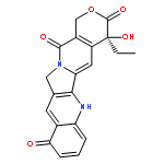

![3H-Indolium,2-[3-(1,3-dihydro-3,3- dimethyl-1-octadecyl-2H-indol-2-ylidene)- 1-propenyl]-3,3-dimethyl-1-octadecyl-,perchlorate](http://img.cochemist.com/ccimg/41100/41085-99-8.png)
![3H-Indolium,2-[3-(1,3-dihydro-3,3- dimethyl-1-octadecyl-2H-indol-2-ylidene)- 1-propenyl]-3,3-dimethyl-1-octadecyl-,perchlorate](http://img.cochemist.com/ccimg/41100/41085-99-8_b.png)