Co-reporter:Cui Liu, Dianhua Ning, Cheng Zhang, Zhengjie Liu, Ruilong Zhang, Jun Zhao, Tingting Zhao, Bianhua Liu, and Zhongping Zhang
ACS Applied Materials & Interfaces June 7, 2017 Volume 9(Issue 22) pp:18897-18897
Publication Date(Web):May 18, 2017
DOI:10.1021/acsami.7b05827
Classical pH test papers are widely used to measure the acid–base degree of media in a qualitative or semiquantitative manner. However, the extension of portable and inexpensive methods to a wide range of analytes so as to eliminate the tediousness of instrumental assays remains unsuccessful. Here, we report a novel kind of dual-colored carbon dot (CD) ratiometric fluorescent test paper for the semiquantitative assay of copper ions (Cu2+) by a dose-sensitive color evolution. The preparation of the test paper is based on the following two interesting findings: on the one hand, residual p-phenylenediamine at the surface of as-synthesized red CDs (r-CDs) efficiently binds Cu2+ ions to produce a strong visible absorption that overlaps the emission of blue CDs (b-CDs); on the other hand, the Cu2+ ions render the adsorption of small b-CDs onto the surface of larger r-CDs through their dual-coordinating interactions with the surface ligands of both r-CDs and b-CDs. These two mechanisms lead to a specific spectral energy transfer to quench the fluorescence of b-CDs with a sensitive detection limit of 8.82 nM Cu2+, whereas the red fluorescence of r-CDs is unaffected as a stable internal standard. Ratiometric fluorescent test papers have been prepared using a mixture of r-CDs and b-CDs (1:7) as ink by jetprinting on a piece of paper. With the addition of Cu2+ ions, the blue test paper produces a consecutive wide-colored evolution from blue to orange-red, with a dose-discerning ability as low as 25 nM.Keywords: carbon dots; copper ions; energy transfer; ratiometric fluorescence; test paper;
Co-reporter:Xinling Yu;Linlin Yang;Tingting Zhao;Ruilong Zhang;Liang Yang;Changlong Jiang;Jun Zhao;Zhongping Zhang
RSC Advances (2011-Present) 2017 vol. 7(Issue 84) pp:53379-53384
Publication Date(Web):2017/11/16
DOI:10.1039/C7RA09972J
Here, a ratiometric fluorescent test paper for the visual and on-site determination of environmental fluoride ions was fabricated by inkjet-printing of the as-prepared “ink” onto a filter paper. The “ink” was prepared by mixing the fluoride-sensitive organic probe (C-TIPS) with red CdTe quantum dots (QDs) in an optimal proportion. The designed fluorescent fluoride probe shows a turn-on effect in the presence of fluoride ions. With the aid of thee red fluorescence of CdTe QDs, the test paper exhibited a distinguishable fluorescence color change from red to purple to blue under a UV lamp. The as-prepared ratiometric-fluorescent test paper displayed a superior sensitivity and visual effectiveness to quantify fluoride ions, with a detection limit of 0.285 μM which is lower than the World Health Organization (WHO) defined limit (79 μM). Moreover, the test paper is highly applicable for the detection of fluoride ions in natural waters in a very simple, cost efficient and on-site way.
Co-reporter:Ruilong Zhang, Shijiang Liu, Jianping Wang, Guangmei Han, Linlin Yang, Bianhua Liu, Guijian Guan and Zhongping Zhang
Analyst 2016 vol. 141(Issue 16) pp:4919-4925
Publication Date(Web):31 May 2016
DOI:10.1039/C6AN00830E
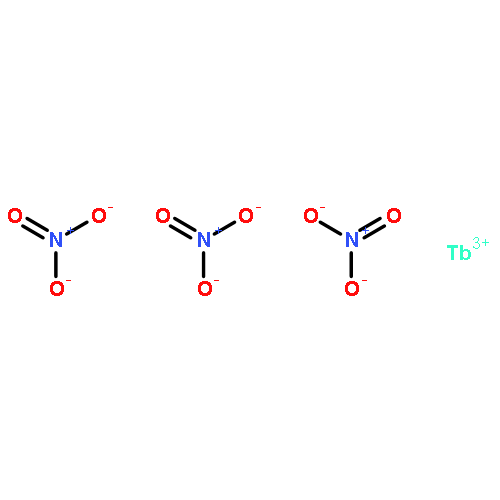
Luminescent chemosensors for hydrogen sulphide (H2S) are of great interest because of the close association of H2S with our health. However, current probes for H2S detection have problems such as low sensitivity/selectivity, poor aqueous-solubility or interference from background fluorescence. This study reports an ultrasensitive and time-gated “switch on” probe for detection of H2S, and its application in test paper for visualization of exhaled H2S. The complex probe is synthesized with a luminescent Tb3+ centre and three ligands of azido (–N3) substituted pyridine-2,6-dicarboxylic acid, giving the probe high hydrophilicity and relatively fast reaction dynamics with H2S because there are three –N3 groups in each molecule. The introduced –N3 group as a strong electron-withdrawing moiety effectively changes the energy level of ligand via intramolecular charge transfer (ICT), and thus breaks the energy transferring from ligand to lanthanide ion, resulting in quenching of Tb3+ luminescence. On addition of H2S, the –N3 group can be reduced to an amine group to break the process of ICT, and the luminescence of Tb3+ is recovered at a nanomolar sensitivity level. With a long lifetime of luminescence of Tb3+ centre (1.9 ms), use of a time-gated technique effectively eliminates the background fluorescence by delaying fluorescence collection for 0.1 ms. The test paper imprinted by the complex probe ink can visualize clearly the trace H2S gas exhaled by mice.
Co-reporter:Cui Liu
The Journal of Physical Chemistry C 2015 Volume 119(Issue 15) pp:8266-8272
Publication Date(Web):March 26, 2015
DOI:10.1021/jp512918h
White-light-emitting materials for the applications in display and lighting have widely been prepared by the rare earth with high photoluminescent efficiency and stability, but the short resource, high cost, and serious environmental concerns are the insurmountable barriers of rare-earth-based materials. Here, we report that strong laser ablation of common organosilica colloid can produce white-light-emitting silica nanoparticles. The instant high temperature and pressure induced by strong laser simultaneously caused the reduction of partial silica and the pyrolysis of carbon chains in the interior of silica nanoparticles, in which the rapid crystallizations of the silicon and carbon vapor led to the formations of silicon dots and diamond-like carbon dots in the silica matrix. Significantly, the resultant silica nanoparticles containing different sized silicon and diamond dots exhibited a wide fluorescence spectrum to display bright white light under UV excitation. Moreover, the light emission diode (LED) device prepared by using the ablated silica nanoparticles as light source gave off the warm white light with Commission Internationale de l’Eclairage (CIE) coordinates (0.34, 0.32). The strategy reported here opens a new window to the exploration of white-light-emitting materials through the very simple, inexpensive, and environment-friendly pathway.
Co-reporter:Chao Yuan, Bianhua Liu, Fei Liu, Ming-Yong Han, and Zhongping Zhang
Analytical Chemistry 2014 Volume 86(Issue 2) pp:1123
Publication Date(Web):December 30, 2013
DOI:10.1021/ac402894z
A new “turn on” fluorescence nanosensor for selective Hg2+ determination is reported based on bis(dithiocarbamato)copper(II) functionalized carbon nanodots (CuDTC2-CDs). The CuDTC2 complex was conjugated to the prepared amine-coated CDs by the condensation of carbon disulfide onto the nitrogen atoms in the surface amine groups, followed by the coordination of copper(II) to the resulting dithiocarbamate groups (DTC) and finally by the additional coordination of ammonium N-(dithicarbaxy) sarcosine (DTCS) to form the CuDTC2-complexing CDs. The CuDTC2 complex at surface strongly quenched the bright-blue fluorescence of the CDs by a combination of electron transfer and energy transfer mechanism. Hg2+ could immediately switch on the fluorescence of the CuDTC2-CDs by promptly displacing the Cu2+ in the CuDTC2 complex and thus shutting down the energy transfer pathway, in which the sensitive limit for Hg2+ as low as 4 ppb was reached. Moreover, a paper-based sensor has been fabricated by printing the CuDTC2-CDs probe ink on a piece of cellulose acetate paper using a commercial inkjet printer. The fluorescence “turn on” on the paper provided the most conveniently visual detection of aqueous Hg2+ ions by the observation with naked eye. The very simple and effective strategy reported here facilitates the development of portable and reliable fluorescence nanosensors for the determination of Hg2+ in real samples.
Co-reporter:Guangmei Han, Renyong Liu, Ming-Yong Han, Changlong Jiang, Jianping Wang, Shuhu Du, Bianhua Liu, and Zhongping Zhang
Analytical Chemistry 2014 Volume 86(Issue 23) pp:11503
Publication Date(Web):November 5, 2014
DOI:10.1021/ac503539w
The molecular processes of drugs from cellular uptake to intracellular distribution as well as the intracellular interaction with the target molecule are critically important for the development of new antitumor drugs. In this work, we have successfully developed a label-free surface-enhanced Raman scattering (SERS) technique to monitor and visualize the metabolism of antitumor drug 6-mercaptopurine in living cells. It has been clearly demonstrated that Au@Ag NPs exhibit an excellent Raman enhancement effect to both 6-mercaptopurine and its metabolic product 6-mercaptopurine-ribose. Their different ways to absorb at the surface of Au@Ag NPs lead to the obvious spectral difference for distinguishing the antitumor drug and its metabolite by SERS spectra. The Au@Ag NPs can easily pass through cell membranes in a large amount and sensitively respond to the biological conversion of 6-mercaptopurine in tumor cells. The Raman imaging can visualize the real-time distribution of 6-mercaptopurine and its biotransformation with the concentrations in tumor cells. The SERS-based method reported here is simple and efficient for the assessments of drug efficacy and the understanding of the molecular therapeutic mechanism of antitumor drugs at the cellular level.
Co-reporter:Benmei Cao, Chao Yuan, Bianhua Liu, Changlong Jiang, Guijian Guan, Ming-Yong Han
Analytica Chimica Acta 2013 Volume 786() pp:146-152
Publication Date(Web):5 July 2013
DOI:10.1016/j.aca.2013.05.015
•The simplicity of the preparation of the ratiometric fluorescence probe.•Visual fluorescence detection of Hg2+ which could be observed with the naked eye.•High selectivity and low detection limit for aqueous Hg2+ sensing.A novel nanohybrid ratiometric fluorescence probe comprised of carbon dots (C-dots) and hydrophilic CdSe@ZnS quantum dots (QDs) has been developed by simply mixing the blue-emission C-dots with red-emission carboxylmethyldithiocarbamate modified CdSe@ZnS QDs (GDTC-QDs). The nanohybrid ratiometric fluorescence probe exhibits dual emissions at 436 nm and 629 nm under a single excitation wavelength. Due to the strong chelating ability of GDTC on the surface of QDs to mercuric ion (Hg2+), the fluorescence of the GDTC-QDs in the nanohybrid system could be selectively quenched in the presence of Hg2+ while the fluorescence of the C-dots remained constant, resulting in an obviously distinguishable fluorescence color evolution (from red to blue) of the nanohybrid system. The detection limit of this method was found to be as low as 0.1 μM. Furthermore, the recovery result for Hg2+ in real samples including tap water and lake water by this method was satisfying, suggesting its potential application for Hg2+ sensing.
Co-reporter:Xinran Liu ; Bianhua Liu ; Zhenyang Wang ; Buchang Zhang ;Zhongping Zhang
The Journal of Physical Chemistry C 2008 Volume 112(Issue 26) pp:9632-9636
Publication Date(Web):June 6, 2008
DOI:10.1021/jp8017088
Highly oriented vaterite CaCO3 tablet-like arrays were formed at the air/water interface through the cooperative mineralization regulated by polypeptide and double hydrophilic block copolymer under ambient conditions. The nearly parallel arrangement of CaCO3 vaterite tablets at the air/water interface shows the remarkable resemblance to the morphology of nacreous layers. The poly(aspartic acid) (PASP) with high molecular weight (Mw = 11 000) and low solubility in water promoted the formation of vaterite tablets at the air/water interface, and stabilized the vaterite tablets by aggregation at the air/water interface and adsorption at the surface of vaterite tablets. Meanwhile, the highly hydrophilic poly(ethylene glycol)-block-poly(methacrylic acid) (PEG-b-PMAA) played an important role in regulating the arrangement and orientation of vaterite CaCO3 tablets, leading to the oriented tablet-like arrays at the air/water interface. Detailed experiments revealed that hydrophilic PEG-b-PMAA alone did not produce any form of CaCO3 crystals at the air/water interface, but could induce the formation of calcite CaCO3 particles in the water phase. However, high molecular weight PASP alone led to the formation of disk-like vaterite particles composed of helically aggregated nanoplates at the air/water interface, suggesting the regulating role of PEG-b-PMAA in the growth of vaterite tablet arrays. These results reported here provide a better understanding of the growth mechanism of nacreous layers and shells in nature.


![3',6'-Dihydroxy-3H-spiro[isobenzofuran-1,9'-xanthen]-3-one](http://img.cochemist.com/ccimg/2400/2321-07-5.png)
![3',6'-Dihydroxy-3H-spiro[isobenzofuran-1,9'-xanthen]-3-one](http://img.cochemist.com/ccimg/2400/2321-07-5_b.png)


