Co-reporter:Nicholas Leioatts, Blake Mertz, Karina Martínez-Mayorga, Tod D. Romo, Michael C. Pitman, Scott E. Feller, Alan Grossfield, and Michael F. Brown
Biochemistry 2014 Volume 53(Issue 2) pp:
Publication Date(Web):December 12, 2013
DOI:10.1021/bi4013947
Rhodopsin, the mammalian dim-light receptor, is one of the best-characterized G-protein-coupled receptors, a pharmaceutically important class of membrane proteins that has garnered a great deal of attention because of the recent availability of structural information. Yet the mechanism of rhodopsin activation is not fully understood. Here, we use microsecond-scale all-atom molecular dynamics simulations, validated by solid-state 2H nuclear magnetic resonance spectroscopy, to understand the transition between the dark and metarhodopsin I (Meta I) states. Our analysis of these simulations reveals striking differences in ligand flexibility between the two states. Retinal is much more dynamic in Meta I, adopting an elongated conformation similar to that seen in the recent activelike crystal structures. Surprisingly, this elongation corresponds to both a dramatic influx of bulk water into the hydrophobic core of the protein and a concerted transition in the highly conserved Trp2656.48 residue. In addition, enhanced ligand flexibility upon light activation provides an explanation for the different retinal orientations observed in X-ray crystal structures of active rhodopsin.
Co-reporter:Joshua N. Horn, Tod D. Romo, and Alan Grossfield
Biochemistry 2013 Volume 52(Issue 33) pp:
Publication Date(Web):July 22, 2013
DOI:10.1021/bi400773q
The emergence of antibiotic resistant pathogens is one of the major medical concerns of the 21st century, prompting renewed interest in the development of novel antimicrobial compounds. Here we use microsecond-scale all-atom molecular dynamics simulations to characterize the structure, dynamics, and membrane-binding mechanism of a synthetic antimicrobial lipopeptide, C16-KGGK. Our simulations suggest that these lipopeptides prefer to aggregate in solution and alter the intrinsic order of the lipid bilayer upon binding. From these results and previous coarse-grained simulations, we have developed a simple model for the binding and insertion process for these lipopeptides.
Co-reporter:Nicholas Leioatts, Tod D. Romo, and Alan Grossfield
Journal of Chemical Theory and Computation 2012 Volume 8(Issue 7) pp:2424-2434
Publication Date(Web):June 5, 2012
DOI:10.1021/ct3000316
Understanding the functions of biomolecules requires insight not only from structures but from dynamics as well. Often, the most interesting processes occur on time scales too slow for exploration by conventional molecular dynamics (MD) simulations. For this reason, alternative computational methods such as elastic network models (ENMs) have become increasingly popular. These simple, coarse-grained models represent molecules as beads connected by harmonic springs; the system’s motions are solved analytically by normal-mode analysis. In the past few years, many different formalisms for performing ENM calculations have emerged, and several have been optimized using all-atom MD simulations. In contrast to other studies, we have compared the various formalisms in a systematic, quantitative way. In this study, we optimize many ENM functional forms using a uniform data set containing only long (>1 μs) all-atom MD simulations. Our results show that all models once optimized produce spring constants for immediate neighboring residues that are orders of magnitude stiffer than more distal contacts. In addition, the statistical significance of ENM performance varied with model resolution. We also show that fitting long trajectories does not improve ENM performance due to a problem inherent in all network models tested: they underestimate the relative importance of the most concerted motions. Finally, we characterize ENMs’ resilience by tessellating the parameter space to show that broad ranges of parameters produce similar quality predictions. Taken together, our data reveal that the choice of spring function and parameters are not vital to the performance of a network model and that simple parameters can by derived “by hand” when no data are available for fitting, thus illustrating the robustness of these models.
Co-reporter:Tod D. Romo and Alan Grossfield
Journal of Chemical Theory and Computation 2011 Volume 7(Issue 8) pp:2464-2472
Publication Date(Web):June 22, 2011
DOI:10.1021/ct2002754
Molecular dynamics (MD) is a powerful tool for understanding the fluctuations of biomolecular systems. It is, however, subject to statistical errors in its sampling of the underlying distribution of states. One must understand these errors in order to draw meaningful conclusions from the simulation. This is becoming ever more critical as MD simulations of even larger systems are attempted. We present here a new method for determining the extent of convergence that relies on measures of the fluctuation space sampled by the simulation without any a priori knowledge of states or partitioning of the configuration space. This method reveals long correlation times, even for simple systems, and suggests caution when interpreting macromolecular simulations. We also compare this method with previous efforts to characterize the sampling of MD simulation.
Co-reporter:Alan Grossfield
Biochimica et Biophysica Acta (BBA) - Biomembranes (July 2011) Volume 1808(Issue 7) pp:
Publication Date(Web):July 2011
DOI:10.1016/j.bbamem.2011.03.010
G protein-coupled receptors (GPCRs) are a large, biomedically important family of proteins, and the recent explosion of new high-resolution structural information about them has provided an enormous opportunity for computational modeling to make major contributions. In particular, molecular dynamics simulations have become a driving factor in many areas of GPCR biophysics, improving our understanding of lipid–protein interaction, activation mechanisms, and internal hydration. Given that computers will continue to get faster and more structures will be solved, the importance of computational methods will only continue to grow, particularly as simulation research is more closely coupled to experiment.Research Highlights► G protein-coupled receptors (GPCRs) are biomedically important. ► Molecular dynamics simulations provide crucial insight into structure and dynamics. ► Recent crystal structures have increased the importance of GPCR simulations.
Co-reporter:Tod D. Romo, Laura A. Bradney, Denise V. Greathouse, Alan Grossfield
Biochimica et Biophysica Acta (BBA) - Biomembranes (August 2011) Volume 1808(Issue 8) pp:
Publication Date(Web):August 2011
DOI:10.1016/j.bbamem.2011.03.017
One approach to the growing health problem of antibiotic resistant bacteria is the development of antimicrobial peptides (AMPs) as alternative treatments. The mechanism by which these AMPs selectively attack the bacterial membrane is not well understood, but is believed to depend on differences in membrane lipid composition. N-acylation of the small amidated hexapeptide, RRWQWR-NH2 (LfB6), derived from the 25 amino acid bovine lactoferricin (LfB25) can be an effective means to improve its antimicrobial properties. Here, we investigate the interactions of C6-LfB6, N-acylated with a 6 carbon fatty acid, with model lipid bilayers with two distinct compositions: 3:1 POPE:POPG (negatively charged) and POPC (zwitterionic). Results from solid-state 2H and 31P NMR experiments are compared with those from an ensemble of all-atom molecular dynamic simulations running in aggregate more than 8.6 ms. 2H NMR spectra reveal no change in the lipid acyl chain order when C6-LfB6 is bound to the negatively charged membrane and only a slight decrease in order when it is bound to the zwitterionic membrane. 31P NMR spectra show no significant perturbation of the phosphate head groups of either lipid system in the presence of C6-LfB6. Molecular dynamic simulations show that for the negatively charged membrane, the peptide's arginines drive the initial association with the membrane, followed by attachment of the tryptophans at the membrane–water interface, and finally by the insertion of the C6 tails deep into the bilayer. In contrast, the C6 tail leads the association with the zwitterionic membrane, with the tryptophans and arginines associating with the membrane–water interface in roughly the same amount of time. We find similar patterns in the order parameters from our simulations. Moreover, we find in the simulations that the C6 tail can insert 1–2 Å more deeply into the zwitterionic membrane and can exist in a wider range of angles than in the negatively charged membrane. We propose this is due to the larger area per lipid in the zwitterionic membrane, which provides more space for the C6 to insert and assume different orientations.Research highlights► Association of small acylated peptide with model bacterial and mammalian membranes. ► Bacterial membrane shows no change in order, and binding is led by arginines. ► Mammalian membrane has decrease in order, and binding is led by C6 tail.
Co-reporter:Tod D. Romo, Alan Grossfield, Michael C. Pitman
Biophysical Journal (6 January 2010) Volume 98(Issue 1) pp:
Publication Date(Web):6 January 2010
DOI:10.1016/j.bpj.2009.09.046
The recently solved crystallographic structures for the A2A adenosine receptor and the β1 and β2 adrenergic receptors have shown important differences between members of the class-A G-protein-coupled receptors and their archetypal model, rhodopsin, such as the apparent breaking of the ionic lock that stabilizes the inactive structure. Here, we characterize a 1.02 μs all-atom simulation of an apo-β2 adrenergic receptor that is missing the third intracellular loop to better understand the inactive structure. Although we find that the structure is remarkably rigid, there is a rapid influx of water into the core of the protein, as well as a slight expansion of the molecule relative to the crystal structure. In contrast to the x-ray crystal structures, the ionic lock rapidly reforms, although we see an activation-precursor-like event wherein the ionic lock opens for ∼200 ns, accompanied by movements in the transmembrane helices associated with activation. When the lock reforms, we see the structure return to its inactive conformation. We also find that the ionic lock exists in three states: closed (or locked), semi-open with a bridging water molecule, and open. The interconversion of these states involves the concerted motion of the entire protein. We characterize these states and the concerted motion underlying their interconversion. These findings may help elucidate the connection between key local events and the associated global structural changes during activation.
Co-reporter:Nicholas Leioatts, Tod D. Romo, Shairy Azmy Danial, Alan Grossfield
Biophysical Journal (4 August 2015) Volume 109(Issue 3) pp:
Publication Date(Web):4 August 2015
DOI:10.1016/j.bpj.2015.06.046
G protein-coupled receptors are vital membrane proteins that allosterically transduce biomolecular signals across the cell membrane. However, the process by which ligand binding induces protein conformation changes is not well understood biophysically. Rhodopsin, the mammalian dim-light receptor, is a unique test case for understanding these processes because of its switch-like activity; the ligand, retinal, is bound throughout the activation cycle, switching from inverse agonist to agonist after absorbing a photon. By contrast, the ligand-free opsin is outside the activation cycle and may behave differently. We find that retinal influences rhodopsin dynamics using an ensemble of all-atom molecular dynamics simulations that in aggregate contain 100 μs of sampling. Active retinal destabilizes the inactive state of the receptor, whereas the active ensemble was more structurally homogenous. By contrast, simulations of an active-like receptor without retinal present were much more heterogeneous than those containing retinal. These results suggest allosteric processes are more complicated than a ligand inducing protein conformational changes or simply capturing a shifted ensemble as outlined in classic models of allostery.
Co-reporter:Dejun Lin, Alan Grossfield
Biophysical Journal (21 October 2014) Volume 107(Issue 8) pp:
Publication Date(Web):21 October 2014
DOI:10.1016/j.bpj.2014.08.026
The development of novel antibiotic drugs is one of the most pressing biomedical problems due to the increasing number of antibiotic-resistant pathogens. Antimicrobial peptides and lipopeptides are a promising category of candidates, but the molecular origins of their antimembrane activity is unclear. Here we explore a series of recently developed antimicrobial lipopeptides, using coarse-grained molecular-dynamics simulations and free energy methods to uncover the thermodynamics governing their binding to membranes. Specifically, we quantify C16-KGGK’s binding affinity to the two types of membrane by umbrella sampling. We also examined the origin of C16-KGGK’s selectivity for bacterial versus mammalian membranes by systematically varying the peptide sequence and salt concentration. Our data showed that the C16 hydrophobic tail is the main contributor to its affinity to lipid membrane, whereas the peptide portion is mainly responsible for its selectivity. Furthermore, the electrostatic interaction between the cationic peptide and anionic bacterial membrane plays a significant role in the selectivity.
Co-reporter:Dejun Lin, Alan Grossfield
Biophysical Journal (18 August 2015) Volume 109(Issue 4) pp:
Publication Date(Web):18 August 2015
DOI:10.1016/j.bpj.2015.07.011
Antimicrobial lipopeptides (AMLPs) are antimicrobial drug candidates that preferentially target microbial membranes. One class of AMLPs, composed of cationic tetrapeptides attached to an acyl chain, have minimal inhibitory concentrations in the micromolar range against a range of bacteria and fungi. Previously, we used coarse-grained molecular dynamics simulations and free energy methods to study the thermodynamics of their interaction with membranes in their monomeric state. Here, we extended the study to the biologically relevant micellar state, using, to our knowledge, a novel reaction coordinate based on hydrophobic contacts. Using umbrella sampling along this reaction coordinate, we identified the critical transition states when micelles insert into membranes. The results indicate that the binding of these AMLP micelles to membranes is thermodynamically favorable, but in contrast to the monomeric case, there are significant free energy barriers. The height of these free energy barriers depends on the membrane composition, suggesting that the AMLPs’ ability to selectively target bacterial membranes may be as much kinetic as thermodynamic. This mechanism highlights the importance of considering oligomeric state in solution as criterion when optimizing peptides or lipopeptides as antibiotic leads.
Co-reporter:Tod D. Romo, Alan Grossfield
Biophysical Journal (17 June 2014) Volume 106(Issue 12) pp:
Publication Date(Web):17 June 2014
DOI:10.1016/j.bpj.2014.05.022
Co-reporter:Joshua N. Horn, Aaron Cravens, Alan Grossfield
Biophysical Journal (1 October 2013) Volume 105(Issue 7) pp:
Publication Date(Web):1 October 2013
DOI:10.1016/j.bpj.2013.08.034
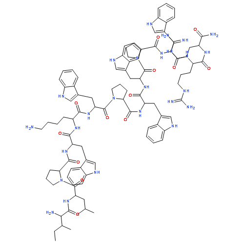
Bacteria, particularly of the genus Bacillus, produce a wide variety of antifungal compounds. They act by affecting the lipid bilayers of fungal membranes, causing curvature-induced strain and eventual permeabilization. One class of these, known as fengycins, has been commercialized for treating agricultural infections and shows some promise as a possible antifungal pharmaceutical. Understanding the mechanism by which fengycins damage lipid bilayers could prove useful to the future development of related antifungal treatments. In this work, we present multi-microsecond-long simulations of fengycin interacting with different lipid bilayer systems. We see fengycin aggregation and uncover a clear aggregation pattern that is partially influenced by bilayer composition. We also quantify some local bilayer perturbations caused by fengycin binding, including curvature of the lipid bilayer and local electrostatic-driven reorganization.

![3,5,8-Trioxa-4-phosphahexacos-17-en-1-aminium,4-hydroxy-N,N,N-trimethyl-9-oxo-7-[[(1-oxohexadecyl)oxy]methyl]-, inner salt,4-oxide, (17Z)-](http://img.cochemist.com/ccimg/26700/26662-91-9.png)
![3,5,8-Trioxa-4-phosphahexacos-17-en-1-aminium,4-hydroxy-N,N,N-trimethyl-9-oxo-7-[[(1-oxohexadecyl)oxy]methyl]-, inner salt,4-oxide, (17Z)-](http://img.cochemist.com/ccimg/26700/26662-91-9_b.png)
![3,5,9-Trioxa-4-phosphapentacosan-1-aminium,4-hydroxy-N,N,N-trimethyl-10-oxo-7-[(1-oxohexadecyl)oxy]-, inner salt, 4-oxide](http://img.cochemist.com/ccimg/2700/2644-64-6.png)
![3,5,9-Trioxa-4-phosphapentacosan-1-aminium,4-hydroxy-N,N,N-trimethyl-10-oxo-7-[(1-oxohexadecyl)oxy]-, inner salt, 4-oxide](http://img.cochemist.com/ccimg/2700/2644-64-6_b.png)
![9-Octadecenoic acid(9Z)-,1-[[[(2-aminoethoxy)hydroxyphosphinyl]oxy]methyl]-2-[(1-oxohexadecyl)oxy]ethylester](http://img.cochemist.com/ccimg/10100/10015-88-0.png)
![9-Octadecenoic acid(9Z)-,1-[[[(2-aminoethoxy)hydroxyphosphinyl]oxy]methyl]-2-[(1-oxohexadecyl)oxy]ethylester](http://img.cochemist.com/ccimg/10100/10015-88-0_b.png)