A re-refinement of 4xan, hen egg-white lysozyme (HEWL) with carboplatin crystallized in NaBr solution, has been made and is published here as an addendum to Tanley et al. [(2014), Acta Cryst. F70, 1135–1142]. This follows a previous re-refinement and PDB deposition (4yem) by Shabalin et al. [(2015), Acta Cryst. D71, 1965–1979]. The critical evaluation of the original PDB deposition (4xan), and the subsequent critical examination of the re-refined structure (4yem), has led to an improved model (PDB code 5hmj).
A re-refinement of 4g4a, the room-temperature X-ray diffraction study of cisplatin and its binding to His15 of HEWL after 14 months chemical exposure in the presence of DMSO is published as an addendum to Tanley et al. [(2012), Acta Cryst. F68, 1300–1306]. This example illustrates the benefits of sharing raw diffraction images, as well as structure factors and molecular coordinates, as the diffraction resolution of the study is now much improved at 1.70 Å.
Cisplatin and carboplatin are platinum anticancer agents that are used to treat a variety of cancers. Previous X-ray crystallographic studies of carboplatin binding to histidine in hen egg-white lysozyme (HEWL) showed a partial chemical conversion of carboplatin to cisplatin owing to the high sodium chloride concentration used in the crystallization conditions. Also, the co-crystallization of HEWL with carboplatin in sodium bromide conditions resulted in the partial conversion of carboplatin to the transbromoplatin form, with a portion of the cyclobutanedicarboxylate (CBDC) moiety still present. The results of the co-crystallization of HEWL with cisplatin or carboplatin in sodium iodide conditions are now reported in order to determine whether the cisplatin and carboplatin converted to the iodo form, and whether this took place in a similar way to the partial conversion of carboplatin to cisplatin in NaCl conditions or to transbromoplatin in NaBr conditions as seen previously. It is reported here that a partial chemical transformation has taken place to a transplatin form for both ligands. The NaI-grown crystals belonged to the monoclinic space group P21 with two molecules in the asymmetric unit. The chemically transformed cisplatin and carboplatin bind to both His15 residues, i.e. in each asymmetric unit. The binding is only at the Nδ atom of His15. A third platinum species is also seen in both conditions bound in a crevice between symmetry-related molecules. Here, the platinum is bound to three I atoms identified based on their anomalous difference electron densities and their refined occupancies, with the fourth bound atom being a Cl atom (in the cisplatin case) or a portion of the CBDC moiety (in the carboplatin case).
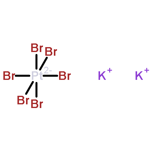
This study examines the binding and chemical stability of the platinum hexahalides K2PtCl6, K2PtBr6 and K2PtI6 when soaked into pre-grown hen egg-white lysozyme (HEWL) crystals as the protein host. Direct comparison of the iodo complex with the chloro and bromo complexes shows that the iodo complex is partly chemically transformed to a square-planar PtI3 complex bound to the Nδ atom of His15, a chemical behaviour that is not exhibited by the chloro or bromo complexes. Each complex does, however, bind to HEWL in its octahedral form either at one site (PtI6) or at two sites (PtBr6 and PtCl6). As heavy-atom derivatives of a protein, the octahedral shape of the hexahalides could be helpful in cases of difficult-to-interpret electron-density maps as they would be recognisable `objects'.
Carboplatin is a second-generation platinum anticancer agent used for the treatment of a variety of cancers. Previous X-ray crystallographic studies of carboplatin binding to histidine (in hen egg-white lysozyme; HEWL) showed the partial conversion of carboplatin to cisplatin owing to the high NaCl concentration used in the crystallization conditions. HEWL co-crystallizations with carboplatin in NaBr conditions have now been carried out to confirm whether carboplatin converts to the bromine form and whether this takes place in a similar way to the partial conversion of carboplatin to cisplatin observed previously in NaCl conditions. Here, it is reported that a partial chemical transformation takes place but to a transplatin form. Thus, to attempt to resolve purely carboplatin binding at histidine, this study utilized co-crystallization of HEWL with carboplatin without NaCl to eliminate the partial chemical conversion of carboplatin. Tetragonal HEWL crystals co-crystallized with carboplatin were successfully obtained in four different conditions, each at a different pH value. The structural results obtained show carboplatin bound to either one or both of the N atoms of His15 of HEWL, and this particular variation was dependent on the concentration of anions in the crystallization mixture and the elapsed time, as well as the pH used. The structural details of the bound carboplatin molecule also differed between them. Overall, the most detailed crystal structure showed the majority of the carboplatin atoms bound to the platinum centre; however, the four-carbon ring structure of the cyclobutanedicarboxylate moiety (CBDC) remained elusive. The potential impact of the results for the administration of carboplatin as an anticancer agent are described.
![2-CYCLOHEXEN-1-ONE, 6-HYDROXY-2,4,4-TRIMETHYL-3-[(1E)-3-OXO-1-BUTENYL]-](http://img.cochemist.com/ccimg/74300/74285-61-3.png)
![2-CYCLOHEXEN-1-ONE, 6-HYDROXY-2,4,4-TRIMETHYL-3-[(1E)-3-OXO-1-BUTENYL]-](http://img.cochemist.com/ccimg/74300/74285-61-3_b.png)