Co-reporter:Shiyi Shao, Bo Chen, Juan Cheng, Chengkun Wang, Yanli Zhang, Lingxiao Shao, Yongzhou Hu, Yifeng Han, Feng Han, Xin Li
Biosensors and Bioelectronics 2017 Volume 94(Volume 94) pp:
Publication Date(Web):15 August 2017
DOI:10.1016/j.bios.2017.02.050
•The first fluorescent probe for imaging protein S-nitrosylation is reported.•The probe features high specificity and sensitivity.•The probe has facilitated, for the first time, direct observation of protein S-nitrosylation in intact live cells.•Robustness of the probe has been exemplified by imaging the dynamic change of GAPDH nitrosylation in live cells.S-nitrosylation is a posttranslational modification of protein cysteine residues leading to the formation of S-nitrosothiols and its detection is crucial to understanding of redox regulation and NO-based signaling. Prototypical detection methods for S-nitrosylation are always carried out ex situ. However, the reversible nature and the tendency of transnitrosylation highlight the necessity of its probing in intact live biological contexts. Herein we provide a fluorogenic chemical probe for the detection of S-nitrosylation in live endothelial cells. The probe is weakly emissive alone and becomes highly fluorescent only after undergoing a reaction with S-nitrosothiols in live cellular environments. This probe features high degrees of specificity and desirable sensitivity. Furthermore, it has been successfully applied to image the dynamic change of protein S-nitrosylation in live endothelial cells. The applicability of the probe in complex biological systems has been additionally verified by imaging a known target of S-nitrosylation, glyceraldehyde-3-phosphate dehydrogenase (GAPDH), in live cells. Due to the versatility exemplified, this probe holds great promise for exploring the role of protein S-nitrosylation in the pathophysiological process of a variety of vascular diseases.Download high-res image (226KB)Download full-size image
Co-reporter:Xiao-Juan Wang, Yin-Ping Gao, Nan-Nan Lu, Wei-Shuo Li, Ji-Fang Xu, Xiao-Ying Ying, Gang Wu, Mei-Hua Liao, Chao Tan, Ling-Xiao Shao, Ying-Mei Lu, Chen Zhang, Kohji Fukunaga, Feng Han, and Yong-Zhong Du
ACS Applied Materials & Interfaces 2016 Volume 8(Issue 51) pp:
Publication Date(Web):October 17, 2016
DOI:10.1021/acsami.6b13052
Clinical treatment for vascular dementia still remains a challenge mainly due to the blood–brain barrier (BBB). Here, a micelle based on polysialic acid (PSA), which is a hydrophilic and endogenous carbohydrate polymer, was designed to deliver calmodulin antagonist for therapy of vascular dementia. PSA was first chemically conjugated with octadecylamine (ODA), and the obtained PSA–ODA copolymer could self-assemble into micelle in aqueous solution with a 120.0 μg/mL critical micelle concentration. The calmodulin antagonist loaded PSA–ODA micelle, featuring sustained drug release behavior over a period of 72 h with a 3.6% (w/w) drug content and a 107.0 ± 4.0 nm size was then fabricated. The PSA–ODA micelle could cross the BBB mainly via active endocytosis by brain endothelial cells followed by transcytosis. In a water maze test for spatial learning, calmodulin antagonist loaded PSA–ODA micelle significantly reduced the escape latencies of right unilateral common carotid arteries occlusion (rUCCAO) mice with dosage significantly reduced versus free drug. The decrease of hippocampal phospho-CaMKII (Thr286/287) and phospho-synapsin I (Ser603) was partially restored in rUCCAO mice following calmodulin antagonist loaded PSA–ODA micelle treatment. Consistent with the restored CaMKII phosphorylation, the elevation of BrdU/NeuN double-positive cells in the same context was also observed. Overall, the PSA–ODA micelle developed from the endogenous material might promote the development of therapeutic approaches for improving the efficacy of brain-targeted drug delivery and have great potential for vascular dementia treatment.Keywords: blood−brain barrier; calmodulin antagonist; drug-delivery system; polymeric micelle; polysialic acid; vascular dementia;
Co-reporter:Huan Wang, Ling-Juan Hong, Ji-Yun Huang, Quan Jiang, Rong-Rong Tao, Chao Tan, Nan-Nan Lu, Cheng-Kun Wang, Muhammad M Ahmed, Ying-Mei Lu, Zhi-Rong Liu, Wei-Xing Shi, En-Yin Lai, Christopher S Wilcox and Feng Han
Cell Research 2015 25(6) pp:674-690
Publication Date(Web):May 22, 2015
DOI:10.1038/cr.2015.61
Septic encephalopathy (SE) is a critical factor determining sepsis mortality. Vascular inflammation is known to be involved in SE, but the molecular events that lead to the development of encephalopathy remain unclear. Using time-lapse in vivo two-photon laser scanning microscopy, we provide the first direct evidence that cecal ligation and puncture in septic mice induces microglial trafficking to sites adjacent to leukocyte adhesion on inflamed cerebral microvessels. Our data further demonstrate that septic injury increased the chemokine CXCL1 level in brain endothelial cells by activating endothelial P2RX7 and eventually enhanced the binding of Mac-1 (CD11b/CD18)-expressing leukocytes to endothelial ICAM-1. In turn, leukocyte adhesion upregulated endothelial CX3CL1, thereby triggering microglia trafficking to the injured site. The sepsis-induced increase in endothelial CX3CL1 was abolished in CD18 hypomorphic mutant mice. Inhibition of the P2RX7 pathway not only decreased endothelial ICAM-1 expression and leukocyte adhesion but also prevented microglia overactivation, reduced brain injury, and consequently doubled the early survival of septic mice. These results demonstrate the role of the P2RX7 pathway in linking neurovascular inflammation to brain damage in vivo and provide a rationale for targeting endothelial P2RX7 for neurovascular protection during SE.
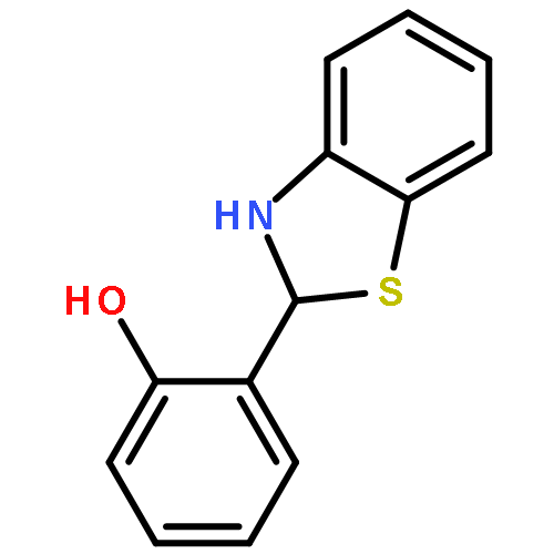
Co-reporter:Xin Li; Rong-Rong Tao; Ling-Juan Hong; Juan Cheng; Quan Jiang; Ying-Mei Lu; Mei-Hua Liao; Wei-Feng Ye; Nan-Nan Lu; Feng Han; Yong-Zhou Hu;You-Hong Hu
Journal of the American Chemical Society 2015 Volume 137(Issue 38) pp:12296-12303
Publication Date(Web):September 9, 2015
DOI:10.1021/jacs.5b06865
Accumulating evidence suggests that formation of peroxynitrite (ONOO–) in the cerebral vasculature contributes to the progression of ischemic damage, while the underlying molecular mechanisms remain elusive. To fully understand ONOO– biology, efficient tools that can realize the real-time tracing of endogenous ONOO– fluxes are indispensable. While a few ONOO– fluorescent probes have been reported, direct visualization of ONOO– fluxes in the cerebral vasculature of live mice remains a challenge. Herein, we present a fluorescent switch-on probe (NP3) for ONOO– imaging. NP3 exhibits good specificity, fast response, and high sensitivity toward ONOO– both in vitro and in vivo. Moreover, NP3 is two-photon excitable and readily blood–brain barrier penetrable. These desired photophysical and pharmacokinetic properties endow NP3 with the capability to monitor brain vascular ONOO– generation after injury with excellent temporal and spatial resolution. As a proof of concept, NP3 has enabled the direct visualization of neurovascular ONOO– formation in ischemia progression in live mouse brain by use of two-photon laser scanning microscopy. Due to these favorable properties, NP3 holds great promise for visualizing endogenous peroxynitrite fluxes in a variety of pathophysiological progressions in vitro and in vivo.
Co-reporter:Ying-mei Lu, Ji-yun Huang, Huan Wang, Xue-fang Lou, Mei-hua Liao, Ling-juan Hong, Rong-rong Tao, Muhammad M. Ahmed, Chun-lei Shan, Xiao-liang Wang, Kohji Fukunaga, Yong-zhong Du, Feng Han
Biomaterials 2014 35(1) pp: 530-537
Publication Date(Web):
DOI:10.1016/j.biomaterials.2013.09.093
Co-reporter:Ji-yun Huang, Ying-mei Lu, Huan Wang, Jun Liu, Mei-hua Liao, Ling-juan Hong, Rong-rong Tao, Muhammad Masood Ahmed, Ping Liu, Shuang-shuang Liu, Kohji Fukunaga, Yong-zhong Du, Feng Han
Biomaterials 2013 34(32) pp: 7960-7970
Publication Date(Web):
DOI:10.1016/j.biomaterials.2013.07.009
Co-reporter:Gen-sheng Zhang, Wei-feng Ye, Rong-rong Tao, Ying-mei Lu, Guo-fang Shen, Kohji Fukunaga, Ji-yun Huang, Yue-long Ji, Feng Han
Experimental and Toxicologic Pathology (September 2012) Volume 64(Issue 6) pp:619-624
Publication Date(Web):1 September 2012
DOI:10.1016/j.etp.2010.12.004
The septal and temporal poles of the hippocampus differ markedly in their anatomical organization, but whether these distinct regions exhibit differential neurochemical profiles underlying lead (Pb2+) neurotoxicity remains to be determined. In the present study, we examined changes in the expression of Ca2+/calmodulin-dependent enzymes, including calpain, calcineurin, phospho-CaMKII (Thr286) and neuronal nitric oxide synthase (nNOS), in the rat dorsal and ventral hippocampus (DH and VH) after acute Pb2+ exposure. Five days after Pb2+ exposure, we observed constitutively active forms of calcineurin (45 kDa and 48 kDa) in ventral portions of the hippocampus, a result consistent with the observed calpain activation that is indicated by the breakdown of spectrin in this region. Our data demonstrate that nNOS expression is significantly higher in the ventral region of the hippocampus when compared to the dorsal region, whereas phosphorylation of CaMKII (Thr286) is less pronounced in the ventral portion of the hippocampus and more pronounced in dorsal regions after acute Pb2+ exposure. Thus, it appears likely that the ventral region of hippocampus is more vulnerable to the neurotoxic effects of Pb2+ than the dorsal region. Taken together, the present data suggest that acute lead exposure leads to differential expression patterns of Ca2+/calmodulin-dependent enzymes along the dorsoventral axis of the hippocampus.
Co-reporter:Qaisar Mahmood, Guang-Fa Wang, Gang Wu, Huan Wang, Chang-Xin Zhou, Hong-Yu Yang, Zhi-Rong Liu, Feng Han, Kui Zhao
Phytomedicine (15 February 2017) Volume 25() pp:8-14
Publication Date(Web):15 February 2017
DOI:10.1016/j.phymed.2016.12.004
BackgroundSalvianolic acid A (SAA) is obtained from Chinese herb Salviae Miltiorrhizae Bunge (Labiatae), has been reported to have the protective effects against cardiovascular and neurovascular diseases.HypothesisThe aim of present study was to investigate the relationship between the effectiveness of SAA against neurovascular injury and its effects on calpain activation and endothelial nitric oxide synthase (eNOS) uncoupling.Study designSAA or vehicle was given to C57BL/6 male mice for seven days before the occlusion of middle cerebral artery (MCAO) for 60 min.MethodsHigh-resolution positron emission tomography scanner (micro-PET) was used for small animal imaging to examine glucose metabolism. Rota-rod time and neurological deficit scores were calculated after 24 h of reperfusion. The volume of infarction was determined by Nissl-staining. The calpain proteolytic activity and eNOS uncoupling were determined by western blot analysis.ResultsSAA administration increased glucose metabolism and ameliorated neuronal damage after brain ischemia, paralleled with decreased neurological deficit and volume of infarction. In addition, SAA pretreatment inhibited eNOS uncoupling and calpain proteolytic activity. Furthermore, SAA inhibited peroxynitrite (ONOO−) generation and upregulates AKT, FKHR and ERK phosphorylation.ConclusionThese findings strongly suggest that SAA elicits a neurovascular protective role through the inhibition of eNOS uncoupling and ONOO− formation. Moreover, SAA attenuates spectrin and calcineurin breakdown and therefore protects the brain against ischemic/reperfusion injury.Download high-res image (71KB)Download full-size image
Co-reporter:Qi-bing Liu, Lu-lu Liu, Ying-mei Lu, Rong-rong Tao, Ji-yun Huang, Feng Han, Yi-jia Lou
Toxicology and Applied Pharmacology (15 August 2010) Volume 247(Issue 1) pp:70
Publication Date(Web):15 August 2010
DOI:10.1016/j.taap.2010.05.014
Co-reporter:Qi-bing Liu, Lu-lu Liu, Ying-mei Lu, Rong-rong Tao, Ji-yun Huang, Norifumi Shioda, Shigeki Moriguchi, Kohji Fukunaga, Feng Han, Yi-jia Lou
Toxicology and Applied Pharmacology (1 May 2010) Volume 244(Issue 3) pp:374-384
Publication Date(Web):1 May 2010
DOI:10.1016/j.taap.2010.02.004
The pathophysiological relevance of S-nitrosoglutathione (GSNO)-induced endothelial cell injury remains unclear. The main objective of this study was to elucidate the molecular mechanisms of GSNO-induced oxidative stress in endothelial cells. Morphological evaluation through DAPI staining and propidium iodide (PI) flow cytometry was used to detect apoptosis. In cultured EA.hy926 endothelial cells, exposure to GSNO led to a time- and dose-dependent apoptotic cascade. When intracellular reactive oxygen species (ROS) production was measured in GSNO-treated cells with the fluorescent probes 5-(and-6)-carboxy-2′,7′-dichlorofluorescein diacetate, we observed elevated ROS levels and a concomitant loss in mitochondrial membrane potential, indicating that GSNO-induced death signaling is mediated through a ROS-mitochondrial pathway. Importantly, we found that peroxynitrite formation and Omi/HtrA2 release from mitochondria were involved in this phenomenon, whereas changes of death-receptor dependent signaling were not detected in the same context. The inhibition of NADPH oxidase activation and Omi/HtrA2 by a pharmacological approach provided significant protection against caspase-3 activation and GSNO-induced cell death, confirming that GSNO triggers the death cascade in endothelial cells in a mitochondria-dependent manner. Taken together, our results indicate that ROS overproduction and loss of mitochondrial Omi/HtrA2 play a pivotal role in reactive nitrogen species-induced cell death, and the modulation of these pathways can be of significant therapeutic benefit.


![1H-Indazole,3-[2-[4-(3-chloro-2-methylphenyl)-1-piperazinyl]ethyl]-5,6-dimethoxy-](http://img.cochemist.com/ccimg/162500/162496-06-2.png)
![1H-Indazole,3-[2-[4-(3-chloro-2-methylphenyl)-1-piperazinyl]ethyl]-5,6-dimethoxy-](http://img.cochemist.com/ccimg/162500/162496-06-2_b.png)


![Benzenamine, 4-[[(1,1-dimethylethyl)dimethylsilyl]oxy]-](http://img.cochemist.com/ccimg/111400/111359-74-1.png)
![Benzenamine, 4-[[(1,1-dimethylethyl)dimethylsilyl]oxy]-](http://img.cochemist.com/ccimg/111400/111359-74-1_b.png)