Co-reporter:Yichi Su; Scott F. Hickey; Samantha G. L. Keyser
Journal of the American Chemical Society 2016 Volume 138(Issue 22) pp:7040-7047
Publication Date(Web):May 18, 2016
DOI:10.1021/jacs.6b01621
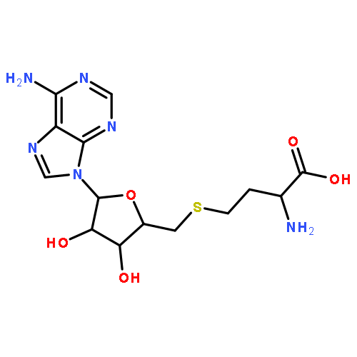
High-throughput enzyme activity screens are essential for target characterization and drug development, but few assays employ techniques or reagents that are applicable to both in vitro and live cell settings. Here, we present a class of selective and sensitive fluorescent biosensors for S-adenosyl-l-homocysteine (SAH) that provide a direct “mix and go” activity assay for methyltransferases (MTases), an enzyme class that includes several cancer therapeutic targets. Our riboswitch-based biosensors required an alternate inverted fusion design strategy, but retained full selectivity for SAH over its close structural analogue, the highly abundant methylation cofactor S-adenosyl-l-methionine (SAM). The level of ligand selectivity for these fluorescent biosensors exceeded that of commercial antibodies for SAH and proved critical to cellular applications, as we employed them to measure methylthioadenosine nucleosidase (MTAN) activity in live Escherichia coli. In particular, we were able to monitor in vivo increase of SAH levels upon chemical inhibition of MTAN using flow cytometry, which demonstrates high-throughput, single cell measurement of an enzyme activity associated with the biosynthesis of quorum sensing signal AI-2. Thus, this study presents RNA-based fluorescent biosensors as promising molecular reagents for high-throughput enzymatic assays that successfully bridge the gap between in vitro and in vivo applications.
Co-reporter:Zachary F. Hallberg;Todd A. Wright;Omer Ad;Jongchan Yeo;Xin C. Wang;Beiyan Nan
PNAS 2016 Volume 113 (Issue 7 ) pp:1790-1795
Publication Date(Web):2016-02-16
DOI:10.1073/pnas.1515287113
Over 30 years ago, GGDEF domain-containing enzymes were shown to be diguanylate cyclases that produce cyclic di-GMP (cdiG),
a second messenger that modulates the key bacterial lifestyle transition from a motile to sessile biofilm-forming state. Since
then, the ubiquity of genes encoding GGDEF proteins in bacterial genomes has established the dominance of cdiG signaling in
bacteria. However, the observation that proteobacteria encode a large number of GGDEF proteins, nearing 1% of coding sequences
in some cases, raises the question of why bacteria need so many GGDEF enzymes. In this study, we reveal that a subfamily of
GGDEF enzymes synthesizes the asymmetric signaling molecule cyclic AMP-GMP (cAG or 3′, 3′-cGAMP). This discovery is unexpected
because GGDEF enzymes function as symmetric homodimers, with each monomer binding to one substrate NTP. Detailed analysis
of the enzyme from Geobacter sulfurreducens showed it is a dinucleotide cyclase capable of switching the major cyclic dinucleotide (CDN) produced based on ATP-to-GTP
ratios. We then establish through bioinformatics and activity assays that hybrid CDN-producing and promiscuous substrate-binding
(Hypr) GGDEF enzymes are found in other deltaproteobacteria. Finally, we validated the predictive power of our analysis by
showing that cAG is present in surface-grown Myxococcus xanthus. This study reveals that GGDEF enzymes make alternative cyclic dinucleotides to cdiG and expands the role of this widely
distributed enzyme family to include regulation of cAG signaling.
Co-reporter:Colleen A. Kellenberger; Chen Chen; Aaron T. Whiteley; Daniel A. Portnoy
Journal of the American Chemical Society 2015 Volume 137(Issue 20) pp:6432-6435
Publication Date(Web):May 12, 2015
DOI:10.1021/jacs.5b00275
Cyclic di-AMP (cdiA) is a second messenger predicted to be widespread in Gram-positive bacteria, some Gram-negative bacteria, and Archaea. In the human pathogen Listeria monocytogenes, cdiA is an essential molecule that regulates metabolic function and cell wall homeostasis, and decreased levels of cdiA result in increased antibiotic susceptibility. We have generated fluorescent biosensors for cdiA through fusion of the Spinach2 aptamer to ligand-binding domains of cdiA riboswitches. The biosensor was used to visualize intracellular cdiA levels in live L. monocytogenes strains and to determine the catalytic domain of the phosphodiesterase PdeA. Furthermore, a flow cytometry assay based on this biosensor was used to screen for diadenylate cyclase activity and confirmed the enzymatic activity of DisA-like proteins from Clostridium difficile and Methanocaldococcus jannaschii. Thus, we have expanded the development of RNA-based biosensors for in vivo metabolite imaging in Gram-positive bacteria and have validated the first dinucleotide cyclase from Archaea.
Co-reporter:Colleen A. Kellenberger;Stephen C. Wilson;Yichi Su;Scott F. Hickey;Zachary F. Hallberg;Thomas F. Brewer;Hans K. Carlson;Anthony T. Iavarone;Tania L. Gonzalez;Yu-Fang Hsieh
PNAS 2015 Volume 112 (Issue 17 ) pp:5383-5388
Publication Date(Web):2015-04-28
DOI:10.1073/pnas.1419328112
Cyclic dinucleotides are an expanding class of signaling molecules that control many aspects of bacterial physiology. A synthase
for cyclic AMP-GMP (cAG, also referenced as 3′-5′, 3′-5′ cGAMP) called DncV is associated with hyperinfectivity of Vibrio cholerae but has not been found in many bacteria, raising questions about the prevalence and function of cAG signaling. We have discovered
that the environmental bacterium Geobacter sulfurreducens produces cAG and uses a subset of GEMM-I class riboswitches (GEMM-Ib, Genes for the Environment, Membranes, and Motility)
as specific receptors for cAG. GEMM-Ib riboswitches regulate genes associated with extracellular electron transfer; thus cAG
signaling may control aspects of bacterial electrophysiology. These findings expand the role of cAG beyond organisms that
harbor DncV and beyond pathogenesis to microbial geochemistry, which is important to environmental remediation and microbial
fuel cell development. Finally, we have developed an RNA-based fluorescent biosensor for live-cell imaging of cAG. This selective,
genetically encodable biosensor will be useful to probe the biochemistry and cell biology of cAG signaling in diverse bacteria.
Co-reporter:Scott F. Hickey, Ming C. Hammond
Chemistry & Biology 2014 Volume 21(Issue 3) pp:345-356
Publication Date(Web):20 March 2014
DOI:10.1016/j.chembiol.2014.01.004
•Synthesis of stable and fluorescent analogs of SAM are described•Sulfone substitution and Cy5 label affect binding to different SAM-I riboswitches•Fluorescent ligand used in direct and competitive fluorescence polarization assays•Quality of the competitive assay for high-throughput screening was validatedMany classes of S-adenosylmethionine (SAM)-binding RNAs and proteins are of interest as potential drug targets in diverse therapeutic areas, from infectious diseases to cancer. In the former case, the SAM-I riboswitch is an attractive target because this structured RNA element is found only in bacterial mRNAs and regulates multiple genes in several human pathogens. Here, we describe the synthesis of stable and fluorescent analogs of SAM in which the fluorophore is introduced through a functionalizable linker to the ribose. A Cy5-labeled SAM analog was shown to bind several SAM-I riboswitches via in-line probing and fluorescence polarization assays, including one from Staphylococcus aureus that controls the expression of SAM synthetase in this organism. A fluorescent ligand displacement assay was developed and validated for high-throughput screening of compounds to target the SAM-I riboswitch class.Figure optionsDownload full-size imageDownload high-quality image (259 K)Download as PowerPoint slide
Co-reporter:Kelly Karns ; Jacob M. Vogan ; Qian Qin ; Scott F. Hickey ; Stephen C. Wilson ; Ming C. Hammond ;Amy E. Herr
Journal of the American Chemical Society 2013 Volume 135(Issue 8) pp:3136-3143
Publication Date(Web):January 23, 2013
DOI:10.1021/ja310742m
Riboswitches are RNA sensors that change conformation upon binding small molecule metabolites, in turn modulating gene expression. Our understanding of riboswitch regulatory function would be accelerated by a high-throughput, quantitative screening tool capable of measuring riboswitch–ligand binding. We introduce a microfluidic mobility shift assay that enables precise and rapid quantitation of ligand binding and subsequent riboswitch conformational change. In 0.3% of the time required for benchtop assays (3.2 versus 1020 min), we screen and validate five candidate SAM-I riboswitches isolated from thermophilic and cryophilic bacteria. The format offers enhanced resolution of conformational change compared to slab gel formats, quantitation, and repeatability for statistical assessment of small mobility shifts, low reagent consumption, and riboswitch characterization without modification of the aptamer structure. Appreciable analytical sensitivity coupled with high-resolution separation performance allows quantitation of equilibrium dissociation constants (Kd) for both rapidly and slowly interconverting riboswitch–ligand pairs as validated through experiments and modeling. Conformational change, triplicate mobility shift measurements, and Kd are reported for both a known and a candidate SAM-I riboswitch with comparison to in-line probing assay results. The microfluidic mobility shift assay establishes a scalable format for the study of riboswitch–ligand binding that will advance the discovery and selection of novel riboswitches and the development of antibiotics to target bacterial riboswitches.
Co-reporter:Colleen A. Kellenberger ; Stephen C. Wilson ; Jade Sales-Lee
Journal of the American Chemical Society 2013 Volume 135(Issue 13) pp:4906-4909
Publication Date(Web):March 14, 2013
DOI:10.1021/ja311960g
Cyclic dinucleotides are an important class of signaling molecules that regulate a wide variety of pathogenic responses in bacteria, but tools for monitoring their regulation in vivo are lacking. We have designed RNA-based fluorescent biosensors for cyclic di-GMP and cyclic AMP-GMP by fusing the Spinach aptamer to variants of a natural GEMM-I riboswitch. In live cell imaging experiments, these biosensors demonstrate fluorescence turn-on in response to cyclic dinucleotides, and they were used to confirm in vivo production of cyclic AMP-GMP by the enzyme DncV.
.jpg)


![Adenosine, 5'-[(3-amino-3-carboxypropyl)sulfonyl]-5'-deoxy-, (S)-](http://img.cochemist.com/ccimg/53200/53199-56-7.png)
![Adenosine, 5'-[(3-amino-3-carboxypropyl)sulfonyl]-5'-deoxy-, (S)-](http://img.cochemist.com/ccimg/53200/53199-56-7_b.png)
![3',6'-Dihydroxy-3H-spiro[isobenzofuran-1,9'-xanthen]-3-one](http://img.cochemist.com/ccimg/2400/2321-07-5.png)
![3',6'-Dihydroxy-3H-spiro[isobenzofuran-1,9'-xanthen]-3-one](http://img.cochemist.com/ccimg/2400/2321-07-5_b.png)


![9,9'-[(2R,3R,3aS,7aR,9R,10R,10aS,14aR)-3,5,10,12-tetrahydroxy-5,12-dioxidooctahydro-2H,7H-difuro[3,2-d:3',2'-j][1,3,7,9,2,8]tetraoxadiphosphacyclododecine-2,9-diyl]bis(2-amino-3,9-dihydro-6H-purin-6-one)](http://img.cochemist.com/ccimg/61100/61093-23-0.png)
![9,9'-[(2R,3R,3aS,7aR,9R,10R,10aS,14aR)-3,5,10,12-tetrahydroxy-5,12-dioxidooctahydro-2H,7H-difuro[3,2-d:3',2'-j][1,3,7,9,2,8]tetraoxadiphosphacyclododecine-2,9-diyl]bis(2-amino-3,9-dihydro-6H-purin-6-one)](http://img.cochemist.com/ccimg/61100/61093-23-0_b.png)