Co-reporter:Min Li;Bingjie Mai;Ao Wang;Yiru Gao;Xiaobing Wang;Xin Liu;Shanshan Song;Quanghong Liu;Shaohua Wei
RSC Advances (2011-Present) 2017 vol. 7(Issue 65) pp:40734-40744
Publication Date(Web):2017/08/18
DOI:10.1039/C7RA06073D
Background and Objectives: Photodynamic antimicrobial chemotherapy (PACT) has emerged as a hopeful method for treating many bacteria-related infections. Phthalocyanines (Pcs) are promising photosensitizers with high photosensitivity. The aim of this study was to investigate the antimicrobial activity of new octa-cationic zinc phthalocyanines bearing 1,2-ethanediamine groups and the quaternized derivatives with different positive charges (ZnPcn+, n = 4 or 8) against Escherichia coli (E. coli) in both planktonic and biofilm states. Methods: The uptake of Pcs in E. coli was evaluated according to photometry after alkaline lysis. The dark-toxicity, light-toxicity and light-mediated antimicrobial effect of the drug were determined by plate count method. Intracellular reactive oxygen species (ROS) production was detected by flow cytometry. Scanning electron microscopy (SEM) and propidium iodide (PI) staining were performed to assess the disruption of the biofilm and membrane integrity, respectively. Results: With the incubation time prolonged, the relative fluorescence intensity of the two Pcs increased and peaked at 40 minutes. Pcs alone and irradiation itself had no evident toxicity to the bacteria while a remarkable survival decrease was observed in the PACT groups in a light dose-dependent manner. ZnPc1 showed a more than 3-log reduction while ZnPc2 caused a nearly 5-log reduction of bacterial counts. Intracellular ROS levels were significantly enhanced by PACT treatment. The disruption of the biofilm and membrane integrity detected by SEM and PI staining suggested that Pcs-PACT can effectively damage the biofilm and cell membrane. Conclusion: All the results indicate that Pcs-PACT presents excellent bactericidal activity.
Co-reporter:Mingxing Zhang;Nan Du;Lu Wang;Xiaobing Wang;Yaping Xiao;Kun Zhang;Quanhong Liu
Food & Function (2010-Present) 2017 vol. 8(Issue 10) pp:3696-3706
Publication Date(Web):2017/10/18
DOI:10.1039/C7FO00712D
Gynostrmma pentaphyllum seed oil (GPSO), extracted from G. pentaphyllum seeds, is rich in conjugated linolenic acid, which is a special fatty acid consisting of cis-9, trans-11, trans-13 isomers. Type 2 diabetes mellitus (T2DM) is characterized by hyperglycemia resulting from insulin resistance, and is usually accompanied by hypertension, hyperlipidemia, atherosclerosis (i.e., the metabolic syndrome, or syndrome X), and polycystic ovarian disease. This study aimed to investigate the effect of GPSO on T2DM hepatic lipid metabolism and the underlying mechanism involving level of protein expression. In the experiment, the model of T2DM was established. Kunming male mice were fed with a high-fat diet and injected with streptozocin, in which the exploration of detailed mechanism in the therapy of T2DM was targeted. The results showed that the ability of oral glucose tolerance was improved in the GPSO group. Biochemical indices also revealed that GPSO had a positive effect on hypoglycemic activity, suggesting that GPSO could promote the expression of glucose transporter 4 in liver and skeletal muscle.
Co-reporter:Jianmin Hu;Xiaobing Wang;Quanhong Liu;Kun Zhang;Wenli Xiong;Chuanshan Xu;Albert Wingnang Leung
Photochemistry and Photobiology 2014 Volume 90( Issue 6) pp:1404-1412
Publication Date(Web):
DOI:10.1111/php.12333
Abstract
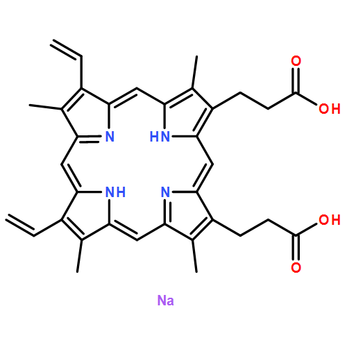
The aim of this study was to evaluate the photodynamic effect of Sinoporphyrin sodium (DVDMS). In this study, Eca-109 cells were treated with DVDMS (5 μg mL−1) and subjected to photodynamic therapy (PDT). The uptake and subcellular localization of DVDMS were monitored by flow cytometry and confocal microscopy. The phototoxicity of DVDMS was studied by MTT assay. The morphological changes were observed by scanning electron microscopy (SEM). DNA damage, reactive oxygen species (ROS) generation and mitochondria membrane potential (MMP) changes were analyzed by flow cytometry. Studies demonstrated maximal uptake of DVDMS occurred within 3 h, with a mitochondrial subcellular localization. MTT assays displayed that DVDMS could be effectively activated by light and the phototoxicity was much higher than photofrin under the same conditions. In addition, SEM observation indicated that cells were seriously damaged after PDT treatment. Furthermore, activation of DVDMS resulted in significant increases in ROS production. The generated ROS played an important role in the phototoxicity of DVDMS. DVDMS-mediated PDT (DVDMS-PDT) also induced DNA damage and MMP loss. It is demonstrated that DVDMS-mediated PDT is an effective approach on cell proliferation inhibition of Eca-109 cells.
Co-reporter:Xiaomin Su;Pan Wang;Shuang Yang;Xiaobing Wang;Quanhong Liu
European Radiology 2014 Volume 24( Issue 11) pp:2739-2753
Publication Date(Web):2014/11/01
DOI:10.1007/s00330-014-3334-3
To prove that DNA damage, intracellular reactive oxygen species (ROS) generation and loss of mitochondrial membrane potential (MMP) are contributing factors for the inhibition of cell proliferation induced by doxorubicin (DOX) administration combined with microbubble-assisted low-level therapeutic ultrasound (US) in K562 cells.3-(4, 5-dimethylthiazol-2-yl)-2, 5-diphenyl tetrazolium bromide assay was adopted to examine cytotoxicity of different treatments. Changes on apoptosis and necrosis rates, DNA fragmentation, intracellular reactive oxygen species production, mitochondrial membrane potential, cellular membrane permeability and DOX-uptake were analysed by flow cytometry. Nuclear morphology changes were observed under a fluorescence microscope. Ultrasonic cavitation was measured by spectrofluorimetry.Under optimal conditions, MB-US significantly aggravated DOX-induced K562 cell death, especially necrosis, when compared with either monotherapy. Synergistic potentiation on DNA damage, ROS generation and MMP loss were observed. Ultrasonic cavitation effects, plasma membrane permeabilization and DOX-uptake were notably improved after MB-US exposure.MB-US could increase the susceptibility of tumours to antineoplastic drugs, suggesting a potential clinical method for US-mediated tumour chemotherapy.• Microbubble-ultrasound (MB-US) aggravated doxorubicin (DOX) induced K562 cell death, especially necrosis• MB-US synergistically potentiated DOX-initiated DNA damage, ROS generation and MMP loss• Ultrasonic cavitation effects, plasma membrane permeabilization and DOX-uptake were improved after treatment• MB-US holds significant potential for improving the efficacy of conventional chemotherapy
Co-reporter:Lijie Wu, Xiaobing Wang, Quanhong Liu, Albert Wingnang Leung, Pan Wang, Chuanshan Xu
Photodiagnosis and Photodynamic Therapy (March 2016) Volume 13() pp:58-65
Publication Date(Web):1 March 2016
DOI:10.1016/j.pdpdt.2015.12.008
•DVDMS-PDT suppressed cell proliferation, triggered collapse of F-actin filaments, inhibited cell migration and initiated cell apoptotic response in MDA-MB-231 cells.•The production of ROS triggered disruption of F-actin filaments in MDA-MB-231 cells.•F-actin filaments contributed to cell migration but produced no obvious effect on cell apoptosis.ObjectiveWe previously demonstrated that the photosensitizer sinoporphyrin sodium (DVDMS) mediated photodynamic therapy (PDT) had potential advantages in inhibiting tumor growth and metastasis. However, details regarding the mechanism of cell migration inhibition remain unclear. Therefore, in this study, we aimed to investigate the effects of DVDMS-PDT on F-actin filaments, cell migration, apoptotic response and the possible interactions between them in human breast cancer MDA-MB-231 cells.Materials and methodsThe cell viability was evaluated by MTT and Guava ViaCount assays. The subcellular localization of DVDMS and reactive oxygen species (ROS) generation were analyzed by fluorescence microscope and flow cytometry. FITC-phalloidin was used to evaluate the changes of F-actin filaments. Cell migration was analyzed by scratch assay and Transwell assay. Cell apoptosis was determined by nuclear TUNEL staining and Annexin V-PE/7-AAD assay. Jasplakinolide, an F-actin stabilizer, was applied to dissect the influences of F-actin filaments disruption on cell migration and apoptosis.ResultsDVDMS-PDT significantly suppressed cell proliferation, promoted early apoptotic response, triggered collapse of F-actin filaments and inhibited cell migration in MDA-MB-231 cells. Cell migration significantly increased when cells were pretreated with F-actin stabilizer jasplakinolide after PDT, while cell apoptosis was not obviously affected. Moreover, ROS was a key factor in causing collapse of F-actin filaments.ConclusionWe demonstrated that DVDMS-PDT triggered cell apoptosis and collapse of F-actin filaments through the induction of ROS in MDA-MB-231 cells. F-actin filaments contributed to cell migration but produced no obvious effect on cell apoptosis.
Co-reporter:Yali Jia, Xiaobing Wang, Quanhong Liu, Albert Wingnang Leung, Pan Wang, Chuanshan Xu
Ultrasonics (January 2017) Volume 73() pp:154-161
Publication Date(Web):1 January 2017
DOI:10.1016/j.ultras.2016.09.013
•HB-SDT suppressed cell viability, potentiated ROS generation in MDA-MB-231 cells.•HB-SDT damaged the structure and function of mitochondria.•The apoptotic response was triggered after HB-SDT treatment.•Caspase pathway was involved in the mechanisms of cell death induced by HB-SDT.ObjectivesThe aim of the present study is to investigate the effects of sonodynamic action of hypocrellin B on human breast cancer cells and further explore its underlying mechanisms.MethodsThe cell viability of breast cancer MDA-MB-231 cells was examined by 3-(4, 5-dimethylthiazol-2-yl)-2, 5-diphenyl tetrazolium bromide (MTT) assay. Alterations on cell apoptosis, intracellular reactive oxygen species generation (ROS), mitochondrial membrane potential, and DNA fragmentation was analyzed by flow cytometer. The subcellular localization of hypocrellin B was assessed by a confocal laser scanning microscope. Mitochondria damage and nuclear morphological changes were observed under a fluorescence microscope. To further explore whether caspase pathway was involved in cell apoptotic induction of sonodynamic action of hypocrellin B, the pan-caspase inhibitor Z-Val-Ala-DL-Asp (ome)-Fluoromethylketone (z-VAD-fmk) was added to the cells one hour prior to loading the sonosensitizer, and then cell viability and apoptosis were analyzed after hypocrellin B treatment.ResultsSonodynamic treatment of hypocrellin B HB significantly suppressed cell viability of MDA-MB-231 cells. Sonodynamic action of hypocrellin B caused excessive ROS accumulation, mitochondrial dysfunction, cell apoptosis, DNA fragmentation and nuclear morphological damage. Moreover, the cytotoxicity and cell apoptosis induced by sonodynamic action of hypocrellin B were remarkably rescued by the caspase spectrum inhibitor z-VAD-fmk.ConclusionsThese results demonstrated that hypocrellin B had significant sonodynamic killing and apoptotic induction effect on breast cancer cells. And cell apoptosis induced by sonodynamic action of hypocrellin B was partly dependent on caspase pathway.


![Butanedinitrile,2,3-bis[amino[(2-aminophenyl)thio]methylene]-](http://img.cochemist.com/ccimg/109600/109511-58-2.png)
![Butanedinitrile,2,3-bis[amino[(2-aminophenyl)thio]methylene]-](http://img.cochemist.com/ccimg/109600/109511-58-2_b.png)
![2-[4-(aminoiminomethyl)phenyl]-1H-Indole-6-carboximidamide](http://img.cochemist.com/ccimg/47200/47165-04-8.png)
![2-[4-(aminoiminomethyl)phenyl]-1H-Indole-6-carboximidamide](http://img.cochemist.com/ccimg/47200/47165-04-8_b.png)


![2H-1-Benzopyran-6-ol,3,4-dihydro-2,5,8-trimethyl-2-[(4R,8R)-4,8,12-trimethyltridecyl]-, (2R)-rel-](http://img.cochemist.com/ccimg/200/148-03-8.png)
![2H-1-Benzopyran-6-ol,3,4-dihydro-2,5,8-trimethyl-2-[(4R,8R)-4,8,12-trimethyltridecyl]-, (2R)-rel-](http://img.cochemist.com/ccimg/200/148-03-8_b.png)