Co-reporter:Hendrik Reinhardt;Christoph Hellmann;Philipp Nürnberger;Stefan Kachel
Advanced Materials Interfaces 2017 Volume 4(Issue 16) pp:
Publication Date(Web):2017/08/01
DOI:10.1002/admi.201700508
Freeform growth of multiwalled carbon nanotubes (MWCNTs) is demonstrated on stainless steel AISI 304 (EN AW 1.4301) modified by pulsed laser irradiation. A nanosecond pulsed laser is utilized as a fast and facile tool for the generation of transition metal oxide precursors that promote spatially selective CNT growth upon chemical vapor deposition at 800 °C in an atmosphere comprising n-hexane and forming gas (5% H2/95% N2). Investigations on a set of 12 precursor oxides indicate that iron-rich transition metal oxides with high granularity provide best conditions for CNT growth. The laser-induced generation of specific oxide precursors facilitates the fabrication of complex CNT microarchitectures. High levels of control over CNT location and height as well as the option to influence CNT alignment provide great flexibilities in design. The introduced technique displays a high degree of automatability and the potential for upscaling thus meeting the increasing demand for large scale fabrication of CNT-enhanced devices.
Co-reporter:Hendrik M. Reinhardt;Dominik Recktenwald;Hee-Cheol Kim
Journal of Materials Science 2016 Volume 51( Issue 22) pp:9971-9978
Publication Date(Web):2016 November
DOI:10.1007/s10853-016-0224-x
A facile route for the synthesis of PHEMA hydrogel incorporating anatase nanoparticles is presented. The hydrogel features a refractive index of 1.527 and high transparency throughout the visual spectral range. Saturn rings have been machined in order to investigate the workability of the material and its potential applicability as intraocular lens implant. The resulting lenses feature high surface quality, foldability, and shape memory which makes them suitable for implantation via extracapsular cataract extraction.
Co-reporter:Annegret P. Busch, Daniel Rhinow, Fang Yang, Hendrik Reinhardt, André Beyer, Armin Gölzhäuser and Norbert Hampp
Journal of Materials Chemistry A 2014 vol. 2(Issue 40) pp:6924-6930
Publication Date(Web):16 Jul 2014
DOI:10.1039/C4TB00468J
Biomineralization of silica precursors, mediated by self-assembled proteins, is performed by many organisms. The silica cell walls of diatoms are perhaps the most stunning biomineral structures. Although the mechanisms of biomineralization are still not fully understood, template-assisted formation of silica nanostructures has gained much attention in the materials science community. Precise control of the location and the shape of structures obtained by biomineralization remains a challenge. This paper introduces a versatile biotechnological process that enables site-selective biomineralization of native biological membranes using genetically modified purple membrane (PM) from Halobacterium salinarum as a template. PM is a two-dimensional crystal consisting of bacteriorhodopsin (BR) and lipids. In this work we study PM-E234R7, a genetically modified PM containing mutated BR, where seven amino acids, starting from E234, were replaced by arginine in the C-terminus. The arginine sequence catalyzes silica formation from a tetraethylorthosilicate (TEOS) precursor. Silicification of the mutated PM variant starts with initial formation of membrane-attached spherical silica nanoparticles, which then fuse to form 2D silica nanoflakes, selectively, on the cytoplasmic side of the PM. Genetical modification of membrane proteins with poly-arginine sequences may be a general route for site-selective biomineralization of native biological membranes.
Co-reporter:Hendrik Reinhardt;Clemens Pietzonka;Bernd Harbrecht
Advanced Materials Interfaces 2014 Volume 1( Issue 2) pp:
Publication Date(Web):
DOI:10.1002/admi.201300060
Co-reporter:Martin Schraub, Hee-Cheol Kim, Norbert Hampp
European Polymer Journal 2014 Volume 51() pp:21-27
Publication Date(Web):February 2014
DOI:10.1016/j.eurpolymj.2013.11.014
•Photorefractive acrylic polymer with phenylcoumarin in the side chain.•High refractive index of n = 1.61.•Light-induced refractive index change of Δn = 0.045.•High lightfastness of the polymer.•Refractive index change due to light-triggered cyclobutan cleavage.A novel photorefractive polymer has been developed where 3-phenyl-coumarin (3PC) is attached via alkyl spacers to a methacrylate backbone. The 3PC is a fusion of the structural motives of coumarin and stilbene. In 3PC the E/Z -isomerization, known from stilbenes, is blocked and a more extended conjugated π-electron system than in coumarin is obtained. A methacrylate based polymeric material comprising 3PC in the side chain has been synthesized and its photorefractive properties have been studied. The initial refractive index is n633o = 1.613 and the maximal light-induced change of the refractive index was found to be Δn633 = −0.045 in spin-coated thin films as well as in bulk material. The glass transition temperature of about 37 °C and the high lightfastness of the polymer make it a candidate material for intraocular lenses with photo-tunable refractive index properties.Graphical abstract
Co-reporter:Hendrik Reinhardt;Hee-Cheol Kim;Clemens Pietzonka;Julia Kruempelmann;Bernd Harbrecht;Bernhard Roling
Advanced Materials 2013 Volume 25( Issue 24) pp:3313-3318
Publication Date(Web):
DOI:10.1002/adma.201205031
Co-reporter:Hendrik Reinhardt;Hee-Cheol Kim;Clemens Pietzonka;Julia Kruempelmann;Bernd Harbrecht;Bernhard Roling
Advanced Materials 2013 Volume 25( Issue 24) pp:
Publication Date(Web):
DOI:10.1002/adma.201370155
Co-reporter:Martin Schraub, Sebastian Soll, Norbert Hampp
European Polymer Journal 2013 Volume 49(Issue 6) pp:1714-1721
Publication Date(Web):June 2013
DOI:10.1016/j.eurpolymj.2013.03.021
•Photorefractive silicones with a refractive index of up to n = 1.603 are presented.•Their refractive index can be photochemically tuned in a range of up to Δn = 0.04.•Wavelength-dependent photodimerization and photocleavage could be shown.•Low Tg values ranging from −2 °C to 35 °C and high thermal stability (T5% > 410 °C).In the past decades the development of photorefractive devices evolved rapidly as can be seen by the increasing number of publications and numerous applications. There is still need of innovative materials which are suitable for a broad range of applications through tailored properties. We present photorefractive linear and crosslinked polysiloxanes with refractive indices of up to n = 1.603 which may be tuned in their refractive index in a range of up to Δn = 0.04 photochemically. The polysiloxanes, more commonly named silicones, have side chains containing coumarin which are attached to the polymer backbone via different spacers ranging in length from 3 to 9 methyl groups. The coumarins undergo wavelength-dependent photodimerization and photocleavage in the polymer which cause the desired refractive index changes. The polysiloxanes have low glass transition temperatures ranging from −2 °C to 35 °C and show a high thermal stability (T5% > 410 °C). These properties make these materials promising candidates for the manufacture of photo tunable polymers, e.g. for the use as optical data storage materials, alignment of liquid crystals, and photorefractive intraocular lenses.Graphical abstract
Co-reporter:Hendrik M. Reinhardt, Hee-Cheol Kim, Norbert A. Hampp
Journal of the European Ceramic Society 2013 Volume 33(Issue 7) pp:1281-1287
Publication Date(Web):July 2013
DOI:10.1016/j.jeurceramsoc.2013.01.005
Anodic aluminum oxide (AAO) stands in the forefront of the most important materials in modern nanotechnology. Its regular nanoporous structure is widely used for template assisted nanopatterning, optical applications, membrane science and plenty more. Our study presents an enhancement on the potential application of AAO by exploiting its inherent nature of polymorphism. Amorphous AAO is selectively transformed to α-Al2O3 by a laser-induced photo-thermal process. Carbon nanotubes (CNT) are employed as a sacrificial laser light absorber to accomplish the phase transformation of AAO to α-Al2O3. Doping of the evolving α-Al2O3 with Cr or Ti enables preparation of free designable photoluminescent surface patterns. The superior properties of α-Al2O3 are utilized to create hierarchically structured systems for optical, biomedical and lithographic applications.
Co-reporter:Philipp J. Behrendt, Hee-Cheol Kim, Norbert Hampp
Chemical Physics Letters 2013 Volume 588() pp:91-96
Publication Date(Web):19 November 2013
DOI:10.1016/j.cplett.2013.09.071
Highlights
- •
Isomer-dependent photochemical cyclobutane cleavage efficiency measured.
- •
Comparison of single-photon-absorption and two-photon-absorption photochemistry.
- •
Significant differences were found, in the range of one order of magnitude.
- •
Isolation of the most light-sensitive isomer is recommended for applications.
- •
Finding is in particular relevant for medical applications, e.g. intraocular lenses.
Co-reporter:Philipp J. Behrendt, Hee-Cheol Kim, Norbert Hampp
Journal of Photochemistry and Photobiology A: Chemistry 2013 Volume 264() pp:67-72
Publication Date(Web):15 July 2013
DOI:10.1016/j.jphotochem.2013.05.006
•Depletion zone photoreaction is a novel process type to run a photoreaction and an enabling tool for crossdimerizations (dimerization between two different components).•Suppression of homodimerization, which is mechanistically always preferred.•Significant improvement in crossdimerization-to-homodimerization ratio by a factor of at least 4 in the example shown.•Yield (crossdimer/substrate) is >90% for photochemically delicate reactions.•Depletion zone photoreaction works in commonly concentrated solutions suitable for lab work.Generally the homodimerization in an intermolecular, photochemical [2+2]-cycloaddition is the dominant reaction over the crossdimerization. Using the example of Coumarin with a 10-fold excess of 5-fluorouracil, just a 2:1 crossdimer-to-homodimer (CD/HD) ratio is obtained in a conventional Rayonet reactor. Here we present an efficient and versatile procedure for the sensitizer-free synthesis of intermolecular [2+2]-cycloaddition crossdimers in solution. Applying a monochromatic pulsed laser system as the light source first enables a highly wavelength selective excitation of only one substrate. Second, the high energy pulses establish a depletion zone inside the reaction vessel, where the concentration of the absorbing substrate in its ground state is very low. This causes the homodimerization to be completely suppressed, while favoring the crossdimerization reaction by increasing the probability to meet a non-absorbing cross-component. In our example the CD/HD-ratio shifts from less than 2 to more than 8. The depletion zone photoreaction is altogether useful for the synthesis of crossdimers.
Co-reporter:Hee-Cheol Kim;Hendrik Reinhardt;Pierre Hillebrecht ;Norbert A. Hampp
Advanced Materials 2012 Volume 24( Issue 15) pp:1994-1998
Publication Date(Web):
DOI:10.1002/adma.201200534
Co-reporter:Daniel Rhinow, Martin Imhof, Ivan Chizhik, Roelf-Peter Baumann, and Norbert Hampp
The Journal of Physical Chemistry B 2012 Volume 116(Issue 25) pp:7455-7462
Publication Date(Web):April 18, 2012
DOI:10.1021/jp2112846
Bacteriorhodopsin (BR) is the key protein of the halobacterial photosynthetic system. BR assembles into two-dimensional crystalline patches, the so-called purple membranes (PM), and acts as a light-driven proton pump converting light energy into the chemical energy of a proton gradient over the cell membrane. The two-photon absorption (TPA) of BR is so far not fully understood. Astonishingly high TPA cross sections have been reported, but the molecular mechanisms have not been elucidated. In this work, we address structural changes in BR and PM upon TPA, investigating its TPA photochemistry by spectroscopy, small-angle X-ray scattering, as well as electron and atomic force microscopy. We observe that TPA of BR leads to formation of an UV-absorbing N-retinyl-bacterioopsin state, which is accompanied by the loss of crystalline order in PM. FTIR and CD spectroscopy confirm that BR trimers as well as the secondary structure of the BR molecules are preserved. We demonstrate that excitation by TPA results in the photochemical reduction of the retinal Schiff base, which in turn causes a permanent asymmetric shape change of BR, similar to the one transiently observed during the photocycle-related opening and closing of the cytoplasmic proton half channel. This shape change causes PM sheets to merely roll up toward the extracellular side and causes the loss of crystallinity of PM. We present a model for the TPA photoresponse of BR, which also explains the irreversibility of the process in terms of a photochemical reduction of the Schiff base.
Co-reporter:Martin Imhof, Jens Pudewills, Daniel Rhinow, Ivan Chizhik, and Norbert Hampp
The Journal of Physical Chemistry B 2012 Volume 116(Issue 32) pp:9727-9731
Publication Date(Web):July 26, 2012
DOI:10.1021/jp3057459
Inkjet printing is a versatile technique widely applied in biological microarray technology. Because of its photochemical and photophysical properties, bacteriorhodopsin (BR) from Halobacterium salinarum holds promise for applications in nanotechnology, and inkjet printing would simplify the transfer of BR to suitable substrates. Surfactants are essential parts of inkjet formulations tuning viscosity, rheology, and spreading behavior of the solution. However, many surfactants destabilize the structure of proteins and often cause denaturation accompanied by a complete loss of function. Inkjet printing of membrane proteins is particularly challenging and special care must be taken in the choice of the surfactant. For BR, the situation is complicated by the fact that the structural integrity of BR depends on its native membrane environment, the so-called purple membrane (PM). PM contains 10 lipid molecules per BR monomer and is very sensitive toward surfactants. In this work, we identified surfactants suitable for inkjet formulations containing PM. Initially, we screened a variety of technically relevant surfactants for compatibility with PM using the UV–vis absorption of the retinal chromophore as a natural probe. Promising candidates were selected, and their impact on the structure of PM and BR was analyzed using UV–vis spectroscopy, CD spectroscopy, and small-angle X-ray scattering (SAXS). We identified two surfactants compatible with PM and suitable for inkjet formulations. An inkjet formulation containing PM as dye component was developed. We demonstrate that the photochromic properties of BR are maintained upon inkjet printing.
Co-reporter:Martin Imhof, Daniel Rhinow, Uwe Linne, and Norbert Hampp
The Journal of Physical Chemistry Letters 2012 Volume 3(Issue 20) pp:2991-2994
Publication Date(Web):October 3, 2012
DOI:10.1021/jz301292n
The interest in microbial opsins stems from their photophysical properties, which are superior to most organic dyes. Microbial rhodopsins like bacteriorhodopsin (BR) from Halobacterium salinarum have an astonishingly high cross-section for two-photon-absorption (TPA), which is of great interest for technological applications such as data storage. Irradiation of BR with intense laser pulses at 532 nm leads to formation of a bathochromic photoproduct, which is further converted to a photochemical species absorbing in the UV range. As demonstrated earlier, the photochemical conversions are induced by resonant TPA. However, the molecular basis of these conversions remained unresolved. In this work we use mass spectroscopy to demonstrate that TPA of BR leads to selective decarboxylation of two aspartic acids in the vicinity of the retinal chromphore. These photochemical conversions are the basis of permanent two-photon data storage in BR and are of critical importance for application of microbial opsins in optogenetics.Keywords: bacteriorhodopsin; channelrhodopsin; data storage; optogenetics; purple membrane; retinal; two-photon;
Co-reporter:Roelf-Peter Baumann, Annegret P. Busch, Björn Heidel, and Norbert Hampp
The Journal of Physical Chemistry B 2012 Volume 116(Issue 14) pp:4134-4140
Publication Date(Web):March 15, 2012
DOI:10.1021/jp210825x
Purple membranes (PM) from Halobacterium salinarum have been discussed for several technical applications. These ideas started just several years after its discovery. The biological function of bacteriorhodopsin (BR), the only protein in PM, is the light-driven proton translocation across the membrane thereby converting light energy into chemical energy. The astonishing physicochemical robustness of this molecular assembly and the ease of its isolation triggered ideas for technical uses. All basic molecular functions of BR, that is, photochromism, photoelectrism, and proton pumping, are key elements for technical applications like optical data processing and data storage, ultrafast light detection and processing, and direct utilization of sunlight in adenosine 5′-triphospate (ATP) generation or seawater desalination. In spite of the efforts of several research groups worldwide, which confirmed the proof-of-principle for all these potential applications, only the photochromism-based applications have reached a technical level. The physical reason for this is that no fixation or orientation of the PMs is required. The situation is quite different for photoelectrism and proton pumping where the macroscopic orientation of PMs is a prerequisite. For proton pumping, in addition, the formation of artificial membranes which prevent passive proton leakage is necessary. In this manuscript, we describe a new class of PM variants with oppositely charged membrane sides which enable an almost 100% orientation on a surface, which is the key element for photoelectric applications of BR. As an example, the mutated BR, BR-E234R7, was prepared and analyzed. A nearly 100% self-orientation on mica was obtained.
Co-reporter:Daniel Kehrloesser, Philipp J. Behrendt, Norbert Hampp
Journal of Photochemistry and Photobiology A: Chemistry 2012 Volume 248() pp:8-14
Publication Date(Web):15 November 2012
DOI:10.1016/j.jphotochem.2012.08.012
Two-photon-absorption (TPA) triggered photochemistry is a versatile tool to photochemically release compounds in an uncaging reaction where the desired reaction should occur behind an UV absorbing barrier, e.g. as in intraocular lenses (IOLs). Nonlinear effects of the UV-absorbing barrier, in this case the cornea, are negligible as long as the laser light passes through it at an intensity low enough that nonlinear effects do not occur. Only in the focus of the beam, i.e. inside the IOL, the required intensities are reached and TPA triggered photo cleavage occurs. The situation becomes complicated as soon as UV-absorbers are admixed to the polymer material. We show that typical concentrations of an UV-absorber only slightly affect the TPA-triggered uncaging reaction rate. As a testbed we used a newly synthetized acrylic polymer derivatized with an o-nitrobenzyl linker group carrying 5-fluorouracil as the model drug to be released. No photochemical decomposition of the UV absorber was observed. The release rate of 5-fluorouracil in presence of the UV absorber was reduced by about 6% only. Further the polymer presented here, is the first to release unmodified 5-fluorouracil without any auxiliary groups attached. This is important for potential applications in humans.Highlights► Synthesis of a photo active monomer with o-nitrobenzyl-5FU moiety for drug delivery. ► SPA quantum yield results to 0.27 and TPA cross-section is calculated to 2.41 GM. ► TPA induced drug delivery in presence of an UV-absorber is shown. ► TPA induced drug delivery from prototype intraocular lens is shown.
Co-reporter:R.-P. Baumann, J. Eussner and N. Hampp
Physical Chemistry Chemical Physics 2011 vol. 13(Issue 48) pp:21375-21382
Publication Date(Web):27 Oct 2011
DOI:10.1039/C1CP22098E
The light-driven proton pump bacteriorhodopsin (BR) embedded in a purple membrane (PM) from Halobacterium salinarum undergoes a series of conformational changes while transporting a proton from the cytoplasmic to the extracellular side over the course of the so-called photocycle. Wild-type BR variant D85T, where aspartic acid 85 is replaced by threonine, allows for the study of structural intermediates of this photocycle that are formed in a light-dependent manner in the wild-type and in thermal equilibrium by tuning the pH of the D85T purple membrane suspension. Especially the last and least studied O-intermediate of the photocycle of bacteriorhodopsin has caught recent attention. First AFM images of D85T under acidic conditions resembling wild-type BR under physiological conditions in the O-photocycle-intermediate are presented. Bacteriorhodopsins embedded in the strongly bent purple membranes were analyzed by single molecule force spectroscopy (SMFS) providing the first single molecule force spectra of BR in the O-intermediate. SMFS was further employed to determine the absolute sign of membrane curvature. Complementary electrostatic force microscopy (EFM) was performed to support PM side discrimination and determination of the bending direction. Bending of PM-D85T was analyzed in more detail providing further insight into the structure–function relationship of the bacteriorhodopsin proton pump as well as PM behaviour at the solid–liquid junction. Findings reported here are of general interest to the field of chemomechanical transducers.
Co-reporter:Martin Schraub, Helen Gray, and Norbert Hampp
Macromolecules 2011 Volume 44(Issue 22) pp:8755-8762
Publication Date(Web):November 1, 2011
DOI:10.1021/ma2015485
Stilbene is known to undergo two different reactions upon photochemical excitation. The first is an E/Z isomerization and the second is a [2 + 2]-cycloaddition of two stilbene molecules. Because both reactions occur in parallel their use is limited. Here we report on photorefractive polymers with a methacrylate backbone and covalently attached 4-hydroxy-(E)-stilbene or 3,5-dimethoxy-4-hydroxy-(E)-stilbene units in the side chain which show [2 + 2]-cycloaddition only. Both polymers, poly(4-methacryloyloxy-(E)-stilbene) (PMAES) and poly(4-methacryloyloxy-3,5-dimethoxy-(E)-stilbene) (PMADMES), show very high initial refractive indices of 1.6533 for PMAES and 1.6288 for PMADMES. The photochemical reaction upon laser irradiation with 355 nm was monitored by UV/vis, fluorescence, and IR spectroscopy. The light-induced changes of the refractive index at 633 nm measured by surface plasmon resonance (SPR) were found to be Δn > 0.05 for PMAES and Δn > 0.04 for PMADMES. The sensitivity of PMADMES is enhanced compared to PMAES due to the electron donating groups (EDG) as the direct comparison of both polymers shows. Both polymers are useful for optical devices because they do not show any absorption in the visible range and are noncrystalline as determined by wide-angle X-ray scattering (WAXS) and differential scanning calorimetry (DSC).
Co-reporter:J. Liese and N. Hampp
The Journal of Physical Chemistry A 2011 Volume 115(Issue 14) pp:2927-2932
Publication Date(Web):March 23, 2011
DOI:10.1021/jp111577j
The nature of the Woodward−Hoffmann-forbidden, thermal activated cycloreversion mechanism of cyclobutane has long been the subject of speculation and intense research. We were now able to prove the theoretically postulated biradicalic mechanism directly from radical scavenging reactions and electron paramagnetic resonance (EPR) experiments on [2 + 2] heterodimers of 5-fluoro-1-heptanoyluracil and 7-methoxy-1,1-dimethylnaphthalenon. The dimers show both the “allowed” photochemically as well as the “forbidden” thermally triggered [2 + 2] cycloreversion of the cyclobutane ring. The quantum efficiency of the photochemical cleavage is about 1%. The thermal cycloreversion reaction is independent from solvent and occurs at low activation energies of about 13 kcal/mol, even in the solid state. The radical scavenger and EPR results are further supported by the finding, that the reaction products are solely the educts for the anti-head-to-tail heterodimer. But for the syn-head-to-head heterodimer two additional products are observed, which require a sufficiently stable biradical intermediate to facilitate the required intramolecular rearrangements. Because of the surprisingly high lifetime of the radical species of these heterodimers it was possible to prove the long-discussed biradical mechanism experimentally.
Co-reporter:Daniel Kehrloesser, Roelf-Peter Baumann, Hee-Cheol Kim, and Norbert Hampp
Langmuir 2011 Volume 27(Issue 7) pp:4149-4155
Publication Date(Web):March 14, 2011
DOI:10.1021/la200238y
We describe the synthesis and photochemistry of coumarin-functionalized silica nanoparticles, which were prepared utilizing 7-[3-(triethoxysilyl)propanyloxy]coumarin (TPC) to attach coumarin as a photoactive group to the silica nanoparticle surface. The nanoparticle size and morphology were investigated by scanning electron microscopy, atomic force microscopy, and dynamic light scattering. The diameter of the spherical nanoparticles was determined by all three methods to be about 40 nm. The surface functionalization was characterized in the bulk by ζ-potential measurements and on the single-nanoparticle level by electrostatic force microscopy, where the difference in surface potential between TPC-modified and unmodified silica nanoparticles is measured. The degree of surface functionalization was determined by thermogravimetric analysis (TGA), and a theoretical limit of about 23 000 coumarin entities per nanoparticle was calculated. The photochemistry, and its reversibility, of the nanoparticle-attached coumarin entities was found to be quite different from the coumarin photochemistry in solution or on flat surfaces. Photodimerization with light of 355 nm and photocleavage with light of 254, 266, and 280 nm were analyzed by absorption and fluorescence spectroscopy. Following several cycles of photodimerization and photocleavage showed that the absorption change at 320 nm decreases from cycle to cycle. The coumarin layer on the nanoparticles was proven to be unchanged by TGA. The apparent loss of absorption change is due to the formation of interlinked nanoparticles during the dimerization−cleavage cycles. Because the coumarin groups on the inside of the obtained nanoparticle clusters are inaccessible to light, the amount of “uncleavable” dicoumarins increases, thus lowering the obtainable absorption change from cycle to cycle.
Co-reporter:Daniel Rhinow, Janet Vonck, Michael Schranz, Andre Beyer, Armin Gölzhäuser and Norbert Hampp
Physical Chemistry Chemical Physics 2010 vol. 12(Issue 17) pp:4345-4350
Publication Date(Web):23 Feb 2010
DOI:10.1039/B923756A
Ultrathin carbon nanomembranes (CNM) have been tested as supports for both cryogenic high-resolution transmission electron microscopy (cryo-EM) as well as atomic force microscopy (AFM) of biological specimens. Purple membrane (PM) from Halobacterium salinarum, a 2-D crystalline monolayer of bacteriorhodopsin (BR) and lipids, was used for this study. Due to their low thickness of just 1.6 nm CNM add virtually no phase contrast to the transmission pattern. This is an important advantage over commonly used amorphous carbon support films which become instable below a thickness of ∼20 nm. Moreover, the electrical conductivity of CNM can be tuned leading to conductive carbon nanomembranes (cCNM). cCNM support films were analyzed for the first time and were found to ideally meet all requirements of cryo-EM of insulating biological samples. A projection map of PM on cCNM at 4 Å resolution has been calculated which proves that the structural integrity of biological samples is preserved up to the high-resolution range. CNM have also proven to be suitable supports for AFM analysis of biological samples. PM on CNM was imaged at molecular resolution and single molecule force spectra were recorded which show no differences compared to force spectra of PM obtained with other substrates. This is the first demonstration of a support film material which meets the requirements of both, cryo-EM and AFM, thus enabling comparative structural studies of biomolecular samples with unchanged sample–substrate interactions. Beyond high-resolution cryo-EM of biological samples, cCNM are attractive new substrates for other biophysical techniques which require conductive supports, i.e. scanning tunneling microscopy (STM) and electrostatic force microscopy (EFM).
Co-reporter:R.-P. Baumann, M. Schranz and N. Hampp
Physical Chemistry Chemical Physics 2010 vol. 12(Issue 17) pp:4329-4335
Publication Date(Web):23 Feb 2010
DOI:10.1039/B919729J
The first AFM images of strongly bent purple membranes as well as the first single molecule force spectra of bacteriorhodopsins embedded therein are presented. AFM images of purple membranes to date always showed a flat membrane topology. Bacteriorhodopsin variants like BR-D85N and BR-D85T resemble an intermediate state of wild-type BR which is slightly ‘wedge’-shaped. Due to the strong interaction within the 2-D crystalline lattice of the purple membrane, the geometrical anisotropy of the individual bacteriorhodopsins adds up to a macroscopic change in the geometry of the purple membranes. Instead of being flat they appear like domes at physiological conditions. Single molecule force spectroscopy was employed to determine the absolute sign of the membrane curvature. As the bacteriorhodopsins in the center of the dome-like purple membranes are not supported by any solid state substrate, the presented force spectra are the first of non-supported bacteriorhodopsin, resembling the natural occurrence in the halobacterial cell.
Co-reporter:Daniel Rhinow, Ivan Chizhik, Roelf-Peter Baumann, Frank Noll, and Norbert Hampp
The Journal of Physical Chemistry B 2010 Volume 114(Issue 46) pp:15424-15428
Publication Date(Web):October 29, 2010
DOI:10.1021/jp108502p
Self-assembly of membrane proteins inside the cell membrane critically depends on specific protein−protein and protein−lipid interactions. Purple membranes (PMs) from Halobacterium salinarum comprise wild-type bacteriorhodopsin (BR) and lipids only and form a 2-D crystalline lattice of P3 symmetry in the cell membrane. It is known that removal of the retinylidene residue as well as the exchange of selected amino acids lead to a loss of crystallinity. In PMs comprising the BR variant D85T, we have observed a tunable tendency to form crystalline domains, which depends on pH-value and chloride ion concentration. BR-D85T resembles the function of the chloride pump halorhodopsin. The protonation state of amino acid residues within the binding pocket and chloride binding in the vicinity of the protonated retinal Schiff base affect the overall shape of BR-D85T molecules in the membrane, thereby changing their interactions and subsequently their tendency to form crystalline areas. The combination of small-angle X-ray scattering, atomic force microscopy, and freeze-fracture electron microscopy enables us to analyze the transitions statistically as well as on the single membrane level. PM-D85T is a model system to study membrane protein association upon substrate binding in a native environment. Furthermore, the ability to reversibly modulate the crystallinity of PMs probably will be useful for the preparation of larger artificial crystalline arrays of BR and its variants.
Co-reporter:Daniel Rhinow and Norbert Hampp
The Journal of Physical Chemistry B 2010 Volume 114(Issue 1) pp:549-556
Publication Date(Web):November 12, 2009
DOI:10.1021/jp908408d
Purple membrane (PM) from Halobacterium salinarum has been studied by many groups and is commonly described as a flat 2-D crystalline membrane microdomain which contains a hexagonal crystalline lattice of bacteriorhodopsin (BR) trimers in a stoichiometric ratio of 10:1 between lipids and BR. BR is the key protein in the halobacterial photosynthetic system and acts as a light-driven proton pump. Upon absorption of a photon, BR undergoes a cyclic series of intramolecular changes, among them a transient “wedge-like” geometrical change of the protein due to a tilt in helix F, one of the seven α-helical domains of BR. Due to the strong coupling between the BRs in the crystalline lattice, this may affect membrane topography. In nature, only low light levels occur and the total number of BRs in the “wedge-shaped” state is negligible. For mutated PMs like PM-D85T and PM-D85N (PM-D85X, X = neutral residue), the change of the membrane topography can be triggered in a pH-dependent manner. PMs containing BR-D85X look like “cups” at certain pH values. How does nature deal with a mutated PM like PM-D96G/F171C/F219L (PM-Tri) which comprises permanently “wedge-shaped” BRs and how does this influence membrane assembly? Astonishingly, we observed that PM-Tri is flat. Obviously, the morphology of Halobacterium salinarum is highly conserved and requires flat PMs to be assembled. We found that the lipid content of PM-Tri is specifically altered to assemble a hexagonal crystalline PM-Tri lattice of flat topography.
Co-reporter:Michael Schranz, Roelf-Peter Baumann, Daniel Rhinow and Norbert Hampp
The Journal of Physical Chemistry B 2010 Volume 114(Issue 27) pp:9047-9053
Publication Date(Web):May 28, 2010
DOI:10.1021/jp102377c
Purple membrane (PM) from Halobacterium salinarum, which comprises bacteriorhodopsin (BR) and lipids only, has been employed by many groups as a model system to study the structure and dynamics of membrane proteins. Although the conformational dynamics of BR within PM has been extensively analyzed with subnanometer resolution by means of diffraction experiments and spectroscopic methods, as well, structural studies of dynamical transitions within single PMs are rare. In this work, we show that tapping-mode atomic force microscopy (TM-AFM) is ideally suited to study dynamical transitions within solid-supported PMs at the nanoscale. Time-dependent AFM analysis of solid-supported PMs shows that redistribution processes take place between a crystalline core region, featuring a height of ∼5 nm, and a highly mobile rim region (∼4 nm in height). Furthermore, we discuss the influence of temperature and substrate on the equilibrium. The experiments are complemented by electrostatic force microscopy (EFM) of PM on mica. Beyond their importance for many physiological processes, dynamical transitions in biological membranes, as observed in this work, are of critical importance for all methods that make use of solid-supported membrane assemblies, either analytical tools or applications.
Co-reporter:Daniel Kehrlösser, Jens Träger, Hee-Cheol Kim and Norbert Hampp
Langmuir 2010 Volume 26(Issue 6) pp:3878-3882
Publication Date(Web):November 9, 2009
DOI:10.1021/la903433r
In this study, we report on a system consisting of self-assembled monolayers (SAMs) formed by 7-(11-trichlorosilylundecyloxy)coumarin on mica and on quartz glass. For the first time, in the absence of an inert atmosphere or a stabilizing matrix, we demonstrate by means of absorption and fluorescence spectroscopy that the photochemical cycloaddition of coumarin head groups is completely reversible in SAMs. Photodimerization and photocleavage were monitored for five cycles of alternating irradiation with light of wavelengths 355 and 254 nm, respectively. SAM formation was analyzed using atomic force microscopy and contact angle measurements. The quantum yield of the single photon absorption induced photocleavage of coumarin dimers in a SAM was determined to be Φ = 0.29. Asymmetrical photocleavage after lactone ring-opening of the coumarin dimer SAM causes a change in contact angle from about 70° to about 55°. This may be observed, for example, as a significant change in surface wettability.
Co-reporter:Jens Träger;Jasmin Heinzer;Hee-Cheol Kim
Macromolecular Bioscience 2008 Volume 8( Issue 2) pp:177-183
Publication Date(Web):
DOI:10.1002/mabi.200700155
Co-reporter:Andreas Schönafinger, Sonja Müller, Frank Noll and Norbert Hampp
Soft Matter 2008 vol. 4(Issue 6) pp:1249-1254
Publication Date(Web):21 Apr 2008
DOI:10.1039/B718150G
Embedding biological compounds, e.g. enzymes or whole cells, in solid host material seems to be a promising approach to widen their field of application far beyond the limits of natural conditions. In fields such as medicine and biotechnology, there is great interest in new methods to produce these types of composite materials in the form of micro- or nanosized particles. Such methods should be applicable to large amounts of substance. Inspired by one of nature's remarkable features—its ability to combine (bio)organic and inorganic components at the nanoscale—we developed a generic silica encapsulation method for biomolecules based on the concepts of polyelectrolyte layer adsorption followed by templated silica mineralization similar to biomineralization in diatoms. Application of this method to the model substance purple membrane (PM) resulted in a defined hybrid material with a nanoscale protective encapsulating silica shell providing a high barrier for the diffusion of low molecular weight molecules.
Co-reporter:J. Träger, S. Härtner, J. Heinzer, H.-C. Kim, N. Hampp
Chemical Physics Letters 2008 Volume 455(4–6) pp:307-310
Publication Date(Web):10 April 2008
DOI:10.1016/j.cplett.2008.02.082
The photocleavage reaction of chalcone photodimers has been studied using a two-photon process. For this purpose, a novel chalcone dimer has been synthesized as a low molecular weight model substance for polymer bound chalcones and its photochemistry triggered by two-photon-absorption (2PA) has been investigated using a pulsed frequency-doubled Nd:YAG-laser. The 2PA-induced cycloreversion reaction selectively leads to the cleavage of the chalcone photodimers resulting in the formation of monomeric chalcone molecules. Hence, as an application chalcones can be used as a photosensitive linker which can be cleaved beyond an UV-absorbing barrier. The 2PA cross section of the chalcone photodimer was determined to be of 1.1 × 10−49 cm4 s photon−1 (11 GM).The two-photon photocleavage of chalcone photodimers is reported. Selective cleavage of the photodimers gives monomeric chalcone as the sole product.
Co-reporter:Daniel Rhinow and Norbert A. Hampp
The Journal of Physical Chemistry B 2008 Volume 112(Issue 41) pp:13116-13120
Publication Date(Web):August 20, 2008
DOI:10.1021/jp803510t
Bacteriorhodopsin (BR) undergoes a conformational change during the photocycle and the proton transport through the membrane. For the first time, we could demonstrate by direct imaging of freely suspended native purple membranes (PMs) that the flat disk-like shape of PMs changes dramatically as soon as most of the BRs are in a state characterized by a deprotonated Schiff base. Light-induced shape changes are easily observed with mutated BRs of the BR-D96N type, i.e., all variants which show an increased M2 lifetime. On the other hand, large-scale shape changes are induced by pH changes with PM containing mutated BRs of the BR-D85T type, where Asp85 is replaced for a neutral amino acid. In such PMs, all BRs are titrated simultaneously and the resulting shape of the membranes depends on the initial shape only. As the majority of PMs in the “flat” state are more or less round disks, the bent membranes often comprise bowl-like and tube-like bent structures. The method presented here enables one to derive size changes of membrane-embedded BRs on the single molecule level from “macroscopic”, easily accessible data like the curvature radii observed in cryo-SEM. The potential of BR as a pH-controlled and/or light-controlled microscaled biological actuator needs further consideration.
Co-reporter:Martin Neebe, Daniel Rhinow, Nina Schromczyk and Norbert A. Hampp
The Journal of Physical Chemistry B 2008 Volume 112(Issue 23) pp:6946-6951
Publication Date(Web):May 21, 2008
DOI:10.1021/jp7111389
Purple membranes (PMs), which consist of the photochromic membrane protein bacteriorhodopsin (BR) and lipids only, show complex thermochromic properties. Three different types of reversible temperature-dependent spectral transitions were found, involving spectral states absorbing at 460, 519, and 630 nm. These thermochromic absorption changes were analyzed in the range from 10 to 80 °C. In dependence on the bulk pH value, hypsochromic or bathochromic shifts in the BR absorption spectra are observed in BR gels as well as in BR films. The thermochromic changes between both purple and blue or purple and red were quantified in the CIE color system. The molecular changes causing these effects are discussed, and a model is presented in terms of intramolecular protonation equilibriums. The thermochromic properties of BR may be of interest in applications like security tags, as this feature may complement the well-known photochromic properties of BR.
Co-reporter:D. Rhinow;N. A. Hampp
Advanced Materials 2007 Volume 19(Issue 15) pp:1967-1972
Publication Date(Web):11 JUL 2007
DOI:10.1002/adma.200602387
Submerged laser ablation is used to obtain multicomponent, patterned alkanethiol monolayers on template-stripped gold. In a decanethiol (DT) self-assembled monolayer first squares of mercaptoundecanoic acid (MUA) and then lines comprising of hexadecanethiol (HDT) were prepared. The patterned monolayers are imaged by scanning electron microscopy (see figure).
Co-reporter:Sebastian Härtner;Hee-Cheol Kim
Journal of Polymer Science Part A: Polymer Chemistry 2007 Volume 45(Issue 12) pp:2443-2452
Publication Date(Web):9 MAY 2007
DOI:10.1002/pola.22007
7-(tert-Butyldimethylsiloxy)-7′-(methacryloxy)dicoumarin (TBS-Cum-D-MA) is introduced as a versatile polymerizable and photocleavable linker system for drug attachment to acrylic polymers. Following the copolymerization of the TBS-Cum-D-MA with monomeric methylmethacrylate, a model drug chlorambucil was attached in a simple reaction. Phototriggered drug release via single- and two-photon absorption induced cycloreversion of the cyclobutane of the dicoumarin part was investigated. The bound drug chlorambucil is sensitive to UV light being required for single-photon induced cleavage of the dicoumarin linker and decomposes upon UV exposure. However, under the conditions of two-photon absorption induced drug release, no photodegradation of chlorambucil was observed. As the only effective two-photon absorber in the molecule is the cyclobutane structure, even high energies of visible light do not cause any degradation of the drug. This suggests that two-photon-triggered drug release may be successfully accomplished even with UV light sensitive drugs. The concept introduced here may be a powerful strategy in polymer design for photocontrolled drug delivery devices. © 2007 Wiley Periodicals, Inc. J Polym Sci Part A: Polym Chem 45: 2443–2452, 2007
Co-reporter:Sebastian Härtner, Hee-Cheol Kim, Norbert Hampp
Journal of Photochemistry and Photobiology A: Chemistry 2007 Volume 187(2–3) pp:242-246
Publication Date(Web):15 April 2007
DOI:10.1016/j.jphotochem.2006.10.015
tert-Butyldimethylsilyl-chloride (TBS) revealed to be a photostable protecting group for the photodimerization of 7-hydroxycoumarin in a [2 + 2]-cycloaddition. TBS-functionalized coumarin dimers show an about 100-fold increased solubility in organic solvents enabling them to be easily incorporated into polymeric films, e.g., PMMA. In the described photochemical dimerization reaction almost pure anti-head-to-head isomer is obtained. The single- and two-photon absorption-induced cycloreversion reactions in acetonitrile as well as in PMMA matrix were investigated and the two-photon absorption cross sections and quantum yields were determined to be around 1 GM and about 0.36, respectively. The only product obtained upon photocleavage of the dimer is the TBS-protected 7-hydroxycoumarin monomer. The TBS-protecting group withstands the high light intensities required for two-photon absorption-induced photocleavage without any noticeable degradation. The mild deprotection conditions for tert-butyldimethylsilyl-ethers (TBS-ethers), the chemical stability of the compound as well as its significantly improved solubility in organic solvents and its miscibility with acrylic polymers, make this a very useful compound for potential applications in 3D volumetric optical data storage and photocontrolled drug delivery.
Co-reporter:Diana Braga, Christof Christophis, Sandra Noll, Norbert Hampp
Journal of Photochemistry and Photobiology A: Chemistry 2005 Volume 172(Issue 2) pp:115-120
Publication Date(Web):31 May 2005
DOI:10.1016/j.jphotochem.2004.11.014
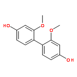
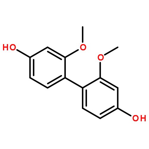
3,3′-Dimethoxy-4,2′-dihydroxybiphenyl (4,2′-DHBP) can be synthesized with high selectivity from 2-methoxyphenol (guaiacol) by photochemical excitation. Alternate methods to synthesize guaiacol dimers, e.g. peroxidase-catalyzed or electrochemically initiated dimerizations, generate a very low yield of 4,2′-DHBP in comparison. Product yield of the photochemical synthesis of 4,2′-DHBP can be enhanced by the addition of benzophenone as an activator without measurable loss in selectivity. Analytical GC–MS as well as a preparative HPLC method for the isolation of 4,2′-DHBP are described. The enzymatic and photochemical approach reveal similar selectivities of about 90%, however the peroxidase reaction leads to 3,3′-dimethoxy-4,4′-dihydroxybiphenyl (4,4′-DHBP) whereas the photochemical reaction affords 3,3′-dimethoxy-4,2′-dihydroxybiphenyl (4,2′-DHBP).
Co-reporter:R.-P. Baumann, J. Eussner and N. Hampp
Physical Chemistry Chemical Physics 2011 - vol. 13(Issue 48) pp:NaN21382-21382
Publication Date(Web):2011/10/27
DOI:10.1039/C1CP22098E
The light-driven proton pump bacteriorhodopsin (BR) embedded in a purple membrane (PM) from Halobacterium salinarum undergoes a series of conformational changes while transporting a proton from the cytoplasmic to the extracellular side over the course of the so-called photocycle. Wild-type BR variant D85T, where aspartic acid 85 is replaced by threonine, allows for the study of structural intermediates of this photocycle that are formed in a light-dependent manner in the wild-type and in thermal equilibrium by tuning the pH of the D85T purple membrane suspension. Especially the last and least studied O-intermediate of the photocycle of bacteriorhodopsin has caught recent attention. First AFM images of D85T under acidic conditions resembling wild-type BR under physiological conditions in the O-photocycle-intermediate are presented. Bacteriorhodopsins embedded in the strongly bent purple membranes were analyzed by single molecule force spectroscopy (SMFS) providing the first single molecule force spectra of BR in the O-intermediate. SMFS was further employed to determine the absolute sign of membrane curvature. Complementary electrostatic force microscopy (EFM) was performed to support PM side discrimination and determination of the bending direction. Bending of PM-D85T was analyzed in more detail providing further insight into the structure–function relationship of the bacteriorhodopsin proton pump as well as PM behaviour at the solid–liquid junction. Findings reported here are of general interest to the field of chemomechanical transducers.
Co-reporter:Daniel Rhinow, Janet Vonck, Michael Schranz, Andre Beyer, Armin Gölzhäuser and Norbert Hampp
Physical Chemistry Chemical Physics 2010 - vol. 12(Issue 17) pp:NaN4350-4350
Publication Date(Web):2010/02/23
DOI:10.1039/B923756A
Ultrathin carbon nanomembranes (CNM) have been tested as supports for both cryogenic high-resolution transmission electron microscopy (cryo-EM) as well as atomic force microscopy (AFM) of biological specimens. Purple membrane (PM) from Halobacterium salinarum, a 2-D crystalline monolayer of bacteriorhodopsin (BR) and lipids, was used for this study. Due to their low thickness of just 1.6 nm CNM add virtually no phase contrast to the transmission pattern. This is an important advantage over commonly used amorphous carbon support films which become instable below a thickness of ∼20 nm. Moreover, the electrical conductivity of CNM can be tuned leading to conductive carbon nanomembranes (cCNM). cCNM support films were analyzed for the first time and were found to ideally meet all requirements of cryo-EM of insulating biological samples. A projection map of PM on cCNM at 4 Å resolution has been calculated which proves that the structural integrity of biological samples is preserved up to the high-resolution range. CNM have also proven to be suitable supports for AFM analysis of biological samples. PM on CNM was imaged at molecular resolution and single molecule force spectra were recorded which show no differences compared to force spectra of PM obtained with other substrates. This is the first demonstration of a support film material which meets the requirements of both, cryo-EM and AFM, thus enabling comparative structural studies of biomolecular samples with unchanged sample–substrate interactions. Beyond high-resolution cryo-EM of biological samples, cCNM are attractive new substrates for other biophysical techniques which require conductive supports, i.e. scanning tunneling microscopy (STM) and electrostatic force microscopy (EFM).
Co-reporter:R.-P. Baumann, M. Schranz and N. Hampp
Physical Chemistry Chemical Physics 2010 - vol. 12(Issue 17) pp:NaN4335-4335
Publication Date(Web):2010/02/23
DOI:10.1039/B919729J
The first AFM images of strongly bent purple membranes as well as the first single molecule force spectra of bacteriorhodopsins embedded therein are presented. AFM images of purple membranes to date always showed a flat membrane topology. Bacteriorhodopsin variants like BR-D85N and BR-D85T resemble an intermediate state of wild-type BR which is slightly ‘wedge’-shaped. Due to the strong interaction within the 2-D crystalline lattice of the purple membrane, the geometrical anisotropy of the individual bacteriorhodopsins adds up to a macroscopic change in the geometry of the purple membranes. Instead of being flat they appear like domes at physiological conditions. Single molecule force spectroscopy was employed to determine the absolute sign of the membrane curvature. As the bacteriorhodopsins in the center of the dome-like purple membranes are not supported by any solid state substrate, the presented force spectra are the first of non-supported bacteriorhodopsin, resembling the natural occurrence in the halobacterial cell.
Co-reporter:Annegret P. Busch, Daniel Rhinow, Fang Yang, Hendrik Reinhardt, André Beyer, Armin Gölzhäuser and Norbert Hampp
Journal of Materials Chemistry A 2014 - vol. 2(Issue 40) pp:NaN6930-6930
Publication Date(Web):2014/07/16
DOI:10.1039/C4TB00468J
Biomineralization of silica precursors, mediated by self-assembled proteins, is performed by many organisms. The silica cell walls of diatoms are perhaps the most stunning biomineral structures. Although the mechanisms of biomineralization are still not fully understood, template-assisted formation of silica nanostructures has gained much attention in the materials science community. Precise control of the location and the shape of structures obtained by biomineralization remains a challenge. This paper introduces a versatile biotechnological process that enables site-selective biomineralization of native biological membranes using genetically modified purple membrane (PM) from Halobacterium salinarum as a template. PM is a two-dimensional crystal consisting of bacteriorhodopsin (BR) and lipids. In this work we study PM-E234R7, a genetically modified PM containing mutated BR, where seven amino acids, starting from E234, were replaced by arginine in the C-terminus. The arginine sequence catalyzes silica formation from a tetraethylorthosilicate (TEOS) precursor. Silicification of the mutated PM variant starts with initial formation of membrane-attached spherical silica nanoparticles, which then fuse to form 2D silica nanoflakes, selectively, on the cytoplasmic side of the PM. Genetical modification of membrane proteins with poly-arginine sequences may be a general route for site-selective biomineralization of native biological membranes.


![1-[4-(3-METHYL-BUTOXY)-PHENYL]-ETHANONE](http://img.cochemist.com/ccimg/30300/30237-26-4.png)
![1-[4-(3-METHYL-BUTOXY)-PHENYL]-ETHANONE](http://img.cochemist.com/ccimg/30300/30237-26-4_b.png)
](http://img.cochemist.com/ccimg/27000/26913-06-4.png)
](http://img.cochemist.com/ccimg/27000/26913-06-4_b.png)


![2H-1-Benzopyran-2-one, 7-[[(1,1-dimethylethyl)dimethylsilyl]oxy]-](http://img.cochemist.com/ccimg/918400/918314-89-3.png)
![2H-1-Benzopyran-2-one, 7-[[(1,1-dimethylethyl)dimethylsilyl]oxy]-](http://img.cochemist.com/ccimg/918400/918314-89-3_b.png)