Co-reporter:Patrick Lo, Christian Crouzet, Vitaly Vasilevko, Bernard Choi
Microvascular Research (May 2016) Volume 105() pp:109-113
Publication Date(Web):1 May 2016
DOI:10.1016/j.mvr.2016.02.002
•We applied optical histology to study of a mouse model of cerebral microbleeds.•Cerebral microbleeds occur in brain regions affected by vascular amyloid deposits.•Microbleeds are associated mainly with leaky/ruptured hemorrhagic rather than microvessels occluded by ischemia.Cerebral amyloid angiopathy (CAA) is a neurovascular disease that is strongly associated with an increase in the number and size of spontaneous microbleeds. Conventional methods of magnetic resonance imaging for detection of microbleeds, and positron emission tomography with Pittsburgh Compound B imaging for amyloid deposits, can separately demonstrate the presence of microbleeds and CAA in affected brains in vivo; however, there still is a critical need for strong evidence that shows involvement of CAA in microbleed formation. Here, we show in a Tg2576 mouse model of Alzheimer's disease, that the combination of histochemical staining and an optical clearing method called optical histology, enables simultaneous, co-registered three-dimensional visualization of cerebral microvasculature, microbleeds, and amyloid deposits. Our data suggest that microbleeds are localized within the brain regions affected by vascular amyloid deposits. All observed microhemorrhages (n = 39) were in close proximity (0 to 144 μm) with vessels affected by CAA. Our data suggest that the predominant type of CAA-related microbleed is associated with leaky or ruptured hemorrhagic microvasculature. The proposed methodological and instrumental approach will allow future study of the relationship between CAA and microbleeds during disease development and in response to treatment strategies.Optical histology enables co-registered, three-dimensional localization of the cerebral vasculature, cerebral amyloid angiopathy (CAA), and microbleeds, in a mouse model of CAA. (Top row, from left to right) 1) Tg2576 mouse brain section (~ 0.5 mm thick) after optical clearing, 2) cerebral blood vessels visualized with DiI fluorescence, 3) amyloid deposits visualized with Thioflavin S fluorescence. (Bottom row, from left to right) 4) microbleeds (located at tips of arrows) visualized with Prussian blue staining for hemosiderin in brightfield images, 5) microbleeds visualized with transmission microscopy, and 6) overlay of DiI, Thioflavin S and Prussian blue monochrome images.Download full-size image
Co-reporter:Patrick Lo, Christian Crouzet, Vitaly Vasilevko, Bernard Choi
Microvascular Research (July 2016) Volume 106() pp:137
Publication Date(Web):1 July 2016
DOI:10.1016/j.mvr.2016.04.004
Co-reporter:Kristen M. Kelly, Wesley J. Moy, Austin J. Moy, Ben S. Lertsakdadet, ... Bernard Choi
Journal of Investigative Dermatology (January 2015) Volume 135(Issue 1) pp:302-304
Publication Date(Web):1 January 2015
DOI:10.1038/jid.2014.304
Co-reporter:Bernard Choi, Wangcun Jia, Jennifer Channual, Kristen M. Kelly, Justin Lotfi
Journal of Investigative Dermatology (February 2008) Volume 128(Issue 2) pp:485-488
Publication Date(Web):1 February 2008
DOI:10.1038/sj.jid.5700991
Co-reporter:Christian Crouzet, John Quan Nguyen, Adrien Ponticorvo, Nicole P. Bernal, Anthony J. Durkin, Bernard Choi
Burns (August 2015) Volume 41(Issue 5) pp:1058-1063
Publication Date(Web):1 August 2015
DOI:10.1016/j.burns.2014.11.018
•We characterized laser speckle imaging (LSI) in a preclinical burn wound model.•LSI obtained blood-flow information of superficial-partial and deep-partial thickness burn wounds.•Blood-flow of superficial-partial and deep-partial burns differed significantly.•Results suggest the use of LSI to clinically evaluating burn wounds.A critical need exists for a robust method that enables early discrimination between superficial-partial and deep-partial thickness burn wounds. In this study, we report on the use of laser speckle imaging (LSI), a simple, non-invasive, optical imaging modality, to measure acute blood flow dynamics in a preclinical burn model. We used a heated brass comb to induce burns of varying severity to nine rats and collected raw speckle reflectance images over the course of three hours after burn. We induced a total of 12 superficial-partial and 18 deep-partial thickness burn wounds. At 3 h after burn we observed a 28% and 44% decrease in measured blood flow for superficial-partial and deep-partial thickness burns, respectively, and that these reductions were significantly different (p = 0.00007). This preliminary data suggests the potential role of LSI in the clinical management of burn wounds.


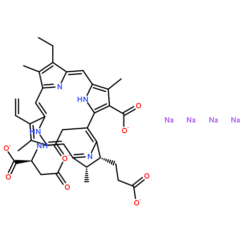
![L-Aspartic acid,N-[2-[(7S,8S)-3-carboxy-7-(2-carboxyethyl)-13-ethenyl-18-ethyl-7,8-dihydro-2,8,12,17-tetramethyl-21H,23H-porphin-5-yl]acetyl]-](http://img.cochemist.com/ccimg/110300/110230-98-3.png)
![L-Aspartic acid,N-[2-[(7S,8S)-3-carboxy-7-(2-carboxyethyl)-13-ethenyl-18-ethyl-7,8-dihydro-2,8,12,17-tetramethyl-21H,23H-porphin-5-yl]acetyl]-](http://img.cochemist.com/ccimg/110300/110230-98-3_b.png)


![[2-(CYCLOPENTYLAMINO)-1-(4-METHOXYPHENYL)-2-OXOETHYL] THIOPHENE-2-CARBOXYLATE](/data/chemimg/55600/40957-95-7.png)
![[2-(CYCLOPENTYLAMINO)-1-(4-METHOXYPHENYL)-2-OXOETHYL] THIOPHENE-2-CARBOXYLATE](/data/chemimg/55600/40957-95-7_b.png)