Co-reporter:Mei-Yun Ye, Rui-Tao Zhu, Xiang Li, Xiao-Shun Zhou, Zheng-Zhi Yin, Qian Li, and Yong Shao
Analytical Chemistry September 5, 2017 Volume 89(Issue 17) pp:8604-8604
Publication Date(Web):August 16, 2017
DOI:10.1021/acs.analchem.7b02467
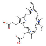
Besides the canonical Watson–Crick (WC) linked antiparallel-stranded duplex (aps-DNA), DNA is also able to form bioactive parallel-stranded duplex (ps-DNA) with the two involving strands adopting the equal 5′-3′ polarity. Discriminating ps-DNA from aps-DNA with an ideal selectivity is more challenging because of their comparable duplex topologies. Herein, we designed a unique probe of HPIN to fluorescently recognize ps-DNA but to keep an almost nonfluorescent response in binding with aps-DNA. The success of the Hoogsteen hydrogen bonding pattern in lighting up the HPIN fluorescence over the reverse Watson–Crick (rWC) one suggests the critical role of HPIN in structurally adaptive recognition to the strand polarity-determined base-pairing peculiarity. The turn-on fluorescence should result from restriction of the HPIN cis/trans isomerization upon the adaptive Hoogsteen base pair binding. Such high performance in recognizing ps-DNA against aps-DNA demonstrates the promising applications of HPIN in developing unique DNA polarity-based sensors.
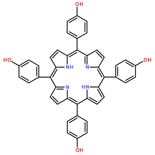
Co-reporter:Fan Lin, Yufeng Zhou, Qiusha Li, Xiaoshun Zhou, Yong Shao, Benoit Habermeyer, Hui Wang, Xinghua Shi, and Zhiai Xu
Analytical Chemistry September 5, 2017 Volume 89(Issue 17) pp:9299-9299
Publication Date(Web):July 25, 2017
DOI:10.1021/acs.analchem.7b02077
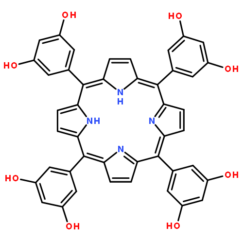
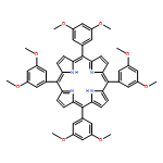
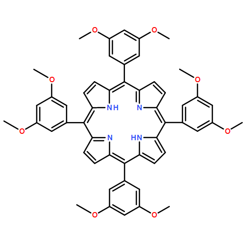
Selective nucleotide recognition for biosensor evolution requires rational probe design toward the binding-pattern-susceptible readout but without serious poison in selectivity from the context sequences. In this work, we synthesized a dual-function (trihydroxyphenyl)porphyrin (POH3) to target the abasic site (AP site) in ds-DNA using the trihydroxyphenyl substituent and the tetrapyrrole macrocycle as the recognition unit (RU) and the fluorescent signal unit (SU), respectively. RU and SU are separated from each other but are prototropically allosteric. We found that an appropriate pH favors formation of the nonfluorescent quinine/pyrrole (O–NH) conformer of POH3. However, the complementary hydrogen bonding of RU in O–NH with the target cytosine opposite the AP site switches on the SU fluorescence through prototropic allostery toward the phenol/isopyrrole (OH–N) conformer, while the bases thymine, guanine, and adenine totally silence this allostery, suggesting a superb selectivity in single-nucleotide polymorphism (SNP) analysis. The role of the prototropic allostery in achieving such SNP selectivity is also evidenced using porphyrins with other hydroxyl substituent patterns. Because of the SU separation from RU, SU is not directly involved in the interaction with the AP site, and thus, the turn-on selectivity is also realized for DNA with flanking guanine, the most easily oxidized base in DNA. This tolerance to the flanking base identity has seldom been achieved in previous studies. Additionally, other DNA structures cannot bring this allostery, indicating that the combination recipe of the AP site design and the prototropically allosteric probe will find wide applications in DNA-based sensors.
Co-reporter:Yuehua Hu;Fan Lin;Tao Wu;Yufeng Zhou;Qiusha Li;Zhiai Xu
Analytical Chemistry February 21, 2017 Volume 89(Issue 4) pp:2181-2185
Publication Date(Web):January 27, 2017
DOI:10.1021/acs.analchem.6b04709
The rapid identification of biomacromolecule structure that has a specific association with chiral enantiomers especially from natural sources will be helpful in developing enantioselective sensor and in speeding up drug exploitation. Herein, owing to its existence also in living cells, apurinic/apyrimidinic site (AP site) was first engineered into ds-DNA duplex to explore its competence in enantiomer selectivity. An AP site-specific fluorophore was utilized as an enantioselective discrimination probe to develop a straightforward chiral sensor using natural tetrahydropalmatine (L- and D-THP) as enantiomer representatives. We found that only L-THP can efficiently replace the prebound fluorophore to cause a significant fluorescence increase due to its specific binding with the AP site (two orders magnitude higher in affinity than binding with D-THP). The AP site binding specificity of L-THP over D-THP was assessed via intrinsic fluorescence, isothermal titration calorimetry, and DNA stability. The enantioselective performance can be easily tuned by the sequences near the AP site and the number of AP sites. A single AP site provides a perfect binding pocket to differentiate the chiral atom-induced structure discrepancy. We expect that our work will inspire interest in engineering local structures into a ds-DNA duplex for developing novel enantioselective sensors.
Co-reporter:Tao Wu, Fan Lin, Yuehua Hu, Ying Wang, Xiaoshun Zhou, Yong Shao
Journal of Luminescence 2016 Volume 179() pp:550-554
Publication Date(Web):November 2016
DOI:10.1016/j.jlumin.2016.08.007
Silver nanoclusters (AgNCs) have attracted wide interests in variant fields due to their easy synthesis and practical tunability in fluorescence properties. DNA has been generally used as the host to grow AgNCs due to the sequence-dependent fluorescence behavior. Actually, in such DNA, various ss-DNA segments that are structurally confined by the rigid ds-DNA counterparts have been used as the AgNCs׳ growth sites. However, whether the ds-DNA structure plays somewhat role in AgNCs׳ creation has not been well elucidated. Herein, we found that ds-DNA can also accommodate the growth of fluorescent AgNCs. The fluorescent AgNCs grown on ds-DNA should be separated each other and the G/C base pairs with right context sequences are the growth sites of fluorescent AgNCs. The intermediate A/T base pair among the continuous G/C ones seems to quench the growth of fluorescent AgNCs. For the repeat sequences, the fluorescence band position of AgNCs is not changed but the intensity is enhanced upon increasing the ds-DNA length, which is different from the results obtained with the previously reported ss-DNAs. AgNCs should be grown on the ds-DNA major groove, as convinced by the cytosine methylation experiment. Our work demonstrates that besides the ss-DNA role in defining AgNCs, one should also take into account the critical role of the ds-DNA segment in tuning the AgNCs׳ fluorescence property.
Co-reporter:Yuehua Hu;Fan Lin;Tao Wu;Ying Wang; Xiao-Shun Zhou ; Yong Shao
Chemistry – An Asian Journal 2016 Volume 11( Issue 14) pp:2041-2048
Publication Date(Web):
DOI:10.1002/asia.201600459
Abstract
DNA triplex assembly has attracted a variety of interest in the regulation of genetic expression, drug screening, molecular switches, and sensors. However, these achievements are essentially dependent on the formation and stability of the triplex assembly. Herein, the recognition of DNA triplex assembly with various isoquinoline alkaloids was investigated. We found that natural chelerythrine (CHE) exhibits the highest selectivity in recognizing the triplex structure. The DNA triplex stability is substantially increased upon CHE binding, as opposed to the invariance in the stability of the duplex counterpart. CHE also favors the assembly of the triplex-forming oligonucleotide (TFO) with its duplex counterpart. The triplex binding switches CHE to a strong fluorescent emitter, which suggests CHE as a useful probe in following triplex assembly. As a unique triplex selector, inducer, and emitter, CHE successfully reports the wide pH- and metal-ion-dependent tunability of the triplex nanoswitch in a label-free manner.
Co-reporter:Ying Wang, Yuehua Hu, Tao Wu, Lihua Zhang, Hua Liu, Xiaoshun Zhou, Yong Shao
Spectrochimica Acta Part A: Molecular and Biomolecular Spectroscopy 2016 Volume 153() pp:645-650
Publication Date(Web):15 January 2016
DOI:10.1016/j.saa.2015.09.038
•The switch-on fluorescence recognition of the DNA abasic nanocavity is investigated with 11 isoquinoline alkaloids.•Palmatine is the most efficient emitter in recognizing the AP nanocavity over the fully matched DNA.•The fluorescence switch-on recognition is suitable for all the nanocavity sequence environments.Removal of a damaged base in DNA produces an abasic site (AP site) nanocavity. If left un-repaired in vivo by the specific enzyme, this nanocavity will result in nucleotide mutation in the following DNA replication. Therefore, selective recognition of AP site nanocavity by small molecules is important for identification of such DNA damage and development of genetic drugs. In this work, we investigate the fluorescence behavior of isoquinoline alkaloids including palmatine (PAL), berberine (BER), epiberberine (EPI), jatrorrhizine (JAT), coptisine (COP), coralyne (COR), worenine (WOR), berberrubine (BEU), sanguinarine (SAN), chelerythrine (CHE), and nitidine (NIT) upon binding with the AP nanocavity. PAL is screened out as the most efficient fluorophore-switched probe to recognize the AP nanocavity over the fully matched DNA. Its fluorescence enhancement occurs for all of the AP nanocavity sequence environments, which has not been achieved by the previously used probes. The bridged π conjugation effect should partially contribute to the AP nanocavity-specific fluorescence, as opposed to the solvent effect. Due to the strong binding with the AP nanocavity, PAL will find wide applications in the DNA damage recognition and sensor development.
Co-reporter:Lihua Zhang, Hua Liu, Yong Shao, Clement Lin, Huan Jia, Gang Chen, Danzhou Yang, and Ying Wang
Analytical Chemistry 2015 Volume 87(Issue 1) pp:730
Publication Date(Web):November 27, 2014
DOI:10.1021/ac503730j
Aptamers, that exist naturally in living cells as functional elements and can switch nonfluorescent natural targets to fluorophores, are very useful in developing highly sensitive and selective biosensors and screening functional agents. This work demonstrates that human telomeric G-quadruplex (HTG) can serve as a potential fluorophore-switching aptamer (FSA) to target a natural isoquinoline alkaloid. We found that, among the G-quadruplexes studied here and the various structurally similar alkaloids including epiberberine (EPI), berberine (BER), palmatine (PAL), jatrorrhizine (JAT), coptisine (COP), worenine (WOR), sanguinarine (SAN), chelerythrine (CHE), and nitidine (NIT), only the HTG DNA, especially with a 5′-TA-3′ residue at the 5′ end of the G-quadruplex tetrad (5′-TAG3(TTAG3)3-3′, TA[Q]) as the minimal sequence, is the most efficient FSA to selectively light up the EPI fluorescence. Compared to the 5′ end flanking sequences, the 3′ end flanking sequences of the tetrad contribute significantly less to the recognition of EPI. The binding affinity of EPI to TA[Q] (Kd = 37 nM) is at least 20 times tighter than those of the other alkaloids. The steady-state absorption, steady-state/time-resolved fluorescence, and NMR studies demonstrate that EPI most likely interact with the 5′ end flanking sequence substructure beyond the core [Q] and the G-quadruplex tetrad in a much more specific manner than the other alkaloids. The highly selective and tight binding of EPI with the FSA and significantly enhanced fluorescence suggest the potential development of a selective EPI sensor (detection limit of 10 nM). More importantly, EPI, as the brightest FSA emitter among the alkaloids, can also serve as an efficient conformation probe for HTG DNA and discriminate the DNA G-quadruplex from the RNA counterpart. Furthermore, EPI can bind stoichiometrically to each G-quadruplex unit of long HTG DNA multimer with the most significant fluorescence enhancement, which has not been achieved by the previously reported probes. Our work suggests the potential use of EPI as a bioimaging probe and a therapeutic DNA binder.
Co-reporter:Ying Wang, Yuehua Hu, Tao Wu, Xiaoshun Zhou, and Yong Shao
Analytical Chemistry 2015 Volume 87(Issue 23) pp:11620
Publication Date(Web):November 10, 2015
DOI:10.1021/acs.analchem.5b02851
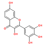
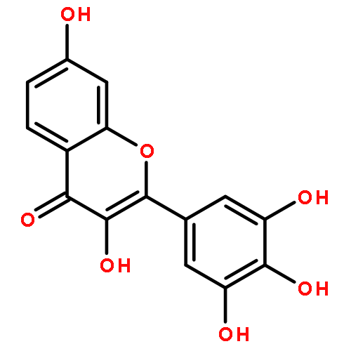
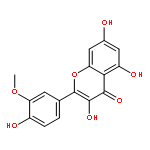
The triplex DNA has received much interest due to its various applications in gene regulation, molecular switch, and sensor development. However, realizing a highly selective recognition using a fluorescence probe specific only for the triplex topology is still a great challenge. Herein, we found that relative to the structural analogues of natural robinetin, myricetin, quercetin, kaempferol, morin, rutin, baicalin, luteolin, naringenin, genistein, chrysin, galangin, isorhamnetin, and several synthetic flavonoids, fisetin (FIS) is the brightest emitter when targeting the triplex DNA in contrast to binding with ss-DNA, ds-DNA (with or without an abasic site), i-motif, and DNA/RNA G-quadruplexes. Only the triplex association triggers the FIS green fluorescence that is relaxed from the tautomer favorable for excited-state intramolecular proton transfer (ESIPT). FIS can stabilize the triplex structure and primarily interact with the two terminals of the triplex via a 2:1 binding mode. This work demonstrates the potential of FIS as a DNA structure-selective switch-on ESIPT probe when evolving the triplex-forming oligonucleotides and developing the novel triplex-based sensors and switches.
Co-reporter:Ying Wang, Yuehua Hu, Tao Wu, Hua Liu, Lihua Zhang, Xiaoshun Zhou and Yong Shao
Analyst 2015 vol. 140(Issue 15) pp:5169-5175
Publication Date(Web):22 May 2015
DOI:10.1039/C5AN00937E
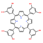
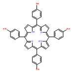
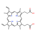
Human telomeric repeat-containing RNA (TERRA), which has recently been found to play as important a role in living cells as its DNA counterpart, solely adopts a parallel G-quadruplex (G4) topology. However, developing a highly selective fluorescent probe specific for the TERRA G4 is a great challenge, since difficulty arises in differentiating it from the DNA G4s that possess polymorphic structures including parallel, (3 + 1) hybrid, basket, and chair topologies. In this work, 5,10,15,20-tetrakis(3,5-dihydroxyphenyl)porphyrin (TOHdPP) was selected out of various porphyrins as the most efficient fluorescent probe in targeting TERRA. We found that only the TERRA binding is effective in activating the hyperporphyrin spectrum of TOHdPP, favoring red-shifted spectral bands and an enhanced fluorescence emission. Following the previous investigations on the TERRA G4 structure and our present experiments, we anticipate that TOHdPP most likely interacts with the 5′ tetrads of two TERRA G4s via a 1:2 sandwich association. The ribose 2′-OH favors the loop adenine residue-extended tetrad G4 plane that is specific for TERRA, thus besides π-stacking with the G4 tetrads, TOHdPP should also interact with this substructure to trigger an efficient electron communication between the tetraphenyl substituents and the porphyrin macrocycle, as required by the hyperporphyrin effect. The hydrogen bonding interactions of the eight hydroxyl substituents in TOHdPP with the backbone phosphate oxygen atoms of TERRA most likely further contribute to the binding selectivity. Our work demonstrates the potential of TOHdPP as a selective TERRA G4 fluorescent probe and a promising TERRA-based sensor reporter.
Co-reporter:Hua Liu, Ying Wang, Lihua Zhang, Yong Shao, Bin Zheng
Materials Letters 2015 Volume 139() pp:265-267
Publication Date(Web):15 January 2015
DOI:10.1016/j.matlet.2014.10.108
•Fluorescent silver nanoclusters (AgNCs) can be grown at CGG trinucleotide repeats.•The growth of fluorescent AgNCs is highly DNA sequence dependent.•Discrimination of CGG trinucleotide repeats from the others can be achieved with a high selectivity.Silver nanoclusters (AgNCs) are an emerging set of fluorescent materials that are advantageous over semiconductor quantum dots and organic fluorophores. Herein, fluorescent AgNCs were used as potential probes for selective recognition of DNA (CGG) trinucleotide repeat that is more related to diseases. In comparison with (CCG), (CAG), and (CTG) trinucleotide repeats, (CGG) repeat is much more efficient to produce fluorescent AgNCs. Thus, AgNCs can serve as useful fluorophores for high selective recognition of (CGG) repeat. Additionally, we used molecular crowding condition to further sensitize the (CGG)-grown AgNCs׳ fluorescence without sacrificing the high sequence selectivity. Unlike the usually used organic fluorophores, AgNCs can be in situ created at the moment of manipulation and the fluorescence is not quenched by guanine in (CGG) repeat, the most easily oxidizable base in nucleic acids.
Co-reporter:Lingling Liu, Yong Shao, Jian Peng, Chaobiao Huang, Hua Liu, and Lihua Zhang
Analytical Chemistry 2014 Volume 86(Issue 3) pp:1622
Publication Date(Web):January 10, 2014
DOI:10.1021/ac403326m
This work demonstrates the significant fluorescence enhancement of thioflavin T (ThT) when binding to G-quadruplexes possessing hybrid structures by using UV–vis absorption spectra, fluorescence spectra, and Tm experiments to confirm the binding events. ThT binding does not disturb native G-quadruplex structures preformed in Na+ and K+ solutions. The fluorescence enhancement is caused by the rotation restriction of benzothiazole (BZT) and dimethylaminobenzene (DMAB) rings in the ThT excited state upon its G-quadruplex binding. This molecular rotor mechanism as a means of fluorescence enhancement is confirmed using a nonrotor analogue of ThT. Hydroxylation and electrolyte experiments demonstrate that ThT stacks on the tetrad of the hybrid G-quadruplexes, whereas electrostatic forces contribute more to ThT binding for other G-quadruplex structures. By stacking on the tetrad, the ThT binding favors selective identification of DNA hybrid G-quadruplex structures with enhanced fluorescence and can serve as a conformation probe to monitor G-quadruplex structure conversion between hybrid and other structures. Using these properties, we developed a selective and label-free fluorescent K+ sensor with a detection limit of 1 mM for K+ in the presence of 100 mM Na+. The coexistence of other metal ions produces a fluorescence response comparable to K+ alone. We believe that ThT can potentially provide structure identification of hybrid G-quadruplexes and aid in the construction of G-quadruplex-based sensors.
Co-reporter:Jian Peng, Yong Shao, Lingling Liu, Lihua Zhang and Hua Liu
Dalton Transactions 2014 vol. 43(Issue 4) pp:1534-1541
Publication Date(Web):08 Oct 2013
DOI:10.1039/C3DT52042K
There is much demand for the development of reliable methods of bulge site recognition in DNA in order to find disease-related mutations and identify bulge sequence environments. Herein, we found that the bulge site in the DNA duplex can be used to selectively grow fluorescent silver nanoclusters (Ag NCs). The as-prepared Ag NCs exhibit an emission behavior dependent on the base environment near the bulge site and can be employed in situ as a readout for specific bulge site recognition. Fluorescent signal-on sensing for the bulge site based on this inorganic fluorophore is substantially advantageous over the organic fluorophores previously used as probes, in which a fluorescent signal-off response was usually observed with context guanines flanking the bulge site. The strong dependence of the fluorescent Ag NCs’ formation on the bases surrounding the bulge site is successfully used to identify the bulge environment near the codon 177 of cancer suppression gene p53. We expect that bulge site-determined Ag NC formation is very likely to be developed into a simple method for bulge site recognition with high selectivity and sensitivity.
Co-reporter:Jian Peng, Yong Shao, Lingling Liu, Lihua Zhang, Hua Liu, Ying Wang
Analytica Chimica Acta 2014 850() pp: 78-84
Publication Date(Web):
DOI:10.1016/j.aca.2014.08.032
Co-reporter:Shujuan Xu, Yong Shao, Fei Wu, Guiying Liu, Lingling Liu, Jian Peng, Yanwei Sun
Journal of Luminescence 2013 Volume 136() pp:291-295
Publication Date(Web):April 2013
DOI:10.1016/j.jlumin.2012.12.016
Many efforts have been made for sequence-specific recognition of DNA by small molecules. In this study, abasic site (AP site) in a DNA duplex was found to be targeted by myricetin (Myr), one of the natural 3-hydroxyflavonols. Steady-state and transient-state fluorescence, FRET, and DNA melting experiments confirmed that the AP site binding of Myr favors an emission from its tautomer that is derived from excited-state intramolecular proton transfer (ESIPT) reaction between the 3-OH and 4-carbonyl moieties. The selective recognition of Myr is less dependent on the flanking bases of the AP site, although the ESIPT emission is more lighting up for the DNAs with cytosine and thymine opposite the AP site. Because Myr alone in aqueous solution is non-fluorescent, this selective lighting-up emission is advantageous for developing a practical sensor to target the DNA AP site with a weak fluorescence background. This selective recognition of the AP site by the lighting-up fluorescence response would find wide applications including efficiently evaluating DNA damage/repair and screening antitumor/antioxidation drugs.Highlights► Myricetin exhibits a strong binding to the DNA abasic site. ► The binding of myricetin at the AP site is sequence dependent. ► The AP site binding of myricetin favors an enhanced ESIPT emission.
Co-reporter:Lingling Liu, Yong Shao, Jian Peng, Hua Liu and Lihua Zhang
Molecular BioSystems 2013 vol. 9(Issue 10) pp:2512-2519
Publication Date(Web):05 Jul 2013
DOI:10.1039/C3MB70201D
Thioflavin T (ThT) has been widely utilized as a fluorescent marker for amyloid fibrils. However, the use of ThT as an efficient reporter for a specific DNA structure still remains in question. Here, we report that the fluorescence intensity of ThT is obviously enhancement in when it binds to ds-DNAs which contain cavity structures such as an abasic site, gap site or mismatch site. Such enhancement in fluorescence cannot be achieved for DNA without these cavity structures. The DNA cavities provide appropriate spaces to accommodate ThT and allow the occurrence of some specific interactions. The stacking interaction of the bound ThT with the cavity context bases is the main driving force for ThT binding to the cavities. This interaction restricts the excited state's rapid torsional rotation around the single C–C bond between the benzothiazole and dimethylaminobenzene moieties and thus results in a decreased population of the nonradiative twisted internal charge-transfer (TICT) state. It is impossible for this stacking interaction to occur in DNA without these cavities. This property can be used to recognize DNA cavities with high selectivity and sensitivity. We expect that the ability of ThT to target these DNA structures has the potential to be developed into practical and functional biomaterials for DNA sensors or devices.
Co-reporter:Yanwei Sun, Yong Shao, Jian Peng, Lingling Liu, Botao Teng
Sensors and Actuators B: Chemical 2013 Volume 188() pp:242-248
Publication Date(Web):November 2013
DOI:10.1016/j.snb.2013.07.034
DNA-binding-switched conversion of small molecule's structure and property provides a promising tool for achieving a high selectivity in identification of a DNA specific site. Herein, the optical properties of neutral red in response to its abasic site (AP site) binding were investigated for this purpose. At pH 8.4, neutral red exists in solution mainly at the neutral form (NR). However, the presence of an AP site in DNA converts NR to a protonated form (NRH+), following a 100-nm red shift in absorption spectra. This conversion is inefficient for the fully matched DNAs without the AP site. Relative to the absorption spectra, the fluorescence in response to the binding-switched conversion is more dependent on the sequences near the AP site. The AP site's hydrophobic microenvironment and electron transfer between the AP site-bound NRH+ in excited state and the nearby context bases govern the emission behavior. It was found that the AP site having a large void space favors occurrence of dipole interaction between the bound NRH+ and the context bases. This AP site-dependent conversion of neutral red in structure and then its optical properties would find wide applications in the field of DNA assay with a high selectivity and sensitivity.
Co-reporter:Qinghua Cui, Kun Ma, Yong Shao, Shujuan Xu, Fei Wu, Guiying Liu, Norio Teramae, Haifeng Bao
Analytica Chimica Acta 2012 Volume 724() pp:86-91
Publication Date(Web):29 April 2012
DOI:10.1016/j.aca.2012.02.044
Silver nanoclusters (Ag NCs) templated with DNAs have attracted much attention as novel fluorophores because of their convenient emission tunability by the sequence and length of the template DNAs. However, the precise production of Ag NCs in a site-specific manner still remains a challenge to attain highly selective and label-free DNA recognition. Here we exploited the availability of a gap site in DNA duplexes as a new scaffold for the synthesis of Ag NCs. Compared to the commonly used DNA templates for the creation of Ag NCs, the gap site in DNA duplexes was found to facilitate the rapid formation of the fluorescent Ag NCs without sacrifice of their bright emission and excellent stability. We found that fluorescent Ag NCs were highly selectively formed when cytosine faced toward the gap site in DNA duplexes, and they were in situ utilized as readout by signal-on manner for the DNA mutation assays. This base-selective growth of the fluorescent Ag NCs at the gap site would find promising applications in practical detection of single nucleotide polymorphism (SNP) and construction of DNA-based functional sensors with label-free and cost-effective merits.Graphical abstractHighlights► A gap site in DNA duplex is exploited as a new scaffold for the rapid synthesis of Ag NCs. ► Fluorescent Ag NCs are highly selectively formed when cytosine faced toward the gap site in DNA duplexes, and they could be utilized as readout with signal-on signaling for DNA single nucleotide polymorphism. ► This base-selective growth of the fluorescent Ag NCs at the gap site would find promising applications in label-free and cost-effective designs of DNA-based functional sensors.
Co-reporter:Fei Wu, Yong Shao, Kun Ma, Qinghua Cui, Guiying Liu and Shujuan Xu
Organic & Biomolecular Chemistry 2012 vol. 10(Issue 16) pp:3300-3307
Publication Date(Web):15 Feb 2012
DOI:10.1039/C2OB00028H
Label-free DNA nucleobase recognition by fluorescent small molecules has received much attention due to its simplicity in mutation identification and drug screening. However, sequence-dependent fluorescence light-up nucleobase recognition and multicolor emission with individual emission energy for individual nucleobases have been seldom realized. Herein, an abasic site (AP site) in a DNA duplex was employed as a binding field for berberine, one of isoquinoline alkaloids. Unlike weak binding of berberine to the fully matched DNAs without the AP site, strong binding of berberine to the AP site occurs and the berberine's fluorescence light-up behaviors are highly dependent on the target nucleobases opposite the AP site in which the targets thymine and cytosine produce dual emission bands, while the targets guanine and adenine only give a single emission band. Furthermore, more intense emissions are observed for the target pyrimidines than purines. The flanking bases of the AP site also produce some modifications of the berberine's emission behavior. The binding selectivity of berberine at the AP site is also confirmed by measurements of fluorescence resonance energy transfer, excited-state lifetime, DNA melting and fluorescence quenching by ferrocyanide and sodium chloride. It is expected that the target pyrimidines cause berberine to be stacked well within DNA base pairs near the AP site, which results in a strong resonance coupling of the electronic transitions to the particular vibration mode to produce the dual emissions. The fluorescent signal-on and emission energy-modulated sensing for nucleobases based on this fluorophore is substantially advantageous over the previously used fluorophores. We expect that this approach will be developed as a practical device for differentiating pyrimidines from purines by positioning an AP site toward a target that is available for readout by this alkaloid probe.
Co-reporter:Qinghua Cui, Yong Shao, Kun Ma, Shujuan Xu, Fei Wu and Guiying Liu
Analyst 2012 vol. 137(Issue 10) pp:2362-2366
Publication Date(Web):15 Mar 2012
DOI:10.1039/C2AN00025C
Due to their good photostability, high quantum yield and low toxicity, fluorescent silver nanoclusters (Ag NCs) have received much attention as novel fluorophores for sensing applications. In this work, we investigate the upconversion emission of Ag NCs templated by single- and double-stranded DNAs. DNA-templated Ag NCs exhibit upconversion emission at wavelengths identical to those observed for the corresponding Stokes emission. Consequently, the Ag NCs' upconversion behavior can be easily tuned by the used DNA sequences. In addition, the Ag NCs are more stable under such an NIR excitation with the upconversion mode relative to the Stokes mode. As a proof-of-concept application, DNA nucleobase recognition with the in situ formed Ag NCs is realized using the Ag NCs' upconversion emission. We expect that the Ag NCs' upconversion emission is more advantageous than the previously used rare-earth materials, at least with respect to easy modulation of the emission energies by DNA sequences, and could find wide application in sensor design.
Co-reporter:Shujuan Xu, Yong Shao, Kun Ma, Qinghua Cui, Guiying Liu, Fei Wu
Sensors and Actuators B: Chemical 2012 s 171–172() pp: 666-671
Publication Date(Web):
DOI:10.1016/j.snb.2012.05.051
Co-reporter:Kun Ma, Yong Shao, Qinghua Cui, Fei Wu, Shujuan Xu, and Guiying Liu
Langmuir 2012 Volume 28(Issue 43) pp:15313-15322
Publication Date(Web):August 11, 2012
DOI:10.1021/la301957m
DNA-templated silver nanoclusters (Ag NCs) are emerging sets of fluorophores that are widely applicable because of high brightness, good photostability, and visible to near-infrared emissions tunable using the DNA sequence and length to change the NC size. We find that fluorescent Ag NCs can be size-selectively grown at DNA abasic sites (AP site) using a constrained duplex environment opposed by a cytosine and flanked by two guanines. The size of the AP site-grown Ag NCs is not affected by the increasing Ag+ concentration. A Job’s plot analysis shows that Ag2 NCs are the species responsible for the observed emissions. Although varying the DNA sequence one base away from the AP site (i.e., the Ag NC growth site) does not alter the size of the fluorescent Ag NCs, the emissions of the formed Ag NCs are still gradually red shifted as the sequence changes from thymine (T) to cytosine (C), adenine (A), and guanine (G). Furthermore, this emission shift is strongly dependent on the base-stacking direction of the 3′-side sequence of the 5′-G stack exactly flanking the AP site, which exhibits a larger emission alteration than altering the 5′-side sequence of the 3′-G stack flanking the AP site on the other side of the site. The excited-state lifetimes of the Ag NCs are inversely proportional to the singlet energies (ΔE0,0) of the Ag NCs relative to their ground state and of the vertical ionization potentials of the guanines directly flanking the AP site as determined by the base stacking. All of these results support the conclusion that the Ag NC excited state becomes more stable by interacting with a guanine base because of the larger electronic dipole moment that can be modified by the stacked sequences. Additionally, the size of the formed Ag NCs seems to be dependent on the consecutive AP site number. Thus, the AP site design in this work provides an easy way to shed light on the role of DNA base stacking in the optical properties of Ag NCs.
Co-reporter:Guiying Liu;Kun Ma;Qinghua Cui;Fei Wu;Shujuan Xu
Gold Bulletin 2012 Volume 45( Issue 2) pp:69-74
Publication Date(Web):2012 July
DOI:10.1007/s13404-012-0049-6
Water-soluble and red-emitting gold nanoclusters (Au NCs) were synthesized with single-stranded DNA as a promising biotemplate and dimethylamine borane as a mild reductant. The fluorescent Au NCs can be formed in a weakly acidic aqueous solution that is free from the simultaneous formation of large nanoparticles. The cluster feature of the formed Au species has been revealed by fluorescence spectra, absorption spectra, and transmission electron microscopy. Additionally, DNA sequences could be used to tune the Au NCs' emissions. The as-prepared Au NCs display high stability at physiological pH condition, and thus, wide potential applications are anticipated for the biocompatible fluorescent Au NCs serving as nanoprobes in bioimaging and related fields.
Co-reporter:Shujuan Xu, Yong Shao, Kun Ma, Qinghua Cui, Guiying Liu, Fei Wu and Minjie Li
Analyst 2011 vol. 136(Issue 21) pp:4480-4485
Publication Date(Web):22 Sep 2011
DOI:10.1039/C1AN15652G
DNA single-nucleotide polymorphism (SNP) detection has attracted much attention due to mutation-related diseases. Various fluorescence methods for SNP detection have been proposed and many are already in use. However, fluorescence enhancement for signal-on SNP identification without label modification still remains a challenge. Here, we find that the abasic site (AP site) in a DNA duplex can be developed as a binding pocket favorable for the occurrence of the excited-state intramolecular proton transfer (ESIPT) of a 3-hydroxyflavone, fisetin, which is used as a proof of concept for effective SNP identification. Fisetin binding at the AP site is highly selective for target thymine or cytosine facing the AP site by observation of a drastic increase in the ESIPT emission band. In addition, the target recognition selectivity based on this ESIPT process is not affected by flanking bases of the AP site. The binding selectivity of fisetin at the AP site is also confirmed by measurements of fluorescence resonance energy transfer, emission lifetime and DNA melting. The fluorescent signal-on sensing for SNP based on this fluorophore is substantially advantageous over the previously used fluorophores such as the AP site-specific signal-off organic ligands with a similar fluorescing mechanism before and after binding to DNA with hydrogen bonding interaction. We expect that this approach will be employed to develop a practical SNP detection method by locating an AP site toward a target and employing an ESIPT probe as readout.
Co-reporter:Yong Shao, Zhenjiang Niu, Shiying Zou
Electrochemistry Communications 2009 Volume 11(Issue 2) pp:417-420
Publication Date(Web):February 2009
DOI:10.1016/j.elecom.2008.12.005
The binding specificity of silver cations to abasic (AP) site-containing DNA was electrochemically investigated by comparison with the fully matched DNA without the AP site. AP site-containing DNA is designed in a way that only the nucleotide opposite the AP site is variable to allow for coexistence of an unpaired nucleotide and a number of DNA base pairs. The surface of a gold electrode was modified by AP site-containing DNA duplex on which Ag+ binding specificity was evaluated. Electrochemical investigations on the AP-DNA-modified electrodes reveal that Ag+ preferentially associates to the unpaired nucleotides instead of the coexisted base pairs and shows sequence-dependant binding, especially stronger for purines than for pyrimidines. Additionally, the hydrogen bond pattern moieties of the unpaired nucleotides should be involved in Ag+ binding evidenced by a decrease of the redox signal when introducing a ligand with its hydrogen bond moiety complementary to the nucleotide deoxycytidine. This is the first attempt to make a comparison in one DNA molecule for metal ion binding to coexisted unpaired nucleotide and DNA base pairs. The present method demonstrates an easy way for investigating binding specificity of heavy metal ions to AP site in the presence of coexisted DNA base pairs.
Co-reporter:Yong SHAO;Zhenjiang NIU;Jianrong CHEN;Liangke ZHANG
Chinese Journal of Chemistry 2009 Volume 27( Issue 6) pp:1117-1122
Publication Date(Web):
DOI:10.1002/cjoc.200990186
Abstract
Intercalators are well known for their DNA binding specificity by inserting between base pairs, whereas the binding event occurring to apurine/apyrimidine site (AP site)-containing DNA for this type of noncovalent interaction is still not highlighted although AP site is frequently in vivo produced in living cells. Here proflavine (PF) as an example is used to investigate the binding specificity of the AP site in DNA for a non-hydrogen bond ligand. Experimental results indicate that the AP site should be the preferential binding site for PF. The intrinsic binding constant of PF for the AP site is one order of magnitude greater than that occurring for PF intercalation. Additionally, the thermostability of the AP site-containing DNA is significantly increased after PF binding. The PF bound to the AP site should adopt a specific binding orientation distinguishable from that by which PF intercalated into base pairs. The results obtained here should be very useful for judging biochemical and biophysical effectiveness of small molecules based on their different binding behavior to DNA.
Co-reporter:Fei Wu, Yong Shao, Kun Ma, Qinghua Cui, Guiying Liu and Shujuan Xu
Organic & Biomolecular Chemistry 2012 - vol. 10(Issue 16) pp:NaN3307-3307
Publication Date(Web):2012/02/15
DOI:10.1039/C2OB00028H
Label-free DNA nucleobase recognition by fluorescent small molecules has received much attention due to its simplicity in mutation identification and drug screening. However, sequence-dependent fluorescence light-up nucleobase recognition and multicolor emission with individual emission energy for individual nucleobases have been seldom realized. Herein, an abasic site (AP site) in a DNA duplex was employed as a binding field for berberine, one of isoquinoline alkaloids. Unlike weak binding of berberine to the fully matched DNAs without the AP site, strong binding of berberine to the AP site occurs and the berberine's fluorescence light-up behaviors are highly dependent on the target nucleobases opposite the AP site in which the targets thymine and cytosine produce dual emission bands, while the targets guanine and adenine only give a single emission band. Furthermore, more intense emissions are observed for the target pyrimidines than purines. The flanking bases of the AP site also produce some modifications of the berberine's emission behavior. The binding selectivity of berberine at the AP site is also confirmed by measurements of fluorescence resonance energy transfer, excited-state lifetime, DNA melting and fluorescence quenching by ferrocyanide and sodium chloride. It is expected that the target pyrimidines cause berberine to be stacked well within DNA base pairs near the AP site, which results in a strong resonance coupling of the electronic transitions to the particular vibration mode to produce the dual emissions. The fluorescent signal-on and emission energy-modulated sensing for nucleobases based on this fluorophore is substantially advantageous over the previously used fluorophores. We expect that this approach will be developed as a practical device for differentiating pyrimidines from purines by positioning an AP site toward a target that is available for readout by this alkaloid probe.
Co-reporter:Jian Peng, Yong Shao, Lingling Liu, Lihua Zhang and Hua Liu
Dalton Transactions 2014 - vol. 43(Issue 4) pp:NaN1541-1541
Publication Date(Web):2013/10/08
DOI:10.1039/C3DT52042K
There is much demand for the development of reliable methods of bulge site recognition in DNA in order to find disease-related mutations and identify bulge sequence environments. Herein, we found that the bulge site in the DNA duplex can be used to selectively grow fluorescent silver nanoclusters (Ag NCs). The as-prepared Ag NCs exhibit an emission behavior dependent on the base environment near the bulge site and can be employed in situ as a readout for specific bulge site recognition. Fluorescent signal-on sensing for the bulge site based on this inorganic fluorophore is substantially advantageous over the organic fluorophores previously used as probes, in which a fluorescent signal-off response was usually observed with context guanines flanking the bulge site. The strong dependence of the fluorescent Ag NCs’ formation on the bases surrounding the bulge site is successfully used to identify the bulge environment near the codon 177 of cancer suppression gene p53. We expect that bulge site-determined Ag NC formation is very likely to be developed into a simple method for bulge site recognition with high selectivity and sensitivity.

![2,6-Dibromobenzo[1,2-b:4,5-b']dithiophene](http://img.cochemist.com/ccimg/909300/909280-97-3.png)
![2,6-Dibromobenzo[1,2-b:4,5-b']dithiophene](http://img.cochemist.com/ccimg/909300/909280-97-3_b.png)

![Propanedinitrile,2-[(2,3,6,7-tetrahydro-1H,5H-benzo[ij]quinolizin-9-yl)methylene]-](http://img.cochemist.com/ccimg/58300/58293-56-4.png)
![Propanedinitrile,2-[(2,3,6,7-tetrahydro-1H,5H-benzo[ij]quinolizin-9-yl)methylene]-](http://img.cochemist.com/ccimg/58300/58293-56-4_b.png)

![1H-Isoindole-1,3(2H)-dione,2-[(3R)-2,6-dioxo-3-piperidinyl]-](http://img.cochemist.com/ccimg/2700/2614-06-4.png)
![1H-Isoindole-1,3(2H)-dione,2-[(3R)-2,6-dioxo-3-piperidinyl]-](http://img.cochemist.com/ccimg/2700/2614-06-4_b.png)
![3',6'-Dihydroxy-3H-spiro[isobenzofuran-1,9'-xanthen]-3-one](http://img.cochemist.com/ccimg/2400/2321-07-5.png)
![3',6'-Dihydroxy-3H-spiro[isobenzofuran-1,9'-xanthen]-3-one](http://img.cochemist.com/ccimg/2400/2321-07-5_b.png)


![Benzo[1,2-b:4,5-b']dithiophene](/data/chemimg/477000/267-65-2.png)
![Benzo[1,2-b:4,5-b']dithiophene](/data/chemimg/477000/267-65-2_b.png)
![9-hydroxy-10-methoxy-5,6-dihydro[1,3]dioxolo[4,5-g]isoquino[3,2-a]isoquinolin-7-ium](http://img.cochemist.com/ccimg/17400/17388-19-1.png)
![9-hydroxy-10-methoxy-5,6-dihydro[1,3]dioxolo[4,5-g]isoquino[3,2-a]isoquinolin-7-ium](http://img.cochemist.com/ccimg/17400/17388-19-1_b.png)
![1,2-dimethoxy-12-methyl[1,3]benzodioxolo[5,6-c]phenanthridinium](http://img.cochemist.com/ccimg/34400/34316-15-9.png)
![1,2-dimethoxy-12-methyl[1,3]benzodioxolo[5,6-c]phenanthridinium](http://img.cochemist.com/ccimg/34400/34316-15-9_b.png)


![Dibenzo[a,g]quinolizinium,2,3,10,11-tetramethoxy-8-methyl-](http://img.cochemist.com/ccimg/6900/6872-73-7.png)
![Dibenzo[a,g]quinolizinium,2,3,10,11-tetramethoxy-8-methyl-](http://img.cochemist.com/ccimg/6900/6872-73-7_b.png)
![[1,3]Dioxolo[4',5':4,5]benzo[1,2-c]phenanthridinium,2,3-dimethoxy-12-methyl-](http://img.cochemist.com/ccimg/6900/6872-57-7.png)
![[1,3]Dioxolo[4',5':4,5]benzo[1,2-c]phenanthridinium,2,3-dimethoxy-12-methyl-](http://img.cochemist.com/ccimg/6900/6872-57-7_b.png)
![2,9,10-trimethoxy-5,6-dihydroisoquinolino[2,1-b]isoquinolin-7-ium-3-ol](http://img.cochemist.com/ccimg/3700/3621-38-3.png)
![2,9,10-trimethoxy-5,6-dihydroisoquinolino[2,1-b]isoquinolin-7-ium-3-ol](http://img.cochemist.com/ccimg/3700/3621-38-3_b.png)
![Bis[1,3]benzodioxolo[5,6-a:4',5'-g]quinolizinium,6,7-dihydro-](http://img.cochemist.com/ccimg/3500/3486-66-6.png)
![Bis[1,3]benzodioxolo[5,6-a:4',5'-g]quinolizinium,6,7-dihydro-](http://img.cochemist.com/ccimg/3500/3486-66-6_b.png)


