Co-reporter:Yan Wei;Shuai Xu;Feng Wang;Aifeng Zou;Shuang Zhang;Yan Xiong;Shilei Cao;Qizhi Zhang;Yajie Wang
Journal of Pharmaceutical Sciences 2015 Volume 104( Issue 1) pp:165-177
Publication Date(Web):
DOI:10.1002/jps.24234
Lapatinib (LPT) could sensitize human epidermal growth factor receptor-2 (HER-2) positive breast cancer to paclitaxel (PTX) and induce synergetic action with PTX in preclinical test and phase II/III trial. In this study, LPT-conjugated poly (ethylene glycol) (PEG) and poly (lactic acid) (PLA) (LPT–PEG–PLA) was first synthesized and confirmed with 1H Nuclear Magnetic Resonance and Matrix-Assisted Laser Desorption/ Ionization Time of Flight Mass Spectrometry, which was used for the preparation of a novel PEG–PLA combined micelles of LPT and PTX (PPM-LP). The obtained PPM-LP exhibited uniform, spherical shape with a size of 25.80 ± 0.47 nm and zeta potential of −3.17 ± 0.15 mv. PTX existed in molecular or amorphous forms in the micelles and superficial LPT could better delay PTX release. The cytotoxicity of PPM-LP with LPT conjugation against SKBr-3 cells (HER-2 positive) was found to be significantly increasing as compared with PPM–PTX, whereas there was no significant difference against MDA-MB-231 cells (HER-2 negative). PPM-LP could escape from endosomes and be distributed into cytoplasm and led to cell arrest in G2/M and G1/S phases simultaneously. Results of nucleus staining and flow cytometry confirmed that LPT could remarkably increase antineoplastic effect of PTX against SKBr-3 cells. All these results demonstrated that PPM-LP may be a promising drug delivery system for HER-2 positive breast cancer. © 2014 Wiley Periodicals, Inc. and the American Pharmacists Association J Pharm Sci 104:165–177, 2015
Co-reporter:Huile Gao, Zhi Yang, Shuang Zhang, Zhiqing Pang, Qingfeng Liu, Xinguo Jiang
Acta Biomaterialia 2014 Volume 10(Issue 2) pp:858-867
Publication Date(Web):February 2014
DOI:10.1016/j.actbio.2013.11.003
Abstract
A dual targeting delivery system was developed to completely conquer the two barriers that glioma treatment faces: the blood–brain barrier (BBB) and the brain–glioma barrier. Recently, a system comprising AS1411 aptamer (for glioma targeting) and TGN peptide (for BBB targeting) modified nanoparticles (AsTNPs) was developed, which can effectively target brain glioma and improve the survival of glioma-bearing mice. However, the in vitro models currently used are far too different from the in vivo tumor microenvironment that the glioma targeting delivery system actually faces. In this study, the pharmacology mechanisms of AsTNPs were explored in several models that imitated the tumor microenvironment. AsTNPs can be selectively taken up by endothelial and glioma cells, effectively penetrating the BBB and brain–glioma barriers to reach glioma cells and display their anti-glioma effect. The cell monolayers, tumor spheroids and coculture systems were more suitable in vitro models for the pharmacology evaluation of targeted drug delivery systems.
Co-reporter:Huile Gao, Shuang Zhang, Shijie Cao, Zhi Yang, Zhiqing Pang, and Xinguo Jiang
Molecular Pharmaceutics 2014 Volume 11(Issue 8) pp:2755-2763
Publication Date(Web):July 1, 2014
DOI:10.1021/mp500113p
Gliomas are hard to treat because of the two barriers involved: the blood–brain barrier and blood–tumor barrier. In this study, a dual-targeting ligand, angiopep-2, and an activatable cell-penetrating peptide (ACP) were functionalized onto nanoparticles for glioma-targeting delivery. The ACP was constructed by conjugating RRRRRRRR (R8) with EEEEEEEE through a matrix metalloproteinase-2 (MMP-2)-sensitive linker. ACP modification effectively enhanced the C6 cellular uptake because of the high expression of MMP-2 on C6 cells. The uptake was inhibited by batimastat, an MMP-2 inhibitor, suggesting that the cell-penetrating property of the ACP was activated by MMP-2. By combining the dual-targeting delivery effect of angiopep-2 and activatable cell-penetrating property of the ACP, the dual-modified nanoparticles (AnACNPs) displayed higher glioma localization than that of single ligand-modified nanoparticles. After loading with docetaxel, a common chemotherapeutic, AnACNPs showed the most favorable antiglioma effect both in vitro and in vivo. In conclusion, a novel drug delivery system was developed for glioma dual targeting and glioma penetrating. The results demonstrated that the system effectively targeted gliomas and provided the most favorable antiglioma effect.Keywords: activatable cell-penetrating peptide; angiopep-2; glioma; systemic targeted delivery;
Co-reporter:Huile Gao, Yang Xiong, Shuang Zhang, Zhi Yang, Shijie Cao, and Xinguo Jiang
Molecular Pharmaceutics 2014 Volume 11(Issue 3) pp:1042-1052
Publication Date(Web):February 12, 2014
DOI:10.1021/mp400751g
As the most common malignant brain tumors, glioblastoma multiforme (GBM) was characterized by angiogenesis and tumor cells proliferation. Dual targeting to neovasculature and GBM cells could deliver cargoes to these two kinds of cells, leading to a combination treatment. In this study, polymeric nanoparticles were functionalized with RGD and interleukin-13 peptide (IRNPs) to construct a neovasculature and tumor cell dual targeting delivery system in which RGD could target αvβ3 on neovasculature and interleukin-13 peptide could target IL13Rα2 on GBM cells. In vitro, interleukin-13 peptide and RGD could enhance the uptake by corresponding cells (C6 and human umbilical vein endothelial cells). Due to the expression of both receptors on C6 cells, RGD also could enhance the uptake by C6 cells. Through receptor labeling, it clearly showed that αvβ3 could mediate the internalization of RGD modified nanoparticles and IL13Rα2 could mediate the internalization of interleukin-13 peptide modified nanoparticles. The ligand functionalization also resulted in a modification on endocytosis pathways, which changed the main endocytosis pathways from macropinocytosis for unmodified nanoparticles to clathrin-mediated endocytosis for IRNPs. IRNPs also displayed the strongest penetration ability according to tumor spheroid analysis. In vivo, IRNPs could effectively deliver cargoes to GBM with higher intensity than monomodified nanoparticles. After CD31-staining, it demonstrated IRNPs could target both neovasculature and GBM cells. In conclusion, IRNPs showed promising ability in dual targeting both neovasculature and GBM cells.Keywords: dual targeting delivery; glioma; interleukin-13 peptide; neovasculature; RGD;
Co-reporter:Huile Gao, Zhi Yang, Shijie Cao, Yang Xiong, Shuang Zhang, Zhiqing Pang, Xinguo Jiang
Biomaterials 2014 35(7) pp: 2374-2382
Publication Date(Web):
DOI:10.1016/j.biomaterials.2013.11.076
Co-reporter:Shun Shen, Jinfeng Ren, Xiaoyan Zhu, Zhiqing Pang, Xiaohui Lu, Chunhui Deng, Ren Zhang and Xinguo Jiang
Journal of Materials Chemistry A 2013 vol. 1(Issue 14) pp:1939-1946
Publication Date(Web):28 Feb 2013
DOI:10.1039/C3TB00543G
In this paper, a facile solvothermal synthesis method was developed to prepare monodisperse magnetites anchored onto multi-walled carbon nanotubes (MWNTs). The pristine MWNTs were treated with a mixture of concentrated sulfuric and nitric acids. The oxidized MWNTs (o-MWNTs) had abundant carboxylic groups on the surface, which have a strong ability to chelate metal ions like Fe3+. The obtained MWNTs–Fe3O4 nanomaterials allowed π–π stacking of fluorescein isothiocyanate (FITC) to monitor inside living cancer cells by fluorescence imaging. Cells labeled with MWNTs–Fe3O4 nanomaterials could be efficiently manipulated by a magnetic field due to the large magnetic moment of the iron oxide domain in the nanocomposites. The MWNTs–Fe3O4 nanomaterials have been demonstrated to be effective for in vivo photothermal treatment of tumors using mice implanted with human U87 tumors as a model.
Co-reporter:Xinguo Jiang
Pharmaceutical Research 2013 Volume 30( Issue 10) pp:2427-2428
Publication Date(Web):2013 October
DOI:10.1007/s11095-013-1148-7
Co-reporter:Huile Gao;Zhiqing Pang
Pharmaceutical Research 2013 Volume 30( Issue 10) pp:2485-2498
Publication Date(Web):2013 October
DOI:10.1007/s11095-013-1122-4
Major central nervous system (CNS) disorders, including brain tumors, Alzheimer’s disease, Parkinson’s disease, and stroke, are significant threats to human health. Although impressive advances in the treatment of CNS disorders have been made during the past few decades, the success rates are still moderate if not poor. The blood–brain barrier (BBB) hampers the access of systemically administered drugs to the brain. The development of nanotechnology provides powerful tools to deliver therapeutics to target sites. Anchoring them with specific ligands can endow the nano-therapeutics with the appropriate properties to circumvent the BBB. In this review, the potential nanotechnology-based targeted drug delivery strategies for different CNS disorders are described. The limitations and future directions of brain-targeted delivery systems are also discussed.
Co-reporter:Shun Shen, Hongyan Tang, Xiaotong Zhang, Jinfeng Ren, Zhiqing Pang, Dangge Wang, Huile Gao, Yong Qian, Xinguo Jiang, Wuli Yang
Biomaterials 2013 34(12) pp: 3150-3158
Publication Date(Web):
DOI:10.1016/j.biomaterials.2013.01.051
Co-reporter:Jinfeng Ren, Shun Shen, Dangge Wang, Zhangjie Xi, Liangran Guo, Zhiqing Pang, Yong Qian, Xiyang Sun, Xinguo Jiang
Biomaterials 2012 33(11) pp: 3324-3333
Publication Date(Web):
DOI:10.1016/j.biomaterials.2012.01.025
Co-reporter:Xiyang Sun, Zhiqing Pang, Hongxing Ye, Bo Qiu, Liangran Guo, Jingwei Li, Jinfeng Ren, Yong Qian, Qizhi Zhang, Jun Chen, Xinguo Jiang
Biomaterials 2012 33(3) pp: 916-924
Publication Date(Web):
DOI:10.1016/j.biomaterials.2011.10.035
Co-reporter:Yuan Yu;Zhiqing Pang;Wei Lu;Qi Yin;Huile Gao
Pharmaceutical Research 2012 Volume 29( Issue 1) pp:83-96
Publication Date(Web):2012 January
DOI:10.1007/s11095-011-0513-7
To develop a novel brain drug delivery system based on self-assembled poly(ethyleneglycol)-poly (D,L-lactic-co-glycolic acid) (PEG-PLGA) polymersomes conjugated with lactoferrin (Lf-POS). The brain delivery properties of Lf-POS were investigated and optimized.Three formulations of Lf-POS, with different densities of lactoferrin on the surface of polymersomes, were prepared and characterized. The brain delivery properties in mice were investigated using 6-coumarin as a fluorescent probe loaded in Lf-POS (6-coumarin-Lf-POS). A neuroprotective peptide, S14G-humanin, was incorporated into Lf-POS (SHN-Lf-POS); a protective effect on the hippocampuses of rats treated by Amyloid-β25-35 was investigated by immunohistochemical analysis.The results of brain delivery in mice demonstrated that the optimized number of lactoferrin conjugated per polymersome was 101. This obtains the greatest blood–brain barrier (BBB) permeability surface area(PS) product and percentage of injected dose per gram brain (%ID/g brain). Immunohistochemistry revealed the SHN-Lf-POS had a protective effect on neurons of rats by attenuating the expression of Bax and caspase-3 positive cells. Meanwhile, the activity of choline acetyltransferase (ChAT) had been increased compared with negative controls.These results suggest that lactoferrin functionalized self-assembled PEG-PLGA polymersomes could be a promising brain-targeting peptide drug delivery system via intravenous administration.
Co-reporter:Jinfeng Ren, Shun Shen, Zhiqing Pang, Xiaohui Lu, Chunhui Deng and Xinguo Jiang
Chemical Communications 2011 vol. 47(Issue 42) pp:11692-11694
Publication Date(Web):26 Sep 2011
DOI:10.1039/C1CC15528H
Superparamagnetic Fe3O4 nanoparticles with positive surface ξ-potential were synthesized via a solvothermal route. After Fe3O4 was mixed with HAuCl4 and NaBH4, the reduced Au nanoparticles could be directly adsorbed onto the surface of Fe3O4 nanoparticles. The as-synthesized nanocomposites were successfully applied to photothermal destruction of cancer cells.
Co-reporter:Zhiqing Pang, Huile Gao, Yuan Yu, Liangran Guo, Jun Chen, Shuaiqi Pan, Jinfeng Ren, Ziyi Wen, and Xinguo Jiang
Bioconjugate Chemistry 2011 Volume 22(Issue 6) pp:1171
Publication Date(Web):May 2, 2011
DOI:10.1021/bc200062q
A brain drug delivery system for glioma chemotherapy based on transferrin-conjugated biodegradable polymersomes, Tf-PO−DOX, was made and evaluated with doxorubicin (DOX) as a model drug. Biodegradable polymersomes (PO) loaded with doxorubicin (DOX) were prepared by the nanoprecipitation method (PO−DOX) and then conjugated with transferrin (Tf) to yield Tf-PO−DOX with an average diameter of 107 nm and surface Tf molecule number per polymersome of approximately 35. Compared with PO−DOX and free DOX, Tf-PO−DOX demonstrated the strongest cytotoxicity against C6 glioma cells and the greatest intracellular delivery. It was shown in pharmacokinetic and brain distribution experiments that Tf-PO significantly enhanced brain delivery of DOX, especially the delivery of DOX into brain tumor cells. Pharmacodynamics results revealed a significant reduction of tumor volume and a significant increase of median survival time in the group of Tf-PO−DOX compared with those in saline control animals, animals treated with PO−DOX, and free DOX solution. By terminal deoxynucleotidyl transferase-mediated dUTP nick-end-labeling, Tf-PO−DOX could extensively make tumor cell apoptosis. These results indicated that Tf-PO−DOX could significantly enhance the intracellular delivery of DOX in glioma and the chemotherapeutic effect of DOX for glioma rats.
Co-reporter:Shun Shen, Jinfeng Ren, Jun Chen, Xiaohui Lu, Chunhui Deng, Xinguo Jiang
Journal of Chromatography A 2011 Volume 1218(Issue 29) pp:4619-4626
Publication Date(Web):22 July 2011
DOI:10.1016/j.chroma.2011.05.060
For the first time, magnetic multiwalled carbon nanotubes (MWNTs) combined with near-infrared radiation-assisted desorption (NIRAD) was successfully developed for the determination of tissue distribution of doxorubicin liposome injects (DOXLI) in rats. The magnetic MWNTs nanomaterials were synthesized via a simple hydrothermal process. Magnetic Fe3O4 beads, with average diameters of ca. 200 nm and narrow size distribution, were decorated along MWNTs to form octopus-like nanostructures. The hybrid nanocomposites provided an efficient way for the extraction and enrichment of doxorubicin (DOX) via π–π stacking of DOX molecules onto the polyaromatic surface of MWNTs. DOX adsorbed with magnetic MWNTs could be simply and rapidly isolated through a magnetic field. In addition, due to the near-infrared radiation (NIR) absorption property of MWNTs, irradiation with NIR laser was employed to induce photothermal conversion, which could trigger rapid DOX desorption from DOX-loaded magnetic MWNTs. Extraction conditions such as amount of magnetic MWNTs added, pH values, adsorption time, desorption solvent and NIR time were investigated and optimized. Method validations including linear range, detection limit, precision, and recovery were also studied. The results showed that the proposed method based on magnetic MWNTs coupled to NIRAD was a simple, rapid and high efficient approach for the analysis of DOXLI in rat tissues.
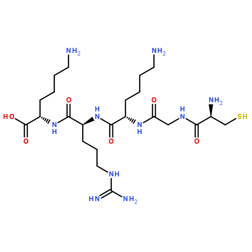
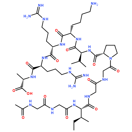
Co-reporter:Jingwei Li, Liang Feng, Li Fan, Yuan Zha, Liangran Guo, Qizhi Zhang, Jun Chen, Zhiqing Pang, Yuchen Wang, Xinguo Jiang, Victor C. Yang, Longping Wen
Biomaterials 2011 32(21) pp: 4943-4950
Publication Date(Web):
DOI:10.1016/j.biomaterials.2011.03.031
Co-reporter:Zhiqing Pang, Liang Feng, Rongrong Hua, Jun Chen, Huile Gao, Shuaiqi Pan, Xinguo Jiang, and Peng Zhang
Molecular Pharmaceutics 2010 Volume 7(Issue 6) pp:1995-2005
Publication Date(Web):October 19, 2010
DOI:10.1021/mp100277h
The blood-brain barrier (BBB) and multidrug resistance (MDR) are the main causes for poor prognosis of glioma patients after chemotherapy. To explore the way for settling this problem, in this study, a novel antitumor agent loaded drug delivery system, lactoferrin-conjugated biodegradable polymersome holding doxorubicin and tetrandrine (Lf-PO-Dox/Tet), integrating both BBB and glioma-targeting moiety and MDR inhibitor, was designed and its chemotherapy for glioma rats was evaluated. Biodegradable polymersome (PO) encapsulating both doxorubicin (Dox) and tetrandrine (Tet) was prepared by the thin-film hydration method (PO-Dox/Tet) and then conjugated with lactoferrin (Lf) to yield Lf-PO-Dox/Tet with an average diameter around 220 nm and surface Lf molecule number per polymersome around 40. Compared with PO-DOX, PO-Dox/Tet, and Lf-PO-Dox, Lf-PO-Dox/Tet demonstrated the strongest cytotoxicity against C6 glioma cells and the greatest uptake index by C6 cells. In vivo imaging analysis indicated that Lf-PO labeled with a near-infrared dye could enter the brain and accumulate at the tumor site. Pharmacokinetics and tissue distribution results also showed that Lf-PO-Dox/Tet accumulated more in the right hemisphere than other groups of polymersomes. Pharmacodynamics results revealed that tumor volume of the Lf-PO-Dox/Tet group was significantly smaller than that of other therapeutic groups, and the median survival time of Lf-PO-Dox/Tet group was longer than that of Lf-PO-Dox group and significantly longer than those of the other three therapeutic groups. These results suggested that Lf-PO-Dox/Tet could have therapeutic potential for gliomas.Keywords: biodegradable polymersome; chemotherapy; doxorubicin; glioma; Lactoferrin; tetrandrine;
Co-reporter:Kaili Hu, Jingwei Li, Yehong Shen, Wei Lu, Xiaoling Gao, Qizhi Zhang, Xinguo Jiang
Journal of Controlled Release 2009 Volume 134(Issue 1) pp:55-61
Publication Date(Web):20 February 2009
DOI:10.1016/j.jconrel.2008.10.016
The lactoferrin (Lf) conjugated poly (ethyleneglycol)–poly (lactide) nanoparticle (Lf–NP) was constructed in this paper as a novel biodegradable brain drug delivery system with evaluation of its in vitro and in vivo delivery properties. Lf was thiolated and conjugated to the distal maleimide functions surrounding on the pegylated nanoparticles to form the Lf–NP. The existence of Lf on the surface of Lf–NP was verified by TEM observation and XPS analysis. The Lf ELISA results confirmed the biorecognitive activity of Lf after the coupling procedure and suggested the average number of Lf conjugated on each nanoparticle was around 55. To evaluate the brain delivery properties of the Lf–NP, a fluorescent probe, coumarin-6 was incorporated into it. The uptake of Lf–NP by bEnd.3 cells was shown significantly higher than that of unconjugated nanoparticle (NP). Following an intravenous administration, a near 3 folds of coumarin-6 were found in the mice brain carried by Lf–NP compared to that carried by NP. Cell viability experiment results confirmed good safety of the biodegradable Lf–NP. The significant in vitro and in vivo results suggest that Lf–NP is a promising brain drug delivery system with low toxicity.A novel brain drug delivery system was constructed by maleimide-mediated conjugation of biodegradable PEG–PLA nanoparticles with lactoferrin (Lf), a newly discovered promising brain delivery targetor. Lf engineering significantly increased the brain uptake of drugs associated to nanoparticles following an intravenous administration, which might provide a novel effective noninvasive technique for brain drug delivery, especially for biotech drugs.
Co-reporter:Zhiqing Pang, Wei Lu, Huile Gao, Kaili Hu, Jun Chen, Chaolin Zhang, Xiaoling Gao, Xinguo Jiang, Cuiqing Zhu
Journal of Controlled Release 2008 Volume 128(Issue 2) pp:120-127
Publication Date(Web):4 June 2008
DOI:10.1016/j.jconrel.2008.03.007
A novel drug carrier for brain delivery, poly(ethyleneglycol)-poly(ε-caprolactone) (PEG-PCL) polymersomes conjugated with mouse-anti-rat monoclonal antibody OX26 (OX26-PO), was developed and its brain delivery property was evaluated. The diblock copolymers of methoxy-PEG-PCL and Maleimide-PEG-PCL were synthesized and applied to prepare polymersomes (PO) which were verified by direct cryogenic temperature transmission electron micrograph (Cryo-TEM) imaging. The TEM examination and dynamic light scattering results showed that OX26-PO had a round and vesicle-like shape with a mean diameter around 100 nm. Coupling of OX26 with PO was confirmed by immuno-gold labeling of OX26 visualized under the TEM and X-ray photoelectron spectroscopy test. The surface OX26 densities were obtained from enzyme-linked immunosorbant assay. The result of brain delivery in rats proved that the increase of surface OX26 density of OX26-PO decreased blood AUC. The optimized OX26 number conjugated per polymersome was 34, which can acquire the greatest blood–brain barrier (BBB) permeability surface area product and percentage of injected dose per gram brain (%ID/g brain). Furthermore, NC-1900, as a model peptide, was encapsulated into OX2634-PO and improved the scopolamine-induced learning and memory impairments in a water maze task via i.v. administration. These results indicated that OX2634-PO is a promising carrier for peptide brain delivery.
Co-reporter:Xiaoling Gao, Jun Chen, Jiyao Chen, Bingxian Wu, Hongzhuan Chen and Xinguo Jiang
Bioconjugate Chemistry 2008 Volume 19(Issue 11) pp:2189
Publication Date(Web):October 15, 2008
DOI:10.1021/bc8002698
Delivery of imaging agents to the brain is highly important for the diagnosis and treatment of central nervous system (CNS) diseases, as well as the elucidation of their pathophysiology. Quantum dots (QDs) provide a novel probe with unique physical, chemical, and optical properties, and become a promising tool for in vivo molecular and cellular imaging. However, their poor stability and low blood−brain barrier permeability severely limit their ability to enter into and act on their target sites in the CNS following parenteral administration. Here, we developed a QDs-based imaging platform for brain imaging by incorporating QDs into the core of poly(ethylene glycol)−poly(lactic acid) nanoparticles, which was then functionalized with wheat germ agglutinin and delivered into the brain via nasal application. The resulting nanoparticles, with high payload capacity, are water-soluble, stable, and showed excellent and safe brain targeting and imaging properties. With PEG functional terminal groups available on the nanoparticles surface, this nanoprobe allows for conjugation of various biological ligands, holding considerable potential for the development of specific imaging agents for various CNS diseases.
Co-reporter:Xiaoling Gao, Bingxian Wu, Qizhi Zhang, Jun Chen, Jianhua Zhu, Weiwei Zhang, Zhengxin Rong, Hongzhuan Chen, Xinguo Jiang
Journal of Controlled Release 2007 Volume 121(Issue 3) pp:156-167
Publication Date(Web):28 August 2007
DOI:10.1016/j.jconrel.2007.05.026
The development of biotech drugs such as peptides and proteins that act in the central nervous system has been significantly impeded by the difficulty of delivering them across the blood-brain barrier. The surface engineering of nanoparticles with lectins opened a novel pathway to the absorption of drugs loaded by biodegradable poly (ethylene glycol)-poly (lactic acid) nanoparticles in the brain following intranasal administration. In the present study, vasoactive intestinal peptide, a neuroprotective peptide, was efficiently incorporated into the poly (ethylene glycol)-poly (lactic acid) nanoparticles modified with wheat germ agglutinin and the biodistribution, brain uptake and neuroprotective effect of the formulation were assessed. The area under the concentration-time curve of intact 125I-vasoactive intestinal peptide in brain of mice following the intranasal administration of 125I-vasoactive intestinal peptide carried by nanoparticles and wheat germ agglutinin-conjugated ones was significantly enlarged by 3.5 ∼ 4.7 folds and 5.6 ∼ 7.7 folds, respectively, compared with that after intranasal application of 125I-vasoactive intestinal peptide solution. The same improvements in spatial memory in ethylcholine aziridium-treated rats were observed following intranasal administration of 25 μg/kg and 12.5 μg/kg of vasoactive intestinal peptide loaded by unmodified nanoparticles and wheat germ agglutinin-modified nanoparticles, respectively. Distribution profiles of wheat germ agglutinin-conjugated nanoparticles in the nasal cavity presented their higher affinity to the olfactory mucosa than to the respiratory one. Inhibition experiment with specific sugars suggested that the interaction between the nasal mucosa and the wheat germ agglutinin-functionalized nanoparticles were due to the immobilization of carbohydrate-binding pockets on the surface of the nanoparticles. The results clearly indicated wheat germ agglutinin-modified nanoparticles might serve as promising carriers especially for biotech drugs such as peptides and proteins.
Co-reporter:Wei Lu, Jin Wan, Zhenjue She, Xinguo Jiang
Journal of Controlled Release 2007 Volume 118(Issue 1) pp:38-53
Publication Date(Web):12 March 2007
DOI:10.1016/j.jconrel.2006.11.015
Cationic bovine serum albumin (CBSA) conjugated poly(ethyleneglycol)-poly(lactide) (PEG-PLA) nanoparticle (CBSA-NP), was designed as a novel drug carrier for brain delivery. In this paper, three formulations of CBSA-NP with different surface CBSA density as well as native bovine serum albumin conjugated nanoparticle (BSA-NP) and CBSA unconjugated pegylated nanoparticle (NP), were formulated. Their brain transcytosis across the blood-brain barrier (BBB) coculture and brain delivery in mice were investigated using 6-coumarin as fluorescent probe. By using free CBSA as specific inhibitor, it was evidenced that CBSA-NP crossed the brain capillary endothelium through absorptive mediated transcytosis. The result of transcytosis across the BBB coculture and brain delivery in mice proved that the increase of surface CBSA density of the nanoparticle enhanced the BBB permeability-surface area but decreased blood AUC. The optimized CBSA number conjugated per averaged nanoparticle was 110, with the maleimide-PEG-PLA/methoxy-PEG-PLA weight ratio 1:10, which can acquire the greatest percentage of injected dose per gram brain (%ID/g brain) by 2.3-fold compared with NP. Besides, “accelerated blood clearance phenomenon” was found through evaluating blood clearance profile of CBSA-NP post-injection of single dose or over a period of successive high doses of CBSA-NP. Understanding these issues is important for the future development of CBSA-NP as a brain delivery carrier and for the attenuation of toxicity or immunological responses to the nanodevice following a consequence of nanomedication.
Co-reporter:Xiaoling Gao, Weixing Tao, Wei Lu, Qizhi Zhang, Yan Zhang, Xinguo Jiang, Shoukuan Fu
Biomaterials 2006 Volume 27(Issue 18) pp:3482-3490
Publication Date(Web):June 2006
DOI:10.1016/j.biomaterials.2006.01.038
In order to improve the absorption of nanoparticles in the brain following nasal administration, a novel protocol to conjugate biorecognitive ligands—lectins to the surface of poly (ethylene glycol)–poly (lactic acid) (PEG–PLA) nanoparticles was established in the study. Wheat germ agglutinin (WGA), specifically binding to N-acetyl-d-glucosamine and sialic acid, both of which were abundantly observed in the nasal cavity, was selected as a model lectin. The WGA-conjugated nanoparticles were prepared by incorporating maleimide in the PLA–PEG molecular and taking advantage of its thiol group binding reactivity to conjugate with 2-iminothialane thiolated WGA. Coupling of WGA with the PEG–PLA nanoparticles was confirmed by the existence of gold-labeled WGA-NP under TEM. The retention of biorecognitive activity of WGA after the covalent coupling procedure was confirmed by haemagglutination test. The resulting nanoparticles presented negligible nasal ciliatoxicity and the brain uptake of a fluorescent marker—coumarin carried by WGA functionized nanoparticles was about 2 folds in different brain tissues compared with that of coumarin incorporated in the unmodified ones. Thus, the technique offered a novel effective noninvasive system for brain drug delivery, especially for brain protein and gene delivery.
Co-reporter:Wei Lu, Yan Zhang, Yu-Zhen Tan, Kai-Li Hu, Xin-Guo Jiang, Shou-Kuan Fu
Journal of Controlled Release 2005 Volume 107(Issue 3) pp:428-448
Publication Date(Web):20 October 2005
DOI:10.1016/j.jconrel.2005.03.027
In this paper, a novel drug carrier for brain delivery, cationic bovine serum albumin (CBSA) conjugated with poly(ethyleneglycol)–poly(lactide) (PEG–PLA) nanoparticle (CBSA–NP), was developed and its effects were evaluated. The copolymers of methoxy-PEG–PLA and maleimide-PEG–PLA were synthesized by ring opening polymerization of d,l-lactide initiated by methoxy-PEG and maleimide-PEG, respectively, which were applied to prepare pegylated nanoparticles by means of double emulsion and solvent evaporation procedure. Native bovine serum albumin (BSA) was cationized and thiolated, followed by conjugation through the maleimide function located at the distal end of PEG surrounding the nanoparticle's surface. Transmission electron micrograph (TEM) and dynamic light scattering results showed that CBSA–NP had a round and regular shape with a mean diameter around 100 nm. Surface nitrogen was detected by X-ray photoelectron spectroscopy (XPS), and colloidal gold stained around the nanoparticle's surface was visualized in TEM, which proved that CBSA was covalently conjugated onto its surface. To evaluate the effects of brain delivery, BSA conjugated with pegylated nanoparticles (BSA–NP) was used as the control group and 6-coumarin was incorporated into the nanoparticles as the fluorescent probe. The qualitative and quantitative results of CBSA–NP uptake experiment compared with those of BSA–NP showed that rat brain capillary endothelial cells (BCECs) took in much more CBSA–NP than BSA–NP at 37 °C, at different concentrations and time incubations. After a dose of 60 mg/kg CBSA–NP or BSA–NP injection in mice caudal vein, fluorescent microscopy of brain coronal sections showed a higher accumulation of CBSA–NP in the lateral ventricle, third ventricle and periventricular region than that of BSA–NP. There was no difference on BCECs' viability between CBSA-conjugated and -unconjugated pegylated nanoparticles. The significant results in vitro and in vivo showed that CBSA–NP was a promising brain drug delivery carrier with low toxicity.
Co-reporter:Yan Wei, Shuai Xu, Feng Wang, Aifeng Zou, ... Xinguo Jiang
Journal of Pharmaceutical Sciences (January 2015) Volume 104(Issue 1) pp:165-177
Publication Date(Web):1 January 2015
DOI:10.1002/jps.24234
Lapatinib (LPT) could sensitize human epidermal growth factor receptor-2 (HER-2) positive breast cancer to paclitaxel (PTX) and induce synergetic action with PTX in preclinical test and phase II/III trial. In this study, LPT-conjugated poly (ethylene glycol) (PEG) and poly (lactic acid) (PLA) (LPT–PEG–PLA) was first synthesized and confirmed with 1H Nuclear Magnetic Resonance and Matrix-Assisted Laser Desorption/ Ionization Time of Flight Mass Spectrometry, which was used for the preparation of a novel PEG–PLA combined micelles of LPT and PTX (PPM-LP). The obtained PPM-LP exhibited uniform, spherical shape with a size of 25.80 ± 0.47 nm and zeta potential of −3.17 ± 0.15 mv. PTX existed in molecular or amorphous forms in the micelles and superficial LPT could better delay PTX release. The cytotoxicity of PPM-LP with LPT conjugation against SKBr-3 cells (HER-2 positive) was found to be significantly increasing as compared with PPM–PTX, whereas there was no significant difference against MDA-MB-231 cells (HER-2 negative). PPM-LP could escape from endosomes and be distributed into cytoplasm and led to cell arrest in G2/M and G1/S phases simultaneously. Results of nucleus staining and flow cytometry confirmed that LPT could remarkably increase antineoplastic effect of PTX against SKBr-3 cells. All these results demonstrated that PPM-LP may be a promising drug delivery system for HER-2 positive breast cancer. © 2014 Wiley Periodicals, Inc. and the American Pharmacists Association J Pharm Sci 104:165–177, 2015
Co-reporter:Shun Shen, Jinfeng Ren, Xiaoyan Zhu, Zhiqing Pang, Xiaohui Lu, Chunhui Deng, Ren Zhang and Xinguo Jiang
Journal of Materials Chemistry A 2013 - vol. 1(Issue 14) pp:NaN1946-1946
Publication Date(Web):2013/02/28
DOI:10.1039/C3TB00543G
In this paper, a facile solvothermal synthesis method was developed to prepare monodisperse magnetites anchored onto multi-walled carbon nanotubes (MWNTs). The pristine MWNTs were treated with a mixture of concentrated sulfuric and nitric acids. The oxidized MWNTs (o-MWNTs) had abundant carboxylic groups on the surface, which have a strong ability to chelate metal ions like Fe3+. The obtained MWNTs–Fe3O4 nanomaterials allowed π–π stacking of fluorescein isothiocyanate (FITC) to monitor inside living cancer cells by fluorescence imaging. Cells labeled with MWNTs–Fe3O4 nanomaterials could be efficiently manipulated by a magnetic field due to the large magnetic moment of the iron oxide domain in the nanocomposites. The MWNTs–Fe3O4 nanomaterials have been demonstrated to be effective for in vivo photothermal treatment of tumors using mice implanted with human U87 tumors as a model.
Co-reporter:Jinfeng Ren, Shun Shen, Zhiqing Pang, Xiaohui Lu, Chunhui Deng and Xinguo Jiang
Chemical Communications 2011 - vol. 47(Issue 42) pp:NaN11694-11694
Publication Date(Web):2011/09/26
DOI:10.1039/C1CC15528H
Superparamagnetic Fe3O4 nanoparticles with positive surface ξ-potential were synthesized via a solvothermal route. After Fe3O4 was mixed with HAuCl4 and NaBH4, the reduced Au nanoparticles could be directly adsorbed onto the surface of Fe3O4 nanoparticles. The as-synthesized nanocomposites were successfully applied to photothermal destruction of cancer cells.