Co-reporter:Yanqing Guan, Jing Dong, Shizhong Chen, Meixian Liu, Daidong Wang, Xiaotian Zhang, Hong Wang, Zongtao Lin
Spectrochimica Acta Part A: Molecular and Biomolecular Spectroscopy 2017 Volume 181(Volume 181) pp:
Publication Date(Web):15 June 2017
DOI:10.1016/j.saa.2017.03.025
•Interactions of 3-, 5-, 4- and 1-CQA to Tf were studied for the first time.•Binding and equilibrium dissociation constants between CQA and Tf are reported.•3-CQA increased α-helix while 1-, 4- and 5-CQA increased β-sheet and random coil of Tf.•Major interactions between 1-, 3-, 5-CQA and Tf are H-bondings.•Electrostatic interactions were likely involved in the binding of 4-CQA and Tf.Transferrin (Tf) is an important protein responsible for circulating and transporting iron into cytoplasm. Tf can be taken into cells through endocytosis mediated by Tf receptor, which usually overexpresses in cancer cells. The Tf-Tf receptor pathway opens a possible avenue for novel targeted cancer therapy by utilizing Tf-binding active compounds. Among which, anti-cancer active caffeoylquinic acids (CQAs) were recently found to be promising Tf-binders by our group. For better understanding the anti-cancer activities of CQAs, it is important to unveil the binding mechanisms between CQAs and Tf. In this study, the fluorescence quenching, surface plasmon resonance (SPR), circular dichroism (CD) and molecular docking were used to investigate the interactions between CQA and Tf. The results showed that the calculated apparent association constants of interactions between 1-, 3-, 4- and 5-CQA and Tf at 298 K were 7.97 × 10⁵ M− 1, 4.36 × 10⁷ M− 1, 6.58 × 10⁵ M− 1 and 4.42 × 10⁶ M− 1, respectively. The thermodynamic parameters indicated that the interaction between 1-, 3-, 5-CQA and Tf is due to H-bonding, and electrostatic interactions were likely involved in the binding of 4-CQA and Tf. The CD results indicated that bindings of 1-CQA, 4-CQA and 5-CQA with Tf resulted in more stretched β-turn and random coil translated from β-sheet. In contrast, 3-CQA led to more stable a-helix conformation. Molecular docking studies of CQAs with Tf further displayed that CQAs were able to interact with residues near Fe3 + binding site. The spectroscopic studies revealed the action mechanisms, thermodynamics and interacting forces between CQAs and Tf, and thus are helpful for future design and discovery of Tf-binders for targeted cancer therapy applying Tf-Tf receptor pathway.Download high-res image (255KB)Download full-size image
Co-reporter:Xiaoxia Zhang, Zongtao Lin, Jinggui Fang, Meixian Liu, Yanyan Niu, Shizhong Chen, Hong Wang
Journal of Chromatography A 2015 Volume 1414() pp:88-102
Publication Date(Web):2 October 2015
DOI:10.1016/j.chroma.2015.08.033
•Optimized on-line HPLC-DAD-ESI-IT-TOF-TACD method was established.•The on-line method obtained DPPH and FRAP antioxidant profiles of Prunus mume flower simultaneously.•Seventy-eight compounds were identified from Prunus mume flowers.•Twenty-one compounds showed DPPH scavenging activity and 32 compounds showed ferric reducing power, respectively.•Total antioxidant activities of 24 Prunus mume flower samples were evaluated in detail.An on-line high-performance liquid chromatography−diode-array detector−electrospray ionization−ion-trap−time-of-flight−mass spectrometry−total antioxidant capacity detection (HPLC−DAD−ESI−IT−TOF−MS−TACD) system was created for identification and evaluation of antioxidants in Prunus (P.) mume flowers. Applying this system, the HPLC fingerprint, ultraviolet (UV) spectra, mass fragmentations, active profiles against 1,1-diphenylpicryl-2-hydrazyl radical (DPPH•) scavenging activity and ferric reducing antioxidant power (FRAP) of each complex sample were obtained simultaneously after one injection. Synchronous structure identification and activities screening of complex samples were thus accomplished. In this study, 78 compounds were identified from P. mume flowers by their chromatographic behaviors, UV spectra and MS data with the assistance of standard compounds and literature reports. The DPPH and FRAP activity of 24 samples (23 different P. mume varieties and 1 related herbal medicine) were then quantified by their detailed activity profiles from the on-line system, and by the total activity of each sample extract from off-line 96-well plate method. As a result, 21 and 32 compounds in the on-line system showed anti-oxidative effects against DPPH and FRAP, respectively. The established on-line system is efficient, sensitive and reliable to tell the DPPH and FRAP antioxidant activities of individual compound in complex samples, and therefore would be a useful and promising technique for antioxidant screening from different food and medicinal matrices.
Co-reporter:Cangman Zhang, Biao Ren, Zongtao Lin, Lingkun Tong, Hong Wang, Shizhong Chen
Journal of Chromatography A 2015 Volume 1381() pp:160-172
Publication Date(Web):13 February 2015
DOI:10.1016/j.chroma.2015.01.019
•A novel online UFLC-DAD-MSn-DFLD method was first established.•Dual-fluorescent probes made the investigation of multiple mechanisms between DNA and small molecule drugs into reality.•Identified compounds with DNA-binding activity fast and accurate from natural products. 62 compounds were identified and 37 were first found in Pyrrosia lingua (Thunb.). 22 constituents showed DNA-binding profile.•Most of the active compounds from Pyrrosia lingua (Thunb.) were found to be DNA-binders via both minor groove-binding and intercalation modes.DNA has been known as the cellular target for many cytotoxic anticancer agents over the years. Discovering DNA-binding compounds has become an active research area, while various DNA-binding mechanisms make the drug discovery even more difficult. In this article, we present a novel analysis method to rapidly identify specific DNA-binding compounds from Pyrrosia lingua (Thunb.) using DNA-dual-fluorescent probes, ethidium bromide and Hoechst 33258, with the technology of ultra-fast liquid chromatography-diode array detector-tandem mass spectrometry and dual-wavelength fluorescence detector (UFLC-DAD-MSn-DFLD). Sixty-two compounds were identified, of which 22 were found to be active in DNA-binding. After investigation of their dose-response behaviors and structure–activity relationships, chlorogenic acids and flavonoid glycosides were found to be DNA-binders via both minor groove-binding and intercalation modes. The precision, reproducibility and stability of this method were validated by vitexin. The established system was sensitive, precise, and reliable to be used for both screening of DNA-binding compounds and investigating of their mechanisms.

![1,3-bis{[(2E)-3-(3,4-dihydroxyphenyl)prop-2-enoyl]oxy}-4,5-dihydroxycyclohexanecarboxylic acid](http://img.cochemist.com/ccimg/251400/251320-66-8.png)
![1,3-bis{[(2E)-3-(3,4-dihydroxyphenyl)prop-2-enoyl]oxy}-4,5-dihydroxycyclohexanecarboxylic acid](http://img.cochemist.com/ccimg/251400/251320-66-8_b.png)
![1H,3H-Pyrano[3,4-c]pyran-1-one, 5-ethenyl-6-(β-D-glucopyranosyloxy)-4,4a,5,6-tetrahydro-3-methoxy-, (3S,4aS,5R,6S)-](http://img.cochemist.com/ccimg/118700/118627-52-4.png)
![1H,3H-Pyrano[3,4-c]pyran-1-one, 5-ethenyl-6-(β-D-glucopyranosyloxy)-4,4a,5,6-tetrahydro-3-methoxy-, (3S,4aS,5R,6S)-](http://img.cochemist.com/ccimg/118700/118627-52-4_b.png)
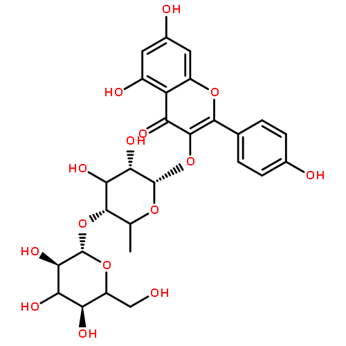
![4H-1-Benzopyran-4-one,3-[[2-O-(6-deoxy-a-L-mannopyranosyl)-b-D-galactopyranosyl]oxy]-2-(3,4-dihydroxyphenyl)-5,7-dihydroxy-](http://img.cochemist.com/ccimg/117700/117611-67-3.png)
![4H-1-Benzopyran-4-one,3-[[2-O-(6-deoxy-a-L-mannopyranosyl)-b-D-galactopyranosyl]oxy]-2-(3,4-dihydroxyphenyl)-5,7-dihydroxy-](http://img.cochemist.com/ccimg/117700/117611-67-3_b.png)
![b-D-Glucopyranoside,2-(3,4-dihydroxyphenyl)ethyl 3-O-(6-deoxy-a-L-mannopyranosyl)-,4-[(2E)-3-(4-hydroxyphenyl)-2-propenoate]](http://img.cochemist.com/ccimg/110400/110326-99-3.png)
![b-D-Glucopyranoside,2-(3,4-dihydroxyphenyl)ethyl 3-O-(6-deoxy-a-L-mannopyranosyl)-,4-[(2E)-3-(4-hydroxyphenyl)-2-propenoate]](http://img.cochemist.com/ccimg/110400/110326-99-3_b.png)


![4H-1-Benzopyran-4-one, 7-[(6-deoxy-α-L-mannopyranosyl)oxy]-3-[(2-O-β-D-glucopyranosyl-β-D-glucopyranosyl)oxy]-5-hydroxy-2-(4-hydroxyphenyl)-](http://img.cochemist.com/ccimg/93100/93098-79-4.png)
![4H-1-Benzopyran-4-one, 7-[(6-deoxy-α-L-mannopyranosyl)oxy]-3-[(2-O-β-D-glucopyranosyl-β-D-glucopyranosyl)oxy]-5-hydroxy-2-(4-hydroxyphenyl)-](http://img.cochemist.com/ccimg/93100/93098-79-4_b.png)


![b-D-Glucopyranoside,2-(3,4-dihydroxyphenyl)ethyl, 4-[(2E)-3-(3,4-dihydroxyphenyl)-2-propenoate]](http://img.cochemist.com/ccimg/84800/84744-28-5.png)
![b-D-Glucopyranoside,2-(3,4-dihydroxyphenyl)ethyl, 4-[(2E)-3-(3,4-dihydroxyphenyl)-2-propenoate]](http://img.cochemist.com/ccimg/84800/84744-28-5_b.png)
![(1S,4aS,6S,7S,7aS)-1-(beta-D-glucopyranosyloxy)-6-hydroxy-7-methyl-1,4a,5,6,7,7a-hexahydrocyclopenta[c]pyran-4-carboxylic acid](http://img.cochemist.com/ccimg/82600/82509-41-9.png)
![(1S,4aS,6S,7S,7aS)-1-(beta-D-glucopyranosyloxy)-6-hydroxy-7-methyl-1,4a,5,6,7,7a-hexahydrocyclopenta[c]pyran-4-carboxylic acid](http://img.cochemist.com/ccimg/82600/82509-41-9_b.png)
![4-[(1S,3aR,4S,6aR)-4-(4-Hydroxy-3-methoxyphenyl)tetrahydro-1H,3H- furo[3,4-c]furan-1-yl]-2-methoxyphenyl β-D-glucopyranoside](http://img.cochemist.com/ccimg/69300/69251-96-3.png)
![4-[(1S,3aR,4S,6aR)-4-(4-Hydroxy-3-methoxyphenyl)tetrahydro-1H,3H- furo[3,4-c]furan-1-yl]-2-methoxyphenyl β-D-glucopyranoside](http://img.cochemist.com/ccimg/69300/69251-96-3_b.png)
![8-[(2s,3r,4s,5s,6r)-4,5-dihydroxy-6-(hydroxymethyl)-3-[(2s,3r,4r,5r,6s)-3,4,5-trihydroxy-6-methyloxan-2-yl]oxyoxan-2-yl]-5,7-dihydroxy-2-(4-hydroxyphenyl)chromen-4-one](http://img.cochemist.com/ccimg/64900/64820-99-1.png)
![8-[(2s,3r,4s,5s,6r)-4,5-dihydroxy-6-(hydroxymethyl)-3-[(2s,3r,4r,5r,6s)-3,4,5-trihydroxy-6-methyloxan-2-yl]oxyoxan-2-yl]-5,7-dihydroxy-2-(4-hydroxyphenyl)chromen-4-one](http://img.cochemist.com/ccimg/64900/64820-99-1_b.png)
![1-O-[alpha-L-Rhamnopyranosyl(1?3)-beta-D-glucopyranoside]-2-(3,4-Dihydroxyphenyl)ethanol](http://img.cochemist.com/ccimg/61600/61548-34-3.png)
![1-O-[alpha-L-Rhamnopyranosyl(1?3)-beta-D-glucopyranoside]-2-(3,4-Dihydroxyphenyl)ethanol](http://img.cochemist.com/ccimg/61600/61548-34-3_b.png)
![8-[(2s,3r,4s,5s,6r)-4,5-dihydroxy-6-(hydroxymethyl)-3-[(2s,3r,4s,5s,6r)-3,4,5-trihydroxy-6-(hydroxymethyl)oxan-2-yl]oxyoxan-2-yl]-5,7-dihydroxy-2-(4-hydroxyphenyl)chromen-4-one](http://img.cochemist.com/ccimg/61400/61360-94-9.png)
![8-[(2s,3r,4s,5s,6r)-4,5-dihydroxy-6-(hydroxymethyl)-3-[(2s,3r,4s,5s,6r)-3,4,5-trihydroxy-6-(hydroxymethyl)oxan-2-yl]oxyoxan-2-yl]-5,7-dihydroxy-2-(4-hydroxyphenyl)chromen-4-one](http://img.cochemist.com/ccimg/61400/61360-94-9_b.png)

![β-D-Glucopyranoside, (1S,4aR,5S,7aS)-7-[(benzoyloxy)methyl]-1,4a,5,7a-tetrahydro-5-hydroxycyclopenta[c]pyran-1-yl](/data/chemimg/1848200/55785-60-9.png)
![β-D-Glucopyranoside, (1S,4aR,5S,7aS)-7-[(benzoyloxy)methyl]-1,4a,5,7a-tetrahydro-5-hydroxycyclopenta[c]pyran-1-yl](/data/chemimg/1848200/55785-60-9_b.png)
![quercetin 3-O-(beta-glucopyranosyl(1->2)[alpha-rhamnopyranosyl(1->6)]-beta-glucopyranoside)](http://img.cochemist.com/ccimg/55700/55696-55-4.png)
![quercetin 3-O-(beta-glucopyranosyl(1->2)[alpha-rhamnopyranosyl(1->6)]-beta-glucopyranoside)](http://img.cochemist.com/ccimg/55700/55696-55-4_b.png)
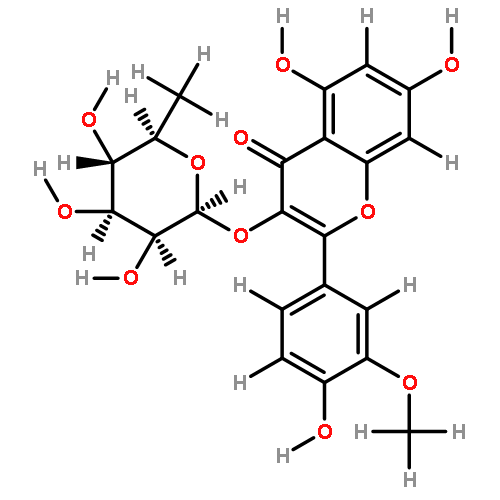
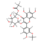
![4H-1-Benzopyran-4-one,3-[[2-O-(6-deoxy-a-L-mannopyranosyl)-b-D-glucopyranosyl]oxy]-5,7-dihydroxy-2-(4-hydroxy-3-methoxyphenyl)-](http://img.cochemist.com/ccimg/55100/55033-90-4.png)
![4H-1-Benzopyran-4-one,3-[[2-O-(6-deoxy-a-L-mannopyranosyl)-b-D-glucopyranosyl]oxy]-5,7-dihydroxy-2-(4-hydroxy-3-methoxyphenyl)-](http://img.cochemist.com/ccimg/55100/55033-90-4_b.png)


![2-METHYL-2-PROPANYL 4-{[(6-CHLORO-3-PYRIDAZINYL)AMINO]METHYL}-1-P<WBR />IPERIDINECARBOXYLATE](http://img.cochemist.com/ccimg/37000/36948-76-2.png)
![2-METHYL-2-PROPANYL 4-{[(6-CHLORO-3-PYRIDAZINYL)AMINO]METHYL}-1-P<WBR />IPERIDINECARBOXYLATE](http://img.cochemist.com/ccimg/37000/36948-76-2_b.png)


![4H-1-Benzopyran-4-one,3-[[6-O-(6-deoxy-a-L-mannopyranosyl)-b-D-glucopyranosyl]oxy]-2-(3,4-dihydroxyphenyl)-7-(b-D-glucopyranosyloxy)-5-hydroxy-](http://img.cochemist.com/ccimg/30400/30311-61-6.png)
![4H-1-Benzopyran-4-one,3-[[6-O-(6-deoxy-a-L-mannopyranosyl)-b-D-glucopyranosyl]oxy]-2-(3,4-dihydroxyphenyl)-7-(b-D-glucopyranosyloxy)-5-hydroxy-](http://img.cochemist.com/ccimg/30400/30311-61-6_b.png)
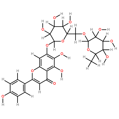
![4H-1-Benzopyran-4-one,7-[[2-O-(6-deoxy-a-L-mannopyranosyl)-b-D-glucopyranosyl]oxy]-2-(3,4-dihydroxyphenyl)-5-hydroxy-](http://img.cochemist.com/ccimg/25700/25694-72-8.png)
![4H-1-Benzopyran-4-one,7-[[2-O-(6-deoxy-a-L-mannopyranosyl)-b-D-glucopyranosyl]oxy]-2-(3,4-dihydroxyphenyl)-5-hydroxy-](http://img.cochemist.com/ccimg/25700/25694-72-8_b.png)
![2(3H)-Furanone, 3-[[4-(b-D-glucopyranosyloxy)-3-methoxyphenyl]methyl]dihydro-4-[(4-hydroxy-3-methoxyphenyl)methyl]-,(3R,4R)-](http://img.cochemist.com/ccimg/23300/23202-85-9.png)
![2(3H)-Furanone, 3-[[4-(b-D-glucopyranosyloxy)-3-methoxyphenyl]methyl]dihydro-4-[(4-hydroxy-3-methoxyphenyl)methyl]-,(3R,4R)-](http://img.cochemist.com/ccimg/23300/23202-85-9_b.png)
![[1S-(1α,4aα,6α,7α,7aα)]-1-(β-D-glucopyranosyloxy)-1,4a,5,6,7,7a-hexahydro-6-hydroxy-7-methylcyclopenta[c]pyran-4-carboxylic acid](http://img.cochemist.com/ccimg/22300/22255-40-9.png)
![[1S-(1α,4aα,6α,7α,7aα)]-1-(β-D-glucopyranosyloxy)-1,4a,5,6,7,7a-hexahydro-6-hydroxy-7-methylcyclopenta[c]pyran-4-carboxylic acid](http://img.cochemist.com/ccimg/22300/22255-40-9_b.png)
![(1S,4aS,5R,7aS)-1-(beta-D-glucopyranosyloxy)-5-hydroxy-7-(hydroxymethyl)-1,4a,5,7a-tetrahydrocyclopenta[c]pyran-4-carboxylic acid](http://img.cochemist.com/ccimg/18900/18842-99-4.png)
![(1S,4aS,5R,7aS)-1-(beta-D-glucopyranosyloxy)-5-hydroxy-7-(hydroxymethyl)-1,4a,5,7a-tetrahydrocyclopenta[c]pyran-4-carboxylic acid](http://img.cochemist.com/ccimg/18900/18842-99-4_b.png)
![4H-1-Benzopyran-4-one,2-(3,4-dihydroxyphenyl)-3-[(2-O-b-D-glucopyranosyl-b-D-glucopyranosyl)oxy]-5,7-dihydroxy-](http://img.cochemist.com/ccimg/18700/18609-17-1.png)
![4H-1-Benzopyran-4-one,2-(3,4-dihydroxyphenyl)-3-[(2-O-b-D-glucopyranosyl-b-D-glucopyranosyl)oxy]-5,7-dihydroxy-](http://img.cochemist.com/ccimg/18700/18609-17-1_b.png)


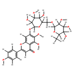
![5,7-dihydroxy-2-(4-hydroxyphenyl)-3-[(2s,3r,4s,5s,6r)-3,4,5-trihydroxy-6-[[(2r,3r,4r,5r,6s)-3,4,5-trihydroxy-6-methyloxan-2-yl]oxymethyl]oxan-2-yl]oxychromen-4-one](http://img.cochemist.com/ccimg/17700/17650-84-9.png)
![5,7-dihydroxy-2-(4-hydroxyphenyl)-3-[(2s,3r,4s,5s,6r)-3,4,5-trihydroxy-6-[[(2r,3r,4r,5r,6s)-3,4,5-trihydroxy-6-methyloxan-2-yl]oxymethyl]oxan-2-yl]oxychromen-4-one](http://img.cochemist.com/ccimg/17700/17650-84-9_b.png)
![3,5-dihydroxy-2-(4-hydroxyphenyl)-7-[(2s,3r,4s,5s,6r)-3,4,5-trihydroxy-6-(hydroxymethyl)oxan-2-yl]oxychromen-4-one](http://img.cochemist.com/ccimg/16300/16290-07-6.png)
![3,5-dihydroxy-2-(4-hydroxyphenyl)-7-[(2s,3r,4s,5s,6r)-3,4,5-trihydroxy-6-(hydroxymethyl)oxan-2-yl]oxychromen-4-one](http://img.cochemist.com/ccimg/16300/16290-07-6_b.png)
![Cyclopenta[c]pyran-4-carboxylicacid, 1-(b-D-glucopyranosyloxy)-1,4a,5,7a-tetrahydro-5-hydroxy-7-(hydroxymethyl)-,(1S,4aS,5S,7aS)-](http://img.cochemist.com/ccimg/14300/14259-55-3.png)
![Cyclopenta[c]pyran-4-carboxylicacid, 1-(b-D-glucopyranosyloxy)-1,4a,5,7a-tetrahydro-5-hydroxy-7-(hydroxymethyl)-,(1S,4aS,5S,7aS)-](http://img.cochemist.com/ccimg/14300/14259-55-3_b.png)
![[(2R,3R,4S,5R)-2,3,4,5-TETRAHYDROXY-6-OXOHEXYL] (E)-3-(3,4-DIHYDROXYPHENYL)PROP-2-ENOATE](http://img.cochemist.com/ccimg/10100/10066-92-9.png)
![[(2R,3R,4S,5R)-2,3,4,5-TETRAHYDROXY-6-OXOHEXYL] (E)-3-(3,4-DIHYDROXYPHENYL)PROP-2-ENOATE](http://img.cochemist.com/ccimg/10100/10066-92-9_b.png)
![(1S,3R,4R,5R)-1,3,4-trihydroxy-5-{[(2E)-3-(4-hydroxyphenyl)prop-2-enoyl]oxy}cyclohexanecarboxylic acid](http://img.cochemist.com/ccimg/1900/1899-30-5.png)
![(1S,3R,4R,5R)-1,3,4-trihydroxy-5-{[(2E)-3-(4-hydroxyphenyl)prop-2-enoyl]oxy}cyclohexanecarboxylic acid](http://img.cochemist.com/ccimg/1900/1899-30-5_b.png)
![4H-1-Benzopyran-4-one,7-[[6-O-(6-deoxy-a-L-mannopyranosyl)-b-D-glucopyranosyl]oxy]-5-hydroxy-2-(4-hydroxyphenyl)-](http://img.cochemist.com/ccimg/600/552-57-8.png)
![4H-1-Benzopyran-4-one,7-[[6-O-(6-deoxy-a-L-mannopyranosyl)-b-D-glucopyranosyl]oxy]-5-hydroxy-2-(4-hydroxyphenyl)-](http://img.cochemist.com/ccimg/600/552-57-8_b.png)
![5,7-dihydroxy-2-(4-hydroxyphenyl)-3-[(2s,3r,4r,5r,6s)-3,4,5-trihydroxy-6-methyloxan-2-yl]oxychromen-4-one](http://img.cochemist.com/ccimg/500/482-39-3.png)
![5,7-dihydroxy-2-(4-hydroxyphenyl)-3-[(2s,3r,4r,5r,6s)-3,4,5-trihydroxy-6-methyloxan-2-yl]oxychromen-4-one](http://img.cochemist.com/ccimg/500/482-39-3_b.png)
![5,7-dihydroxy-2-(4-hydroxyphenyl)-3-[(2s,3r,4s,5s,6r)-3,4,5-trihydroxy-6-(hydroxymethyl)oxan-2-yl]oxychromen-4-one](http://img.cochemist.com/ccimg/500/480-10-4.png)
![5,7-dihydroxy-2-(4-hydroxyphenyl)-3-[(2s,3r,4s,5s,6r)-3,4,5-trihydroxy-6-(hydroxymethyl)oxan-2-yl]oxychromen-4-one](http://img.cochemist.com/ccimg/500/480-10-4_b.png)
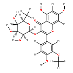
![3-O-[beta-D-Glucopyranosyl-(1?2)-beta-D-galactopyranoside]-3,4',5,7-Tetrahydroxy-3'-methoxyflavone](http://img.cochemist.com/ccimg/74200/74137-43-2.png)
![3-O-[beta-D-Glucopyranosyl-(1?2)-beta-D-galactopyranoside]-3,4',5,7-Tetrahydroxy-3'-methoxyflavone](http://img.cochemist.com/ccimg/74200/74137-43-2_b.png)
![methyl (1S,4aS,5S,7aS)-5-hydroxy-7-(hydroxymethyl)-1-(((2S,3R,4S,5S,6R)-3,4,5-trihydroxy-6-(hydroxymethyl)tetrahydro-2H-pyran-2-yl)oxy)-1,4a,5,7a-tetrahydrocyclopenta[c]pyran-4-carboxylate](http://img.cochemist.com/ccimg/52700/52613-28-2.png)
![methyl (1S,4aS,5S,7aS)-5-hydroxy-7-(hydroxymethyl)-1-(((2S,3R,4S,5S,6R)-3,4,5-trihydroxy-6-(hydroxymethyl)tetrahydro-2H-pyran-2-yl)oxy)-1,4a,5,7a-tetrahydrocyclopenta[c]pyran-4-carboxylate](http://img.cochemist.com/ccimg/52700/52613-28-2_b.png)
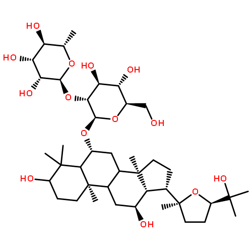
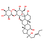
![(3WEI )-3-{[6-DEOXY-WEI -L-MANNOPYRANOSYL-(1->2)-[WEI -D-XYLOPYRANOSYL-(1->3)]-WEI -L-ARABINOPYRANOSYL]OXY}-20-HYDROXY-19-OXODAMMAR-24-EN-21-YL WEI -D-GLUCOPYRANOSIDE](http://img.cochemist.com/ccimg/33300/33228-65-8.png)
![(3WEI )-3-{[6-DEOXY-WEI -L-MANNOPYRANOSYL-(1->2)-[WEI -D-XYLOPYRANOSYL-(1->3)]-WEI -L-ARABINOPYRANOSYL]OXY}-20-HYDROXY-19-OXODAMMAR-24-EN-21-YL WEI -D-GLUCOPYRANOSIDE](http://img.cochemist.com/ccimg/33300/33228-65-8_b.png)


![methyl 1-(beta-D-glucopyranosyloxy)-6-hydroxy-7-methyl-1,4a,5,6,7,7a-hexahydrocyclopenta[c]pyran-4-c](http://img.cochemist.com/ccimg/18600/18524-94-2.png)
![methyl 1-(beta-D-glucopyranosyloxy)-6-hydroxy-7-methyl-1,4a,5,6,7,7a-hexahydrocyclopenta[c]pyran-4-c](http://img.cochemist.com/ccimg/18600/18524-94-2_b.png)
![Cyclohexanecarboxylicacid, 1,3,4-trihydroxy-5-[[(2E)-3-(4-hydroxyphenyl)-1-oxo-2-propen-1-yl]oxy]-,(1S,3R,4R,5R)-](http://img.cochemist.com/ccimg/5800/5746-55-4.png)
![Cyclohexanecarboxylicacid, 1,3,4-trihydroxy-5-[[(2E)-3-(4-hydroxyphenyl)-1-oxo-2-propen-1-yl]oxy]-,(1S,3R,4R,5R)-](http://img.cochemist.com/ccimg/5800/5746-55-4_b.png)
![(1S,3S,4S,5S)-1,3,4-trihydroxy-5-{[(2E)-3-(4-hydroxy-3-methoxyphenyl)prop-2-enoyl]oxy}cyclohexanecarboxylic acid](http://img.cochemist.com/ccimg/1900/1899-29-2.png)
![(1S,3S,4S,5S)-1,3,4-trihydroxy-5-{[(2E)-3-(4-hydroxy-3-methoxyphenyl)prop-2-enoyl]oxy}cyclohexanecarboxylic acid](http://img.cochemist.com/ccimg/1900/1899-29-2_b.png)
![2-(3,4-dihydroxyphenyl)-5,7-dihydroxy-3-[(2R,3S,4R,5S,6S)-3,4,5-trihydroxy-6-(hydroxymethyl)tetrahydropyran-2-yl]oxy-chromen-4-one](http://img.cochemist.com/ccimg/500/482-35-9.png)
![2-(3,4-dihydroxyphenyl)-5,7-dihydroxy-3-[(2R,3S,4R,5S,6S)-3,4,5-trihydroxy-6-(hydroxymethyl)tetrahydropyran-2-yl]oxy-chromen-4-one](http://img.cochemist.com/ccimg/500/482-35-9_b.png)


![Phenol,4-[5-(4-methyl-1-piperazinyl)[2,5'-bi-1H-benzimidazol]-2'-yl]-](http://img.cochemist.com/ccimg/23500/23491-44-3.png)
![Phenol,4-[5-(4-methyl-1-piperazinyl)[2,5'-bi-1H-benzimidazol]-2'-yl]-](http://img.cochemist.com/ccimg/23500/23491-44-3_b.png)


![(2-ISOPROPYL-3-INDOLIZINYL)(4-{3-[(2-METHYL-2-PROPANYL)AMINO]PROP<WBR />OXY}PHENYL)METHANONE](http://img.cochemist.com/ccimg/3600/3546-21-2.png)
![(2-ISOPROPYL-3-INDOLIZINYL)(4-{3-[(2-METHYL-2-PROPANYL)AMINO]PROP<WBR />OXY}PHENYL)METHANONE](http://img.cochemist.com/ccimg/3600/3546-21-2_b.png)


![2(3H)-Furanone,dihydro-3,4-bis[(4-hydroxy-3-methoxyphenyl)methyl]-, (3R,4R)-](http://img.cochemist.com/ccimg/600/580-72-3.png)
![2(3H)-Furanone,dihydro-3,4-bis[(4-hydroxy-3-methoxyphenyl)methyl]-, (3R,4R)-](http://img.cochemist.com/ccimg/600/580-72-3_b.png)


![b-D-Glucopyranose,1-[3-(3,4-dihydroxyphenyl)-2-propenoate]](http://img.cochemist.com/ccimg/14400/14364-08-0.png)
![b-D-Glucopyranose,1-[3-(3,4-dihydroxyphenyl)-2-propenoate]](http://img.cochemist.com/ccimg/14400/14364-08-0_b.png)


![2-[4-(aminoiminomethyl)phenyl]-1H-Indole-6-carboximidamide](http://img.cochemist.com/ccimg/47200/47165-04-8.png)
![2-[4-(aminoiminomethyl)phenyl]-1H-Indole-6-carboximidamide](http://img.cochemist.com/ccimg/47200/47165-04-8_b.png)


![1-[3-(4-hydroxyphenyl)-2-propenoate]-beta-D-glucopyranoside](http://img.cochemist.com/ccimg/7200/7139-64-2.png)
![1-[3-(4-hydroxyphenyl)-2-propenoate]-beta-D-glucopyranoside](http://img.cochemist.com/ccimg/7200/7139-64-2_b.png)