Co-reporter:Prem Kumar Sinha, Norma Castro-Guerrero, Gaurav Patki, Motoaki Sato, Jesus Torres-Bacete, Subhash Sinha, Hideto Miyoshi, Akemi Matsuno-Yagi, and Takao Yagi
Biochemistry 2015 Volume 54(Issue 3) pp:753-764
Publication Date(Web):December 29, 2014
DOI:10.1021/bi501403t
The NuoD segment (homologue of mitochondrial 49 kDa subunit) of the proton-translocating NADH:quinone oxidoreductase (complex I/NDH-1) from Escherichia coli is in the hydrophilic domain and bears many highly conserved amino acid residues. The three-dimensional structural model of NDH-1 suggests that the NuoD segment, together with the neighboring subunits, constitutes a putative quinone binding cavity. We used the homologous DNA recombination technique to clarify the role of selected key amino acid residues of the NuoD segment. Among them, residues Tyr273 and His224 were considered candidates for having important interactions with the quinone headgroup. Mutant Y273F retained partial activity but lost sensitivity to capsaicin-40. Mutant H224R scarcely affected the activity, suggesting that this residue may not be essential. His224 is located in a loop near the N-terminus of the NuoD segment (Gly217–Phe227) which is considered to form part of the quinone binding cavity. In contrast to the His224 mutation, mutants G217V, P218A, and G225V almost completely lost the activity. One region of this loop is positioned close to a cytosolic loop of the NuoA subunit in the membrane domain, and together they seem to be important in keeping the quinone binding cavity intact. The structural role of the longest helix in the NuoD segment located behind the quinone binding cavity was also investigated. Possible roles of other highly conserved residues of the NuoD segment are discussed.
Co-reporter:Takao Yagi;Akemi Matsuno-Yagi
Journal of Bioenergetics and Biomembranes 2014 Volume 46( Issue 4) pp:245-246
Publication Date(Web):2014 August
DOI:10.1007/s10863-014-9560-1
Co-reporter:Tetsuo Yamashita;So Iwata;Momi Iwata;Alexander D. Cameron;Megan J. Maher;Yang Lee
PNAS 2012 Volume 109 (Issue 38 ) pp:15247-15252
Publication Date(Web):2012-09-18
DOI:10.1073/pnas.1210059109
Bioenergy is efficiently produced in the mitochondria by the respiratory system consisting of complexes I–V. In various organisms,
complex I can be replaced by the alternative NADH-quinone oxidoreductase (NDH-2), which catalyzes the transfer of an electron
from NADH via FAD to quinone, without proton pumping. The Ndi1 protein from Saccharomyces cerevisiae is a monotopic membrane protein, directed to the matrix. A number of studies have investigated the potential use of Ndi1
as a therapeutic agent against complex I disorders, and the NDH-2 enzymes have emerged as potential therapeutic targets for
treatments against the causative agents of malaria and tuberculosis. Here we present the crystal structures of Ndi1 in its
substrate-free, NAD+- and ubiquinone- (UQ2) complexed states. The structures reveal that Ndi1 is a peripheral membrane protein forming an intimate
dimer, in which packing of the monomeric units within the dimer creates an amphiphilic membrane-anchor domain structure. Crucially,
the structures of the Ndi1–NAD+ and Ndi1–UQ2 complexes show overlapping binding sites for the NAD+ and quinone substrates.
Co-reporter:Norma Castro-Guerrero, Prem Kumar Sinha, Jesus Torres-Bacete, Akemi Matsuno-Yagi, and Takao Yagi
Biochemistry 2010 Volume 49(Issue 47) pp:
Publication Date(Web):October 27, 2010
DOI:10.1021/bi100885v
The prokaryotic proton-translocating NADH-quinone oxidoreductase (NDH-1) is an L-shaped membrane-bound enzyme that contains 14 subunits (NuoA−NuoN or Nqo1−Nqo14). All subunits have their counterparts in the eukaryotic enzyme (complex I). NDH-1 consists of two domains: the peripheral arm (NuoB, -C, -D, -E, -F, -G, and -I) and the membrane arm (NuoA, -H, -J, -K, -L, -M, and -N). In Escherichia coli NDH-1, the hydrophilic subunits NuoC/Nqo5/30k and NuoD/Nqo4/49k are fused together in a single polypeptide as the NuoCD subunit. The NuoCD subunit is the only subunit that does not bear a cofactor in the peripheral arm. While some roles for inhibitor and quinone association have been reported for the NuoD segment, structural and functional roles of the NuoC segment remain mostly elusive. In this work, 14 highly conserved residues of the NuoC segment were mutated and 21 mutants were constructed using the chromosomal gene manipulation technique. From the enzymatic assays and immunochemical and blue-native gel analyses, it was found that residues Glu-138, Glu-140, and Asp-143 that are thought to be in the third α-helix are absolutely required for the energy-transducing NDH-1 activities and the assembly of the whole enzyme. Together with available information for the hydrophobic subunits, we propose that Glu-138, Glu-140, and Asp-143 of the NuoC segment may have a pivotal role in the structural stability of NDH-1.
Co-reporter:Mathieu Marella, Gaurav Patki, Akemi Matsuno-Yagi, Takao Yagi
Neurobiology of Disease (October 2013) Volume 58() pp:281-288
Publication Date(Web):1 October 2013
DOI:10.1016/j.nbd.2013.06.010
•Complex I inhibition of the visual pathway leads to nodal disorganization.•The anchorage proteins of the nods were impacted.•Disorganization of the nodal structure was tightly linked to visual impairment.•The present study may lead to therapeutic interventions for optic neuropathy.Mitochondrial defects can have significant consequences on many aspects of neuronal physiology. In particular, deficiencies in the first enzyme complex of the mitochondrial respiratory chain (complex I) are considered to be involved in a number of human neurodegenerative diseases. The current work highlights a tight correlation between the inhibition of complex I and the state of axonal myelination of the optic nerve. Exposing the visual pathway of rats to rotenone, a complex I inhibitor, resulted in disorganization of the node of Ranvier. The structure and function of the node depend on specific cell adhesion molecules, among others, CASPR (contactin associated protein) and contactin. CASPR and contactin are both on the axonal surfaces and need to be associated to be able to anchor their myelin counterpart. Here we show that inhibition of mitochondrial complex I by rotenone in rats induces reactive oxygen species, disrupts the interaction of CASPR and contactin couple, and thus damages the organization and function of the node of Ranvier. Demyelination of the optic nerve occurs as a consequence which is accompanied by a loss of vision. The physiological impairment could be reversed by introducing an alternative NADH dehydrogenase to the mitochondria of the visual system. The restoration of the nodal structure was specifically correlated with visual recovery in the treated animal.
.jpg)
![1H-Tetrazole, 5-[[(3E)-4-iodo-3-methyl-3-butenyl]sulfonyl]-1-phenyl-](http://img.cochemist.com/ccimg/871400/871316-22-2.png)
![1H-Tetrazole, 5-[[(3E)-4-iodo-3-methyl-3-butenyl]sulfonyl]-1-phenyl-](http://img.cochemist.com/ccimg/871400/871316-22-2_b.png)
![1H-TETRAZOLE, 5-[[(3E)-4-IODO-3-METHYL-3-BUTENYL]THIO]-1-PHENYL-](http://img.cochemist.com/ccimg/871400/871316-21-1.png)
![1H-TETRAZOLE, 5-[[(3E)-4-IODO-3-METHYL-3-BUTENYL]THIO]-1-PHENYL-](http://img.cochemist.com/ccimg/871400/871316-21-1_b.png)
![Pyridine, 2,3-dimethoxy-5-methyl-6-[[[tris(1-methylethyl)silyl]oxy]methyl]-](http://img.cochemist.com/ccimg/871400/871316-16-4.png)
![Pyridine, 2,3-dimethoxy-5-methyl-6-[[[tris(1-methylethyl)silyl]oxy]methyl]-](http://img.cochemist.com/ccimg/871400/871316-16-4_b.png)


![Silane,(1,1-dimethylethyl)[[(2E)-3-iodo-2-methyl-2-propenyl]oxy]dimethyl-](http://img.cochemist.com/ccimg/180600/180590-91-4.png)
![Silane,(1,1-dimethylethyl)[[(2E)-3-iodo-2-methyl-2-propenyl]oxy]dimethyl-](http://img.cochemist.com/ccimg/180600/180590-91-4_b.png)


![Benzoic acid,4-[[[(E)-[(1,3-dimethyl-5-phenoxy-1H-pyrazol-4-yl)methylene]amino]oxy]methyl]-](http://img.cochemist.com/ccimg/149100/149054-56-8.png)
![Benzoic acid,4-[[[(E)-[(1,3-dimethyl-5-phenoxy-1H-pyrazol-4-yl)methylene]amino]oxy]methyl]-](http://img.cochemist.com/ccimg/149100/149054-56-8_b.png)


![4-[3-(Trifluoromethyl)-3H-diazirin-3-yl]benzaldehyde](http://img.cochemist.com/ccimg/128900/128886-88-4.png)
![4-[3-(Trifluoromethyl)-3H-diazirin-3-yl]benzaldehyde](http://img.cochemist.com/ccimg/128900/128886-88-4_b.png)
![2-Pyrazinecarboxamide,3-amino-N-(aminoiminomethyl)-6-chloro-5-[methyl(2-methylpropyl)amino]-](http://img.cochemist.com/ccimg/96900/96861-65-3.png)
![2-Pyrazinecarboxamide,3-amino-N-(aminoiminomethyl)-6-chloro-5-[methyl(2-methylpropyl)amino]-](http://img.cochemist.com/ccimg/96900/96861-65-3_b.png)


![Benzene, [[(1Z)-1-propenyloxy]methyl]-](http://img.cochemist.com/ccimg/32500/32426-80-5.png)
![Benzene, [[(1Z)-1-propenyloxy]methyl]-](http://img.cochemist.com/ccimg/32500/32426-80-5_b.png)


![2-Pyrazinecarboxamide,3,5-diamino-6-chloro-N-[imino[(phenylmethyl)amino]methyl]-](http://img.cochemist.com/ccimg/2900/2898-76-2.png)
![2-Pyrazinecarboxamide,3,5-diamino-6-chloro-N-[imino[(phenylmethyl)amino]methyl]-](http://img.cochemist.com/ccimg/2900/2898-76-2_b.png)


![2-Pyrazinecarboxamide,3-amino-N-(aminoiminomethyl)-6-chloro-5-[ethyl(1-methylethyl)amino]-](http://img.cochemist.com/ccimg/1200/1154-25-2.png)
![2-Pyrazinecarboxamide,3-amino-N-(aminoiminomethyl)-6-chloro-5-[ethyl(1-methylethyl)amino]-](http://img.cochemist.com/ccimg/1200/1154-25-2_b.png)


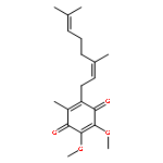
![2,5-Cyclohexadiene-1,4-dione,2-[(2E,6E,10E,14E,18E)-3,7,11,15,19,23-hexamethyl-2,6,10,14,18,22-tetracosahexaen-1-yl]-5,6-dimethoxy-3-methyl-](http://img.cochemist.com/ccimg/1100/1065-31-2.png)
![2,5-Cyclohexadiene-1,4-dione,2-[(2E,6E,10E,14E,18E)-3,7,11,15,19,23-hexamethyl-2,6,10,14,18,22-tetracosahexaen-1-yl]-5,6-dimethoxy-3-methyl-](http://img.cochemist.com/ccimg/1100/1065-31-2_b.png)


![[(3-chlorophenyl)hydrazono]malononitrile](http://img.cochemist.com/ccimg/600/555-60-2.png)
![[(3-chlorophenyl)hydrazono]malononitrile](http://img.cochemist.com/ccimg/600/555-60-2_b.png)


![4-Pyridinol,2-[(2E,5E,7E,9R,10R,11E)-10-hydroxy-3,7,9,11-tetramethyl-2,5,7,11-tridecatetraen-1-yl]-5,6-dimethoxy-3-methyl-](http://img.cochemist.com/ccimg/2800/2738-64-9.png)
![4-Pyridinol,2-[(2E,5E,7E,9R,10R,11E)-10-hydroxy-3,7,9,11-tetramethyl-2,5,7,11-tridecatetraen-1-yl]-5,6-dimethoxy-3-methyl-](http://img.cochemist.com/ccimg/2800/2738-64-9_b.png)


![2(5H)-Furanone,3-[(2R,13R)-2,13-dihydroxy-13-[(2R,2'R,5R,5'R)-octahydro-5'-[(1R)-1-hydroxyundecyl][2,2'-bifuran]-5-yl]tridecyl]-5-methyl-,(5S)-](http://img.cochemist.com/ccimg/103000/102989-24-2.png)
![2(5H)-Furanone,3-[(2R,13R)-2,13-dihydroxy-13-[(2R,2'R,5R,5'R)-octahydro-5'-[(1R)-1-hydroxyundecyl][2,2'-bifuran]-5-yl]tridecyl]-5-methyl-,(5S)-](http://img.cochemist.com/ccimg/103000/102989-24-2_b.png)