Co-reporter:Changrong Wang, Lili Du, Junhui Zhou, Lingwei Meng, Qiang Cheng, Chun Wang, Xiaoxia Wang, Deyao Zhao, Yuanyu Huang, Shuquan Zheng, Huiqing Cao, Jianhua Zhang, Liandong Deng, Zicai Liang, and Anjie Dong
ACS Applied Materials & Interfaces September 27, 2017 Volume 9(Issue 38) pp:32463-32463
Publication Date(Web):September 1, 2017
DOI:10.1021/acsami.7b07337
Hydrophobization of cationic polymers, as an efficient strategy, had been widely developed in the structure of cationic polymer micelles to improve the delivery efficiency of nucleic acids. However, the distribution of hydrophobic segments in the polymer chains is rarely considered. Here, we have elaborated three types of hydrophobized polyethylene glycol (PEG)-blocked cationic polymers with different distributions of the hydrophobic segments in the polymer chains PEG–PAM–PDP (E–A–D), PEG–PDP–PAM (E–D–A), and PEG–P(AM/DP) (E–(A/D)), which were synthesized by reversible addition–fragmentation chain transfer polymerization of methoxy PEG, cationic monomer aminoethyl methacrylate, and pH-sensitive hydrophobic monomer 2-diisopropylaminoethyl methacrylate, respectively. In aqueous solution, all of the three copolymers, E–A–D, E–D–A, and E–(A/D), were able to spontaneously form nanosized micelles (100–150 nm) (ME–A–D, ME–D–A, and ME–(A/D)) and well-incorporated small interfering RNA (siRNA) into complex micelles (CMs). The effect of distributions of the hydrophobic segments on siRNA delivery had been evaluated in vitro and in vivo. Compared with ME–D–A and ME–(A/D), ME–A–D showed the best siRNA binding capacity to form stable ME–A–D/siRNA CMs less than 100 nm, mediated the best gene-silencing efficiency and inhibition effect of tumor cell growth in vitro, and showed better liver gene-silencing effect in vivo. In the case of ME–(A/D) with a random distribution of cationic and hydrophobic segments, a gene-silencing efficiency higher than Lipo2000 but lesser than ME–A–D and ME–D–A was obtained. As the mole ratio of positive and negative charges increased, ME–D–A/siRNA and ME–A–D/siRNA showed similar performances in size, zeta potential, cell uptake, and gene silencing, but ME–(A/D)/siRNA showed reversed performances. In addition, ME–A–D as the best siRNA carrier was evaluated in the tumor tissue in the xenograft murine model and showed good anticancer capacity. Obviously, the distribution of the hydrophobic segments in the amphiphilic cationic polymer chains should be seriously considered in the design of siRNA vectors.Keywords: amphiphilic cationic polymers; delivery efficiency; distribution of the hydrophobic segments; gene silence; siRNA delivery;
Co-reporter:Dong Huang, Deyao Zhao, Jinhui Li, Yuting Wu, Wenbo Zhou, Wei Wang, Zicai Liang, Zhihong Li
Sensors and Actuators B: Chemical 2017 Volume 250(Volume 250) pp:
Publication Date(Web):1 October 2017
DOI:10.1016/j.snb.2017.04.085
•Hydrodynamics strategy of Dean vortex was utilized to isolate the cells from electrode enhance the cell viability.•An optimized flow rate for cell electroporation was obtained.•The device realized successful electroporation on several cell types with high viability and high transfection rate.•The high-throughput reaching 2.2 ml/min and continuous electroporation were achieved.In this paper, by utilizing a curved channel, where the Dean flow is generated, the cell viability in the microfluidic electroporation chip has been significantly increased. The Dean vortex plays two major roles. Firstly, in the middle region of the curved channel, the cell solution is enveloped by the protective buffer, which keeps cells away from the metal electrodes throughout the electroporation process. Therefore, distasteful effects due to water electrolysis, such as air bubbles, Joule heating and pH changing, are mitigated or avoided. Secondly, in the ending region of the curved channel, the Dean vortex will recombine the split protective flow and neutralize positive and negative ions. Consequently, several hard-to-transfect cell types were successfully transferred with this device. Compared to conventional microfluidic electroporation devices, our electroporation chip can achieve flow-through electroporation with high viability and high transfection rate, while providing a continuous electroporation environment to address large doses of biological transfection requirement.
Co-reporter:Yuanyu Huang, Qiang Cheng, Xingyu Jin, Jia-Li Ji, Shutao Guo, Shuquan Zheng, Xiaoxia Wang, Huiqing Cao, Shan Gao, Xing-Jie Liang, Quan Du and Zicai Liang
Biomaterials Science 2016 vol. 4(Issue 3) pp:494-510
Publication Date(Web):19 Jan 2016
DOI:10.1039/C5BM00429B
The drug development of siRNA has been seriously hindered by the lack of an effective, safe and clinically applicable delivery system. The cyclic NGR motif and its isomerization product isoDGR recruit CD13 and integrin as their specific receptors, both of which are overexpressed by tumor and neovascular cells. In this study, a bi-functional peptide, named NGR-10R, was designed and tested for siRNA delivery in vitro and in vivo. Through the formation of peptide/siRNA nanoparticles, RNase resistance was greatly enhanced for the siRNAs. Both FACS and confocal assays revealed that the peptide/siRNA complexes were effectively internalized by MDA-MB-231 cells. Gene silencing assays indicated that anti-Lamin A/C siRNA delivered by NGR-10R robustly repressed gene expression in MDA-MB-231 and HUVEC (a CD13+/αvβ3+ cell). Importantly, the siRNAs were efficiently delivered into tumor tissues and localized around the nuclei, as revealed by in vivo imaging and cryosection examination. In summary, NGR-10R not only efficiently delivered siRNAs into MDA-MB-231 cells in vitro but also delivered siRNAs into tumor cells in vivo, taking advantage of its specific binding to CD13 (neovascular) or αvβ3 (MDA-MB-231). Therefore, the NGR-10R peptide provides a promising siRNA delivery reagent that could be used for drug development, particularly for anti-tumor therapeutics.
Co-reporter:Yu Sun;Junhui Zhou;Qiang Cheng;Daoshu Lin;Qian Jiang;Anjie Dong;Liong Deng
Journal of Applied Polymer Science 2016 Volume 133( Issue 16) pp:
Publication Date(Web):
DOI:10.1002/app.43303
ABSTRACT
Graphene oxide (GO)-based nanohybrids were designed for small interfering RNA (siRNA) delivery for their high water dispensability, good biocompatibility, easily tunable surface functionalization, and particular optical properties. In this study, novel nanohybrids based on GO were fabricated. Methoxypoly(ethylene glycol) (mPEG) was covalently conjugated to GO via amide bonds. Then, poly(2-dimethyl aminoethyl methacrylate) (PDMAEMA), which was synthesized via reversible addition–fragmentation chain transfer polymerization (RAFT) with 2-(dodecyl thiocarbonothioyl thio)-2-methyl propionic acid (DTM) as the RAFT agent, was attached onto GO via physical interaction between DTM and GO. Compared with Lipofectamine 2000, the novel mPEG–GO/PDMAEMA nanohybrids showed comparable gene transfection efficiency and a low cytotoxicity. Moreover, the mPEG–GO/PDMAEMA nanohybrids showed enhanced optical properties compared to the original GO because of the presence of mPEG and PDMAEMA. Our work encouraged further exploration of the novel nanovector for combined photothermal and siRNA delivery. © 2015 Wiley Periodicals, Inc. J. Appl. Polym. Sci. 2016, 133, 43303.
Co-reporter:Lei Zhang;Ye Xu;Xingyu Jin;Zengwu Wang;Yidi Wu;Deyao Zhao;Gang Chen;Deyu Li;Xiaoxia Wang;Huiqing Cao;Yuntao Xie;Zicai Liang
Breast Cancer Research and Treatment 2015 Volume 154( Issue 2) pp:423-434
Publication Date(Web):2015/11/01
DOI:10.1007/s10549-015-3591-0
Novel, non-invasive biomarkers to diagnose breast cancer with high sensitivity and specificity are greatly desired. Circulating microRNAs (miRNAs) show potential for breast cancer detection, but the existing results appear to be mixed. Using microscale serum, we established a novel serum-direct multiplex detection assay based on RT-PCR (SdM-RT-PCR). Ninety-three miRNAs dysregulated or with functions in breast cancer were selected as candidates, and additional 3 miRNAs were chosen as endogenous controls. We first conducted miRNA profiling of these 96 miRNAs by SdM-RT-PCR using the sera of 25 breast cancer patients at diagnosis prior to treatment and 20 age-matched healthy controls. miRNAs showing significantly different expression levels between patients and controls were further analyzed using a logistic regression model. A miRNA signature was validated in an independent set of 128 serum samples composed of 76 breast cancer patients and 52 healthy controls. In the discovery stage, we identified 23 miRNAs as significantly dysregulated in breast cancer patients compared with healthy controls. Of these, 10 miRNAs were previously identified as dysregulated in breast cancer; 14 miRNAs remained significant after P-values were adjusted by both correction methods. Principal component analysis and hierarchical clustering of these miRNAs separated patients from controls. Furthermore, the 3-miRNA signature (miR-199a, miR-29c, and miR-424) with the highest diagnostic accuracy for distinguishing breast cancer patients from controls by ROC curve analysis (AUC = 0.888) was successfully confirmed in the validation set (AUC = 0.901). Our data demonstrate that the SdM-RT-PCR assay is an effective breast cancer profiling method that utilizes very small volumes and is compatible with Biobank. Furthermore, the identified 3-miRNA signature is a promising circulating biomarker for breast cancer diagnosis.
Co-reporter:Zewen Wei, Xueming Li, Deyao Zhao, Hao Yan, Zhiyuan Hu, Zicai Liang, and Zhihong Li
Analytical Chemistry 2014 Volume 86(Issue 20) pp:10215
Publication Date(Web):September 24, 2014
DOI:10.1021/ac502294e
Microfluidics based continuous cell electroporation is an appealing approach for high-throughput cell transfection, but cell viability of existing methods is usually compromised by adverse electrical or hydrodynamic effects. Here we present the validation of a flow-through cell electroporation microchip, in which dielectrophoretic force was employed to sort viable cells. By integrating parallel electroporation electrodes and dielectrophoresis sorting electrodes together in a simple straight microfluidic channel, sufficient electrical pulses were applied for efficient electroporation, and a proper sinusoidal electrical field was subsequently utilized to exclude damaged cells by dielectrophoresis. Thus, the difficulties for seeking the fine balance between electrotransfection efficiency and cell viability were steered clear. After careful investigation and optimization of the DEP behaviors of electroporated cells, efficient electrotransfection of plasmid DNA was demonstrated in vulnerable neuron cells and several hard-to-transfect primary cell types with excellent cell viability. This microchip constitutes a novel way of continuous cell transfection to significantly improve the cell viability of existing methodologies.
Co-reporter:Yanyan Li, Shuquan Zheng, Xiaolong Liang, Yushen Jin, Yidi Wu, Huichen Bai, Renfa Liu, Zhifei Dai, Zicai Liang, and Tiejun Shi
Bioconjugate Chemistry 2014 Volume 25(Issue 11) pp:2055
Publication Date(Web):September 26, 2014
DOI:10.1021/bc500414e
The therapeutic application of small interfering RNA (siRNA) requires safe nanocarriers for specific and efficient delivery in vivo. Herein, PEGylated cationic cerasomes (PCCs) were fabricated by doping a cationic lipid with a hydroxyl group into nanohybrid cerasomes. Multiple properties of PCCs provide a solution to many of the limitations associated with current platforms for the delivery of siRNA. The polyorganosiloxane surface imparts PCCs with higher morphological stability than conventional liposomes. The PEGylation of the cationic cerasome could protect the cerasome nanoparticles from agglomeration and macrophage capture, reduce protein absorption, and consequently prolong the blood circulating time and enhance the siRNA delivery efficiency. In addition, incorporation of the lipid containing a hydroxyl group further facilitates endosome release. Moreover, PCCs were further used to transport siRNA into the cytosol primarily via endocytosis. When applied to systemic administration, PCCs have demonstrated effective delivery into the liver and preferential uptake by hepatocytes in mice, thereby leading to high siRNA gene-silencing activity. All these results show potential therapeutic applications of PCCs-mediated delivery of siRNA for liver diseases.
Co-reporter:Daoshu Lin, Qian Jiang, Qiang Cheng, Yuanyu Huang, Pingsheng Huang, Shangcong Han, Shutao Guo, Zicai Liang, Anjie Dong
Acta Biomaterialia 2013 Volume 9(Issue 8) pp:7746-7757
Publication Date(Web):August 2013
DOI:10.1016/j.actbio.2013.04.031
Abstract
Long circulation, cell internalization, endosomal escape and small interfering RNA (siRNA) release to the cytoplasm are the prerequisite considerations for siRNA delivery vectors. Herein, a kind of sheddable nanoparticles (NPs) with micelle architecture for siRNA delivery were fabricated by using an intracellular-activated polycation-detachable copolymer (PECssD), which was prepared by introducing highly reducing environment-responsive disulfide linkages between PEGylated polycaprolactone (PCL) and the grafted polycation, poly(2-dimethylaminoethyl methacrylate) (PDMAEMA). The architecture of PECssD self-assembled NPs includes a biodegradable hydrophobic PCL core, a PEG shield and a detachable comb-like polycation surface. The stable nanosized complexes of PECssD NPs with siRNA, termed PECssD/siRNA micelleplexes, were formed, which could prolong circulation, improve accumulation and retention in tumor tissue, and be favorable for internalization. In particular, the cleavage of the disulfide linkages in the intracellular microenvironment and the subsequent dissociation of the PDMAEMA/siRNA polyplexes from the PEGylated PCL cores of PECssD/siRNA micelleplexes were also confirmed, which facilitated the endosomal escape and the efficient release of siRNA. As a result, the distribution of siRNA in cytoplasm was enhanced and subsequently promoted the efficiency of siRNA in gene silencing. Furthermore, systemic administration of the NPs carrying siPlk1 (polo-like kinase 1 specific siRNA) induced a tumor-suppressing effect in the HeLa-Luc xenograft murine model. Therefore, the devised strategy of the polycation-detachable copolymer PECssD NPs could address the requirements of the multistep systemic delivery process of siRNA. The hydrophobic core of the PECssD/siRNA micelleplexes is expected to entrap antitumor drugs or other therapeutic agents for combined therapies.
Co-reporter:Mengxi Wu, Deyao Zhao, Zewen Wei, Wenfeng Zhong, Hao Yan, Xiaoxia Wang, Zicai Liang, and Zhihong Li
Analytical Chemistry 2013 Volume 85(Issue 9) pp:4483
Publication Date(Web):April 2, 2013
DOI:10.1021/ac400017x
We have developed a rapid method to optimize the electric parameters of cell electroporation. In our design, a pair of ring-dot formatted electrodes was used to generate a radial distribution of electric field from the center to the periphery. Varied electric field intensity was acquired in different annulus when an electric pulse was applied. Cells were cultured on the microchips for adherent cell electroporation and in situ observation. The electroporation parameters of electric field intensity were explored and evaluated in terms of cell viability and transfection efficiency. The optimization was performed in consideration of both cell viability, which was investigated to decrease as electric field increases, and the transfection rate, which normally increases at stronger electric field. The electroporation characteristics HEK-293A and Hela cells were investigated, and the optimum parameters were obtained. Verified by a commercial electroporation system as well as self-made microchips endowed the optimization with wider meaning. At last, as applications, we acquired the optimal electroporation pulse intensity of Neuro-2A cells and a type of primary cell (human umbilical vein endothelial cell, HUVEC) by one time electroporation using the proposed method.
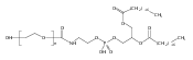
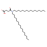
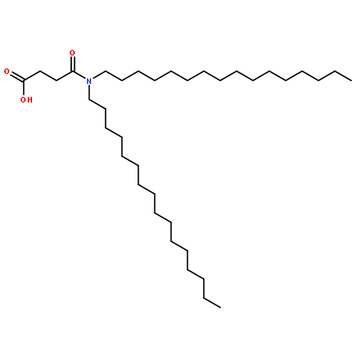
Co-reporter:Qiang Cheng, Yuanyu Huang, Hua Zheng, Tuo Wei, Shuquan Zheng, Shuaidong Huo, Xiaoxia Wang, Quan Du, Xiaoning Zhang, Hong-Yan Zhang, Xing-Jie Liang, Chun Wang, Rupei Tang, Zicai Liang
Biomaterials 2013 34(12) pp: 3120-3131
Publication Date(Web):
DOI:10.1016/j.biomaterials.2013.01.043
Co-reporter:Wendi Zhang, Qiang Cheng, Shutao Guo, Daoshu Lin, Pingsheng Huang, Juan Liu, Tuo Wei, Liandong Deng, Zicai Liang, Xing-Jie Liang, Anjie Dong
Biomaterials 2013 34(27) pp: 6495-6503
Publication Date(Web):
DOI:10.1016/j.biomaterials.2013.04.030
Co-reporter:Yuanyu Huang, Daoshu Lin, Qian Jiang, Wendi Zhang, Shutao Guo, Ping Xiao, Shuquan Zheng, Xiaoxia Wang, Hongbo Chen, Hong-Yan Zhang, Liandong Deng, Jinfeng Xing, Quan Du, Anjie Dong, Zicai Liang
Biomaterials 2012 33(18) pp: 4653-4664
Publication Date(Web):
DOI:10.1016/j.biomaterials.2012.02.052
Co-reporter:Zewen Wei, Deyao Zhao, Xueming Li, Mengxi Wu, Wei Wang, Huang Huang, Xiaoxia Wang, Quan Du, Zicai Liang, and Zhihong Li
Analytical Chemistry 2011 Volume 83(Issue 15) pp:5881
Publication Date(Web):June 17, 2011
DOI:10.1021/ac200625b
By introducing a hydrodynamic mechanism into a microfluidics-based electroporation system, we developed a novel laminar flow electroporation system with high performance. The laminar buffer flow implemented in the system separated the cell suspension flow from the electrodes, thereby excluding many unfavorable effects due to electrode reaction during electroporation, such as hydrolysis, bubble formation, pH change, and heating. Compared to conventional microfluidic electroporation systems, these improvements significantly enhanced transfection efficiency and cell viability. Furthermore, successful electrotransfection of plasmid DNA and, more importantly, synthetic siRNA, was demonstrated in several hard-to-transfect cell types using this system.
Co-reporter:Shutao Guo, Yuanyu Huang, Wendi Zhang, Weiwei Wang, Tuo Wei, Daoshu Lin, Jinfeng Xing, Liandong Deng, Quan Du, Zicai Liang, Xing-Jie Liang, Anjie Dong
Biomaterials 2011 32(18) pp: 4283-4292
Publication Date(Web):
DOI:10.1016/j.biomaterials.2011.02.034
Co-reporter:Daoshu Lin, Yuanyu Huang, Qian Jiang, Wendi Zhang, Xinye Yue, Shutao Guo, Ping Xiao, Quan Du, Jinfeng Xing, Liandong Deng, Zicai Liang, Anjie Dong
Biomaterials 2011 32(33) pp: 8730-8742
Publication Date(Web):
DOI:10.1016/j.biomaterials.2011.07.089
Co-reporter:Huang Huang;Na Wei;Yingfei Xiong;Feng Yang;Huaqiang Fang
Frontiers in Biology 2010 Volume 5( Issue 3) pp:272-281
Publication Date(Web):2010 June
DOI:10.1007/s11515-010-0047-0
To modulate gene expression in research studies or in potential clinical therapies, transfection of exogenous nucleic acids including plasmid DNA and small interference RNA (siRNA) are generally performed. However, the cellular processing and the fate of these nucleic acids remain elusive. By investigating the cellular behavior of transfected nucleic acids using confocal imaging, here we show that when siRNA was co-transfected into cultured cells with other nucleic acids, including single-stranded RNA oligonucleotides, single and double-stranded DNA oligonucleotides, as well as long double-stranded plasmid DNA, they all aggregate in the same cytoplasmic granules. Interestingly, the amount of siRNA aggregating in granules was found not to correlate with the gene silencing activity, suggesting that assembly of cytoplasmic granules triggered by siRNA transfection may be separable from the siRNA silencing event. Our results argue against the claim that the siRNA-aggregating granules are the functional site of RNA interference (RNAi). Taken together, our studies suggest that, independent of their types or forms, extraneously transfected nucleic acids are processed through a common cytoplasmic pathway and trigger the formation of a new type of cytoplasmic granules “transfection granules”.
Co-reporter:J Xiong;Q Du;Z Liang
Oncogene 2010 29(35) pp:4980-4988
Publication Date(Web):2010-06-21
DOI:10.1038/onc.2010.241
Oncogenic c-Myc has been described to modulate the expression of a subset of microRNAs (miRNAs), which include miR-22; however, the mechanism through which a miRNA controls c-Myc activity remains unclear. Here we report a novel anti-c-Myc function mediated by miR-22. Ectopically expressed miR-22 inhibited cell proliferation and anchorage-independent growth of human cancer cell lines. Microarray screening and western analyses revealed that miR-22 repressed the c-Myc-binding protein MYCBP, a positive regulator of c-Myc. Consistent with this, reporter assays showed that miR-22-mediated MYCBP gene suppression largely depends on the conserved miR-22 target site within the MYCBP 3′-untranslational region (3′UTR), implying that MYCBP mRNA is a direct miR-22 target. Depletion of MYCBP using small interfering RNA (siRNA) recapitulated the miR-22-induced anti-growth effect on tumor cells, whereas ectopically expressed MYCBP rescued cells from the growth suppression mediated by miR-22. Moreover, repression of MYCBP by miR-22 downregulated a panel of E-box-containing c-Myc target genes. Our results suggest that miR-22 acts as a tumor suppressor through direct repression of MYCBP expression and subsequent reduction of oncogenic c-Myc activities. As c-Myc inhibits the expression of miR-22, we propose a novel positive feedback loop formed by oncogenic c-Myc to accelerate cell proliferation by suppressing miR-22, a potent inhibitor of MYCBP.
Co-reporter:Xiaoxia Wang, Feng Yan, Quan Du, Zicai Liang
Drug Discovery Today: Technologies (Summer 2010) Volume 7(Issue 2) pp:e125-e130
Publication Date(Web):1 June 2010
DOI:10.1016/j.ddtec.2010.11.011
siRNA technology has not only greatly facilitated functional characterization of new genes, but also presented tremendous potential for becoming the next generation of blockbuster drugs. The technology has penetrated life science research in an extremely fast speed, thus providing a huge knowledge base for siRNA drug development. Significant technical progresses have been achieved over the past ten years towards the research and development of siRNA therapeutics, and several compounds have been put into clinical trials. This review would provide a summary of such progresses.
Co-reporter:Zicai Liang, Xiu-Jie Wang
Journal of Genetics and Genomics (20 April 2013) Volume 40(Issue 4) pp:141-142
Publication Date(Web):20 April 2013
DOI:10.1016/j.jgg.2013.03.007
Co-reporter:Feng Yang, Fan Yi, Xiaorui Han, Quan Du, Zicai Liang
FEBS Letters (1 October 2013) Volume 587(Issue 19) pp:3175-3181
Publication Date(Web):1 October 2013
DOI:10.1016/j.febslet.2013.07.048
•Translocation of MALAT-1 from the nucleus to the cytoplasm during G2/M cell cycle phase.•Down-regulation of MALAT-1 expression compromises the cytoplasmic translocation of hnRNP C and leads to cell cycle arrest.•MALAT-1 interacts with hnRNP C to function in cell cycle regulation.As a conserved non-coding RNA gene, transcripts of MALAT-1 localize predominately in the nucleus. However in G2/M cell cycle phase, MALAT-1 transcripts were surprisingly found to translocate from the nucleus into the cytoplasm. Investigation also found that in this process MALAT-1 interacts with an abundant nuclear factor, hnRNP C protein. Using a loss-of-function assay, we found that down-regulation of MALAT-1 expression compromised the cytoplasmic translocation of hnRNP C in the G2/M phase and resulted in G2/M arrest. In addition to characterize the physiological interaction between MALAT-1 and hnRNP C, our study also highlights the role of MALAT-1 in cell cycle regulation.
Co-reporter:Yuanyu Huang, Qiang Cheng, Xingyu Jin, Jia-Li Ji, Shutao Guo, Shuquan Zheng, Xiaoxia Wang, Huiqing Cao, Shan Gao, Xing-Jie Liang, Quan Du and Zicai Liang
Biomaterials Science (2013-Present) 2016 - vol. 4(Issue 3) pp:
Publication Date(Web):
DOI:10.1039/C5BM00429B