Co-reporter:Yuqi Yang, Shizhen Chen, Lianhua Liu, Sha Li, Qingbin Zeng, Xiuchao Zhao, Haidong Li, Zhiying Zhang, Louis-S. Bouchard, Maili Liu, and Xin Zhou
ACS Applied Materials & Interfaces July 19, 2017 Volume 9(Issue 28) pp:23400-23400
Publication Date(Web):June 26, 2017
DOI:10.1021/acsami.7b05463
Currently, the potential of cancer therapy is compromised by a variety of problems related to tumor specificity, drug access, and limited efficacy. We report a novel approach to improve the effectiveness of cancer treatment utilizing a light-responsive nanoconstruct. Effectiveness is increased by enhancing drug absorption through heating and the production of free radicals. Treatment specificity is increased through chemical targeting of the nanoconstruct and localization of light delivery to the tumor. When reaching the tumor, magnetic resonance imaging is enhanced and near-infrared fluorescence is activated upon drug release, making it possible to visualize the localized treatment at both the tissue and cellular levels. This dual-modality imaging nanoconstruct enables the synergistic treatment and observable evaluation of solid tumors with dramatically improved efficacy, giving rise to a promising new approach for cancer therapy and evaluation.Keywords: biological redox therapy; dual-modality imaging; graphene quantum dots; photodynamic therapy; photothermal therapy;
Co-reporter:Qingbin Zeng, Qianni Guo, Yaping Yuan, Yuqi Yang, Bin Zhang, Lili Ren, Xiaoxiao Zhang, Qing Luo, Maili Liu, Louis-S. Bouchard, and Xin Zhou
Analytical Chemistry 2017 Volume 89(Issue 4) pp:
Publication Date(Web):January 30, 2017
DOI:10.1021/acs.analchem.6b03742
Biothiols such as gluthathione (GSH), cysteine (Cys), homocysteine (Hcy), and thioredoxin (Trx) play vital roles in cellular metabolism. Various diseases are associated with abnormal cellular biothiol levels. Thus, the intracellular detection of biothiol levels could be a useful diagnostic tool. A number of methods have been developed to detect intracellular thiols, but sensitivity and specificity problems have limited their applications. To address these limitations, we have designed a new biosensor based on hyperpolarized xenon magnetic resonance detection, which can be used to detect biothiol levels noninvasively. The biosensor is a multimodal probe that incorporates a cryptophane-A cage as 129Xe NMR reporter, a naphthalimide moiety as fluorescence reporter, a disulfide bond as thiol-specific cleavable group, and a triphenylphosphonium moiety as mitochondria targeting unit. When the biosensor interacts with biothiols, disulfide bond cleavage leads to enhancements in the fluorescence intensity and changes in the 129Xe chemical shift. Using Hyper-CEST (chemical exchange saturation transfer) NMR, our biosensor shows a low detection limit at picomolar (10–10 M) concentration, which makes a promise to detect thiols in cells. The biosensor can detect biothiol effectively in live cells and shows good targeting ability to the mitochondria. This new approach not only offers a practical technique to detect thiols in live cells, but may also present an excellent in vivo test platform for xenon biosensors.
Co-reporter:Lili Ren, Shizhen Chen, Haidong Li, Zhiying Zhang, Jianping Zhong, Maili Liu, Xin Zhou
Acta Biomaterialia 2016 Volume 35() pp:260-268
Publication Date(Web):15 April 2016
DOI:10.1016/j.actbio.2016.02.011
Abstract
Liposomes are effective drug delivery systems that can be functionalized with imaging contrast agents, providing both diagnosis and monitoring of disease treatment. Here we describe the design of a theranostic liposomal drug delivery system whose biodistribution can be real time imaged by contrast enhanced MRI and can achieve tandem chemotherapy drug delivery. Because T1 relaxation of MRI depends upon the chemical structure of contrast agent as well as its interaction with neighbor environment, we rationally designed a functional liposome for in vivo T1 enhanced MRI. The liposome shows a 36-fold higher T1 relaxation rate over the commercial MRI contrast agent Omniscan® and a long circulation time up to 300 min in vivo. Moreover, the multifunctional liposome carries both hydrophobic and hydrophilic chemotherapeutic drugs, can synergistically enhance therapeutic effects of multiple drugs and selectively deliver them to lung tumors, leading to lower doses, toxicity and sustained release. The nanoparticles, which exhibit favorable biodistributions to tumors, offer new possibilities for the simultaneous delivery of more than one drug and the evaluation of therapeutic response in vivo by T1 enhanced MRI.
Statement of Significance
Cancer cells invoke different mechanisms to resist cancer therapies, particularly when delivering a single agent in a given therapy. The combination of two (or more) thermotherapy agents provides a promising way to circumvent such situations of drug resistance, due to a favorable synergistic effect that “tricks” the drug resistance mechanism.
However, challenges to the simultaneous delivery of two drugs prevail, especially with regards to the simultaneous delivery of hydrophobic and hydrophobic drugs. Furthermore, non-invasive in vivo imaging of drug distribution enables the real-time monitoring and prediction of therapeutic responses to treatment.
In this study, we rationally designed a theranostic liposomal drug delivery system whose biodistribution can be imaged via T1-weighted MRI in real-time and can achieve tandem chemotherapy drug delivery. This original study will be of considerable use to the wider drug delivery community.
Co-reporter:Shengjun Yang, Weiping Jiang, Lili Ren, Yaping Yuan, Bin Zhang, Qing Luo, Qianni Guo, Louis-S. Bouchard, Maili Liu, and Xin Zhou
Analytical Chemistry 2016 Volume 88(Issue 11) pp:5835
Publication Date(Web):April 29, 2016
DOI:10.1021/acs.analchem.6b00403
Biothiols such as cysteine (Cys), homocysteine (Hcy), and glutathione (GSH) play an important role in regulating the vital functions of living organisms. Knowledge of their biodistribution in real-time could help diagnose a variety of conditions. However, existing methods of biothiol detection are invasive and require assays. Herein we report a molecular biosensor for biothiol detection using the nuclear spin resonance of 129Xe. The 129Xe biosensor consists of a cryptophane cage encapsulating a xenon atom and an acrylate group. The latter serves as a reactive site to covalently bond biothiols through a thiol-addition reaction. The biosensor enables discrimination of Cys from Hcy and GSH through the chemical shift and average reaction rate. This biosensor can be detected at a concentration of 10 μM in a single scan and it has been applied to detect biothiols in bovine serum solution. Our results indicate that this biosensor is a promising tool for the real-time imaging of biothiol distributions.
Co-reporter:Haidong Li;Zhiying Zhang;Xiuchao Zhao;Xianping Sun;Chaohui Ye
Magnetic Resonance in Medicine 2016 Volume 76( Issue 2) pp:408-416
Publication Date(Web):
DOI:10.1002/mrm.25894
Purpose
To demonstrate the feasibility of quantitative and comprehensive global evaluation of pulmonary function and microstructural changes in rats with radiation-induced lung injury (RILI) using hyperpolarized xenon MR.
Methods
Dissolved xenon spectra were dynamically acquired using a modified chemical shift saturation recovery pulse sequence in five rats with RILI (bilaterally exposed by 6-MV x-ray with a dose of 14 Gy 3 mo. prior to MR experiments) and five healthy rats. The dissolved xenon signals were quantitatively analyzed, and the pulmonary physiological parameters were extracted with the model of xenon exchange.
Results
The obtained pulmonary physiological parameters and the ratio of 129Xe signal in red blood cells (RBCs) versus barrier showed a significant difference between the groups. In RILI rats versus controls, the exchange time increased from 44.5 to 112 ms, the pulmonary capillary transit time increased from 0.51 to 1.48 s, and the ratio of 129Xe spectroscopic signal in RBCs versus barrier increased from 0.294 to 0.484.
Conclusion
Hyperpolarized xenon MR is effective for quantitative and comprehensive global evaluation of pulmonary function and structural changes without the use of radiation. This may open the door for its use in the diagnosis of lung diseases that are related to gas exchange. Magn Reson Med 76:408–416, 2016. © 2015 Wiley Periodicals, Inc.
Co-reporter:Dr. Qianni Guo;Qingbin Zeng;Weiping Jiang;Xiaoxiao Zhang;Dr. Qing Luo;Dr. Xu Zhang;Dr. Louis-S. Bouchard;Dr. Maili Liu;Dr. Xin Zhou
Chemistry - A European Journal 2016 Volume 22( Issue 12) pp:3967-3970
Publication Date(Web):
DOI:10.1002/chem.201600193
Abstract
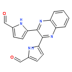
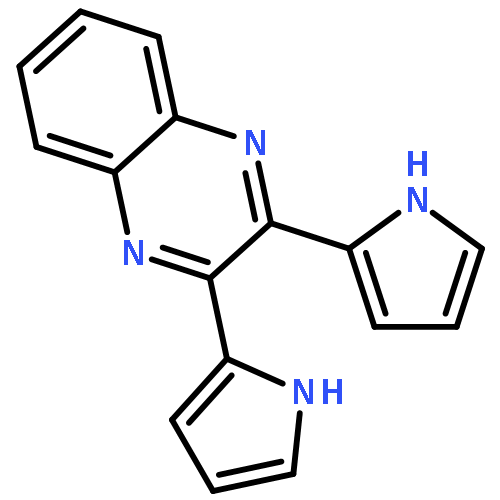
Mercury pollution, in the form of mercury ions (Hg2+), is a major health and environmental hazard. Commonly used sensors are invasive and limited to point measurements. Fluorescence-based sensors do not provide depth resolution needed to image spatial distributions. Herein we report a novel sensor capable of yielding spatial distributions by MRI using hyperpolarized 129Xe. A molecular clamp probe was developed consisting of dipyrrolylquinoxaline (DPQ) derivatives and twocryptophane-A cages. The DPQ derivatives act as cation receptors whereas cryptophane-A acts as a suitable host molecule for xenon. When the DPQ moiety interacts with mercury ions, the molecular clamp closes on the ion. Due to overlap of the electron clouds of the two cryptophane-A cages, the shielding effect on the encapsulated Xe becomes important. This leads to an upfield change of the chemical shift of the encapsulated Xe. This sensor exhibits good selectivity and sensitivity toward the mercury ion. This mercury-activated hyperpolarized 129Xe-based chemosensor is a new concept method for monitoring Hg2+ ion distributions by MRI.
Co-reporter:Lili Ren, Shizhen Chen, Haidong Li, Zhiying Zhang, Chaohui Ye, Maili Liu and Xin Zhou
Nanoscale 2015 vol. 7(Issue 30) pp:12843-12850
Publication Date(Web):11 May 2015
DOI:10.1039/C5NR02144H
Real-time diagnosis and monitoring of disease development, and therapeutic responses to treatment, are possible by theranostic magnetic resonance imaging (MRI). Here we report the synthesis of a multifunctional liposome, which contains Gd-DOTA (an MRI probe), paclitaxel and c(RGDyk) (a targeted peptide). This nanoparticle overcame the insolubility of paclitaxel, reduced the side effects of FDA-approved formulation of PTX-Cre (Taxol®) and improved drug delivery efficiency to the tumor. c(RGDyk) modification greatly enhanced the cytotoxicity of the drug in tumor cells A549. The T1 relaxivity in tumor cells treated with the targeted liposome formulation was increased 16-fold when compared with the non-targeted group. In vivo, the tumors in mice were visualized using T1-weighted imaging after administration of the liposome. Also the tumor growth could be inhibited well after the treatment. Fluorescence images in vitro and ex vivo also showed the targeting effect of this liposome in tumor cells, indicating that this nanovehicle could limit the off-target side effects of anticancer drugs and contrast agents. These findings lay the foundation for further tumor inhibition study and application of this delivery vehicle in cancer therapy settings.
Co-reporter:Xiaolei Zhu, Shizhen Chen, Qing Luo, Chaohui Ye, Maili Liu and Xin Zhou
Chemical Communications 2015 vol. 51(Issue 44) pp:9085-9088
Publication Date(Web):02 Apr 2015
DOI:10.1039/C5CC02587G
A novel thermo-sensitive micelle contrast agent and its enhancement of MRI contrast with temperature are reported. The morphology changes sharply near 37 °C, resulting in a significant amplification of the CEST signal. This enables detection of small changes in body temperature.
Co-reporter:Qi Wang, Shizhen Chen, Qing Luo, Maili Liu and Xin Zhou
RSC Advances 2015 vol. 5(Issue 3) pp:1808-1811
Publication Date(Web):28 Nov 2014
DOI:10.1039/C4RA12201A
A novel reconstituted high-density lipoprotein (rHDL) nanocomposite has been prepared for highly-sensitive magnetic resonance (MR)-fluorescence multimodal imaging. Such a nanocomposite is able to enhance the MR sensitivity up to 129 fold in comparison to the traditional small molecule MRI agent based on paramagnetic chemical exchange saturation transfer (PARACEST). It has also demonstrated specific targeting to macrophage cells, which shows great potential for the detection of atherosclerosis.
Co-reporter:Shizhen Chen, Yuqi Yang, Haidong Li, Xin Zhou and Maili Liu
Chemical Communications 2014 vol. 50(Issue 3) pp:283-285
Publication Date(Web):16 Oct 2013
DOI:10.1039/C3CC47324D
Novel pH-triggered nanoprobe were fabricated for 19F MRI and fluorescence imaging (MRI-FI) of cancer cells. The biocompatibility, durability, high internalizing efficiency and pore architecture justify the Au-fluorescent mesoporous silica nanoparticles as ideal, highly sensitive and highly specific vectors for 19F MRI and FI of cancer cells.
Co-reporter:Ji Zhang, Weiping Jiang, Qing Luo, Xiaoxiao Zhang, Qianni Guo, Maili Liu, Xin Zhou
Talanta 2014 Volume 122() pp:101-105
Publication Date(Web):May 2014
DOI:10.1016/j.talanta.2014.01.023
•Ultrahigh NMR signal sensitivity: Utilization of hyperpolarized Xe NMR, which enhances the signal more than 10,000 times over the conventional NMR at thermal equilibrium.•Large chemical shift response: Novelty designed supermolecule sensor enables 129Xe NMR spectroscopy was shifted up to 6.4 ppm in the presence of Zn2+ ions, which is 4 times larger than that of the reported similar sensor.•High selectivity to Zn2+ions: The response of Xe NMR exhibited high selectivity to Zn2+ ions as discriminated from other six potentially competing metal ions.•Fast and non-destructive analytical techniques: Take the advantage of NMR technique, the determination of Zn2+ ions was allowed in solution and in solid state, as well as to enable the study of biological tissues.Although Zn2+ ions are involved in large numbers of physiopathological processes, non-invasive detection of Zn2+ ions in opaque biological samples remains a huge challenge. Here, we developed a novel zinc-responsive hyperpolarized (HP) 129Xe-based NMR molecular sensor. This HP 129Xe-based NMR molecular sensor was synthesized by attaching 2-(diphenylphosphino) benzenamine as ligand for zinc ions to the xenon-binding supramolecular cage, cryptophane. The 129Xe NMR spectroscopy of such molecular sensor was shifted up to 6.4 ppm in the presence of Zn2+ ions, which was nearly four times larger than that of the reported similar sensor. The application of the sensor would benefit low concentration detection by using indirect NMR/MRI method. The response exhibited high sensitivity and selectivity as discriminated from other six potentially competing metal ions. The application of this sensor in the analysis of zinc ions in the rat serum samples was also evaluated. The strategy is generally applicable in developing sensitive and selective sensors for quantitative determination of zinc ions.We developed a novel hyperpolarized 129Xe-based NMR molecular sensor for detection of Zn2+ ions. The 129Xe NMR spectroscopy of such molecular sensor was shifted up to 6.4 ppm in the presence of Zn2+ ions, which was nearly four times larger than that of the reported similar sensor. This zinc-activated HP 129Xe-based NMR molecular sensor suggests a great potential to be used in the monitoring of Zn2+ ions and investigating Zn2+ ions related physiopathological processes in biological organisms in the foreseeable future.
Co-reporter:Guobin Liu, Xiaofeng Li, Xianping Sun, Jiwen Feng, Chaohui Ye, Xin Zhou
Journal of Magnetic Resonance 2013 237() pp: 158-163
Publication Date(Web):
DOI:10.1016/j.jmr.2013.10.008
Co-reporter:
Science 1920 Vol 51(1315) pp:270-271
Publication Date(Web):12 Mar 1920
DOI:10.1126/science.51.1315.270
Co-reporter:He Deng, Xianping Sun, Maili Liu, Chaohui Ye, Xin Zhou
Pattern Recognition (January 2017) Volume 61() pp:66-77
Publication Date(Web):1 January 2017
DOI:10.1016/j.patcog.2016.07.036
•We present an adaptive entropy-based window selection scheme.•The novel local difference measure map can keep low false alarm rates under the same probability of detection.•The proposed method is simple and effective with regard to detection accuracy.Dim and small target detection in complex background is considered a difficult and challenging problem. Conventional algorithms using the local difference/mutation possibly produce high missed or mistaken detection rates. In this paper, we propose an effective algorithm for detecting dim and small infrared targets. In order to synchronously enhance targets and suppress complex background clutters, we adopt an adaptive entropy-based window selection technique to construct a novel local difference measure (LDM) map of an input image, which measures the dissimilarity between the current region and its neighboring ones. In this way, the window size can be adaptively regulated according to local statistical properties. Compared with the original image, the LDM map has less background clutters and noise residual. This guarantees the lower false alarm rates under the same probability of detection. Subsequently, a simple threshold is used to segment the target. More than 600 dim and small infrared target images against different complex and noisy backgrounds were utilized to validate the detection performance of the proposed approach. Extensive experimental results demonstrate that the proposed method not only works more stably for different target movements and signal-to-clutter ratio values, but also has a better performance compared with classical baseline methods. The evaluation results suggest that the proposed method is simple and effective with regard to detection accuracy.
Co-reporter:Shizhen Chen, Yuqi Yang, Haidong Li, Xin Zhou and Maili Liu
Chemical Communications 2014 - vol. 50(Issue 3) pp:NaN285-285
Publication Date(Web):2013/10/16
DOI:10.1039/C3CC47324D
Novel pH-triggered nanoprobe were fabricated for 19F MRI and fluorescence imaging (MRI-FI) of cancer cells. The biocompatibility, durability, high internalizing efficiency and pore architecture justify the Au-fluorescent mesoporous silica nanoparticles as ideal, highly sensitive and highly specific vectors for 19F MRI and FI of cancer cells.
Co-reporter:Xiaolei Zhu, Shizhen Chen, Qing Luo, Chaohui Ye, Maili Liu and Xin Zhou
Chemical Communications 2015 - vol. 51(Issue 44) pp:NaN9088-9088
Publication Date(Web):2015/04/02
DOI:10.1039/C5CC02587G
A novel thermo-sensitive micelle contrast agent and its enhancement of MRI contrast with temperature are reported. The morphology changes sharply near 37 °C, resulting in a significant amplification of the CEST signal. This enables detection of small changes in body temperature.


![Gadolinate(1-),[1,4,7,10-tetraazacyclododecane-1,4,7,10-tetraacetato(4-)-kN1,kN4,kN7,kN10,kO1,kO4,kO7,kO10]-, sodium (9CI)](http://img.cochemist.com/ccimg/93000/92923-44-9.png)
![Gadolinate(1-),[1,4,7,10-tetraazacyclododecane-1,4,7,10-tetraacetato(4-)-kN1,kN4,kN7,kN10,kO1,kO4,kO7,kO10]-, sodium (9CI)](http://img.cochemist.com/ccimg/93000/92923-44-9_b.png)