Co-reporter:Zeng-Ying Qiao, Wen-Jing Zhao, Yu-Juan Gao, Yong Cong, Lina Zhao, Zhiyuan Hu, and Hao Wang
ACS Applied Materials & Interfaces September 13, 2017 Volume 9(Issue 36) pp:30426-30426
Publication Date(Web):August 22, 2017
DOI:10.1021/acsami.7b09033
Peptide nanomaterials have recently attracted considerable interest in the biomedical field. However, their poor bioavailability and less powerful therapeutic efficacy hamper their further applications. Herein, we discovered reconfigurable and activated nanotherapeutics in the tumor microenvironment. Two peptides, that is, a pH-responsive peptide HLAH and a matrix metalloprotease-2 (MMP2)-sensitive peptide with a poly(ethylene glycol) (PEG) terminal were conjugated onto the hydrophobic poly(β-thioester)s backbones to gain the copolymer P–S–H. The therapeutic activity of the HLAH peptide could be activated in tumors owing to its reconfiguration under microenvironmental pH. The resultant copolymers self-assembled into nanoparticles under physiological condition, with HLAH in cores protected by PEG shells. The moderate size (∼100 nm) and negative potential enabled the stable circulation of P–S–H in the bloodstream. Once arrived at the tumor site, the P–S–H nanoparticles were stimulated by overexpressed MMP2 and acidic pH, and subsequently the shedding of the PEG shell and protonation of the HLAH peptide induced the reassembly of nanoparticles, resulting in the formation of nanoparticles with activated cytotoxic peptides on the surface. In vivo experiments demonstrated that the reorganized nanoassembly contained three merits: (1) effective accumulation in the tumor site, (2) enhanced antitumor capacity, and (3) no obvious toxic effect at the treatment dose. This on-site reorganization strategy provides an avenue for developing high-performance peptide nanomaterials in cancer treatment.Keywords: MMP; nanotherapeutics; peptide; pH-sensitive; reconfiguration;
Co-reporter:Zeng-Ying Qiao, Wen-Jia Lai, Yao-Xin Lin, Dan Li, Xiao-Hui Nan, Yi Wang, Hao Wang, and Qiao-Jun Fang
Bioconjugate Chemistry June 21, 2017 Volume 28(Issue 6) pp:1709-1709
Publication Date(Web):May 9, 2017
DOI:10.1021/acs.bioconjchem.7b00176
Nanoscaled polymer–peptide conjugates (PPCs) containing both functional peptides and synthetic polymer comprise a new family of biomaterials that can circumvent the limitation of peptides alone. Our previous work showed that PPCs with the therapeutic peptide KLAK, especially PPCs with shorter PEG spacers and a higher degree of polymerization, exhibit enhanced antitumor effects through disrupting mitochondrial membranes. However, as PPCs have a spherical nanostructure (45–60 nm), this may have other effects besides the conjugated therapeutic peptide KLAK itself when they enter cancer cells. In this research, we compared the proteome differences of U87 cells treated with KLAK, polymer, and their conjugates (P–KLAK) through quantitative proteomics technology. The result reveals that proteins involved in oxidative stress response and the Nrf2/ARE pathway were significantly up-regulated after P–KLAK treatment. Moreover, the overexpression of sequestosome 1, a protein substrate that is selectively incorporated into the formation of autophagosome and degraded by autophagy, is found in our study and has not been reported previously in the study of KLAK toxicity. Additional experiments suggest that upon endocytosis, P–KLAK causes lysosome impairment and results in autophagosomes accumulation. Hence, P–KLAK might induce U87 cell death by autophagy blockage due to lysosome impairment as well as mitochondria damage synergistically.
Co-reporter:Xiao-Xue Hu, Ping-Ping He, Guo-Bin Qi, Yu-Juan Gao, Yao-Xin Lin, Chao Yang, Pei-Pei Yang, Hongxun Hao, Lei Wang, and Hao Wang
ACS Nano April 25, 2017 Volume 11(Issue 4) pp:4086-4086
Publication Date(Web):March 23, 2017
DOI:10.1021/acsnano.7b00781
Tumor metastasis is one of the big challenges in cancer treatment and is often associated with high patient mortality. Until now, there is an agreement that tumor invasion and metastasis are related to degradation of extracellular matrix (ECM) by enzymes. Inspired by the formation of natural ECM and the in situ self-assembly strategy developed in our group, herein, we in situ constructed an artificial extracellular matrix (AECM) based on transformable Laminin (LN)-mimic peptide 1 (BP-KLVFFK-GGDGR-YIGSR) for inhibition of tumor invasion and metastasis. The peptide 1 was composed of three modules including (i) the hydrophobic bis-pyrene (BP) unit for forming and tracing nanoparticles; (ii) the KLVFF peptide motif that was inclined to form and stabilize fibrous structures through intermolecular hydrogen bonds; and (iii) the Y-type RGD-YIGSR motif, derived from LN conserved sequence, served as ligands to bind cancer cell surfaces. The peptide 1 formed nanoparticles (1-NPs) by the rapid precipitation method, owing to strong hydrophobic interactions of BP. Upon intravenous injection, 1-NPs effectively accumulated in the tumor site due to the enhanced permeability and retention (EPR) effect and/or targeting capability of RGD-YIGSR. The accumulated 1-NPs simultaneously transformed into nanofibers (1-NFs) around the solid tumor and further entwined to form AECM upon binding to receptors on the tumor cell surfaces. The AECM stably existed in the primary tumor site over 72 h, which consequently resulted in efficiently inhibiting the lung metastasis in breast and melanoma tumor models. The inhibition rates in two tumor models were 82.3% and 50.0%, respectively. This in vivo self-assembly strategy could be widely utilized to design effective drug-free biomaterials for inhibiting the tumor invasion and metastasis.Keywords: metastasis; peptide; self-assembly; therapy; tumor;
Co-reporter:Dong-Bing Cheng, Guo-Bin Qi, Jing-Qi Wang, Yong Cong, Fu-Hua Liu, Haijun Yu, Zeng-Ying Qiao, and Hao Wang
Biomacromolecules April 10, 2017 Volume 18(Issue 4) pp:1249-1249
Publication Date(Web):March 7, 2017
DOI:10.1021/acs.biomac.6b01922
The stimuli-responsive polymeric nanocarriers have been studied extensively, and their structural changes in cells are important for the controlled intracellular drug release. The present work reported RGD-dextran/purpurin 18 conjugates with pH-responsive phenylboronate as spacer for monitoring the structural change of nanovehicles through ratiometric photoacoustic (PA) signal. Phenylboronic acid modified purpurin 18 (NPBA-P18) could attach onto the RGD-decorated dextran (RGD-Dex), and the resulting RGD-Dex/NPBA-P18 (RDNP) conjugates with different molar ratios of RGD-Dex and NPBA-P18 were prepared. When the moles of NPBA-P18 were equivalent to more than triple of RGD-Dex, the single-stranded RDNP conjugates could self-assemble into nanoparticles in aqueous solution due to the fairly strong hydrophobicity of NPBA-P18. The pH-responsive aggregations of NPBA-P18 were investigated by UV–vis, fluorescence, and circular dichroism spectra, as well as transmission electron microscope. Based on distinct PA signals between monomeric and aggregated state, ratiometric PA signal of I750/I710 could be presented to trace the structural change progress. Compared with RDNP single chains, the nanoparticles exhibited effective cellular internalization through endocytosis pathway. Furthermore, the nanoparticles could form well-ordered aggregates responding to intracellular acidic environment, and the resulting structural change was also monitored by ratiometric PA signal. Therefore, the noninvasive PA approach could provide a deep insight into monitoring the intracellular structural change process of stimuli-responsive nanocarriers.
Co-reporter:Kuo Zhang, Pei-Pei Yang, Jing-Ping Zhang, Lei Wang, Hao Wang
Chinese Chemical Letters 2017 Volume 28, Issue 9(Volume 28, Issue 9) pp:
Publication Date(Web):1 September 2017
DOI:10.1016/j.cclet.2017.07.001
In recent years, various transformable nanoparticles (NPs) were successfully prepared and widely utilized for biomedical applications. The sizes, surface charges or morphologies of transformable NPs would affect their behavior in physiological/pathological conditions including circulation, penetration, accumulation and retention etc. The other way round, the NPs could be precisely modulated in the specific physiological/pathological condition for precision theranostics of diseases. Herein, we summarized recent advances of transformable NPs for disease diagnostics and therapy. In this review, the transformation of NPs was divided into three groups including changes in size, surface charge and morphology, which was induced by internal stimuli, such as pH, enzyme, receptor or external stimuli, such as light, temperature etc. Moreover, we focused on the characterization of structural transformation in vivo, as well as the transformation-induced biological effects for theranostics of disease.We summary recent advances of transformable NPs for nanomedicine. In this review, the transformation of NPs is divided into three groups including changes in size, surface charge and morphology, which is induced by internal stimuli, such as pH, enzyme, receptor or external stimuli, such as light, temperature.Download high-res image (131KB)Download full-size image
Co-reporter:Dong-Bing Cheng;Pei-Pei Yang;Yong Cong;Fu-Hua Liu;Zeng-Ying Qiao
Polymer Chemistry (2010-Present) 2017 vol. 8(Issue 16) pp:2462-2471
Publication Date(Web):2017/04/18
DOI:10.1039/C7PY00101K
Nanoparticles as drug-delivery systems have received significant attention due to their merits such as prolonged circulation time and passive targeting of a tumor site. Polymer–peptide conjugates (PPCs) tend to self-assemble into nanoparticles in an aqueous solution, and the resulting nanoparticles as drug carriers combine the virtues of both the polymers and peptides. In this study, a simple synthetic method based on a thiol–acrylate Michael addition reaction was used for the one-pot synthesis of amphiphilic hyperbranched poly(β-thioester)s (PPHD-PK) conjugated with cytotoxic peptide (KLAKLAK)2 (denoted as KLAK) and poly(ethylene glycol) (PEG). In aqueous media, PPHD-PK self-assembled into nanoparticles, and the hyperbranched poly(β-thioester)s (PPHD) containing acid-labile β-thiopropionate group acted as the interior of the nanoparticles, whereas PEG and KLAK were employed as the outer shell. The PPHD-PK nanoparticles showed enhanced cellular uptake and favorable antitumor activity, which was attributed to the spherical structure with superficial positive charges and mitochondria-regulated apoptosis of the KLAK peptide. Compared with linear PPCs, the stability of the nanoparticles and the drug-loading efficiency of PPHD-PK were significantly improved, implying that a stronger intermolecular interaction was generated by intertwisting of the branched networks in the nanoparticle core region. Doxorubicin (DOX), as a typical chemotherapeutic drug, was readily released from PPHD-PK under the acidic environment of lysosome, thus leading to efficient nuclear drug translocation and resultant potent drug efficacy. Furthermore, DOX-loaded PPHD-PK nanoparticles showed higher cytotoxic activity than DOX-loaded PPHD-P (without KLAK) and blank PPHD-PK nanoparticles, indicating that the combined treatment of DOX and KLAK was most effective to kill HeLa cells. Therefore, DOX-loaded PPHD-PK nanoparticles with enhanced stability and loading efficiency exhibit great potential as antitumor nanodrugs for efficient cancer therapy.
Co-reporter:Yao-Xin Lin, Sheng-Lin Qiao, Yi Wang, Ruo-Xin Zhang, Hong-Wei An, Yang Ma, R. P. Yeshan J. Rajapaksha, Zeng-Ying Qiao, Lei Wang, and Hao Wang
ACS Nano 2017 Volume 11(Issue 2) pp:
Publication Date(Web):January 23, 2017
DOI:10.1021/acsnano.6b07843
Autophagy plays a crucial role in the metabolic process. So far, conventional methods are incapable of rapid, precise, and real-time monitoring of autophagy in living objects. Herein, we describe an in situ intracellular self-assembly strategy for quantitative and temporal determination of autophagy in living objectives. The intelligent building blocks (DPBP) are composed by a bulky dendrimer as a carrier, a bis(pyrene) derivative (BP) as a signal molecule, and a peptide linker as a responsive unit that can be cleaved by an autophagy-specific enzyme, i.e., ATG4B. DPBP maintains the quenched fluorescence with monomeric BP. However, the responsive peptide is specifically tailored upon activation of autophagy, resulting in self-aggregation of BP residues which emit a 30-fold enhanced fluorescence. By measuring the intensity of fluorescent signal, we are able to quantitatively evaluate the autophagic level. In comparison with traditional techniques, such as TEM, Western blot, and GFP-LC3, the reliability and accuracy of this method are finally validated. We believe this in situ intracellular self-assembly strategy provides a rapid, effective, real-time, and quantitative method for monitoring autophagy in living objects, and it will be a useful tool for autophagy-related fundamental and clinical research.Keywords: ATG4B; autophagy; dendrimer; fluorescence; self-assembly;
Co-reporter:Xuefeng Hu;Peipei Yang;Jianping He;Ruijie Liang;Dechao Niu;Yongsheng Li
Journal of Materials Chemistry B 2017 vol. 5(Issue 30) pp:5931-5936
Publication Date(Web):2017/08/02
DOI:10.1039/C7TB01268C
We present a simple route to fabricate peptide modified spherical gold nanoparticles (AuNPs@Pep1/Pep2) with enhanced retention performance in tumor sites for improved photothermal treatment (PTT), which was achieved through its in vivo self-assembly triggered by matrix metalloproteinase-2 (MMP-2).
Co-reporter:Yao-Xin Lin, Yi Wang, Sheng-Lin Qiao, Hong-Wei An, Jie Wang, Yang Ma, Lei Wang, Hao Wang
Biomaterials 2017 Volume 141(Volume 141) pp:
Publication Date(Web):1 October 2017
DOI:10.1016/j.biomaterials.2017.06.042
Autophagic therapy is regarded as a promising strategy for disease treatment. Appropriate autophagy regulations in vivo play a crucial role in translating this new concept from benchside to bedside. So far, emerging technologies are required to spatially and quantitatively monitor autophagic process in vivo in order to minimize the cytotoxity concerns associated with autophagy-mediated therapy. We successfully demonstrate the “proof-of-concept” study on autophagy-mediated chemotherapy in mice. Here, we describe a photoacoustic (PA) nanoprobe based on “in vivo self-assembly” idea for real-time and quantitative detection of autophagy in mice for the first time. The purpurin-18 (P18) monomer is connected to hydrophilic poly(amidoamine) dendrimer (4th generation) through a peptide (GKGSFGFTG) that can be cleaved by an autophagy-specific enzyme, i.e., ATG4B, consequently resulting in aggregation of P18 and enhanced PA signals. Based on this aggregation-induced “turn-on” PA signals, we noninvasively determine the ATG4B activity for monitoring autophagy of tumor in vivo. According to the results of PA imaging, we could optimize chemotherapy efficacy through precisely modulating autophagy, which thereby decrease systemic toxicity from chemotherapeutics and autophagy inhibitors. We envision it will pave the way for developing autophagy-based treatment of diseases in the future.
Co-reporter:Guo-Bin Qi;Di Zhang;Fu-Hua Liu;Zeng-Ying Qiao
Advanced Materials 2017 Volume 29(Issue 36) pp:
Publication Date(Web):2017/09/01
DOI:10.1002/adma.201703461
To date, numerous nanosystems have been developed as antibiotic replacements for bacterial infection treatment. However, these advanced systems are limited owing to their nontargeting accumulation and the consequent side effects. Herein, transformable polymer–peptide biomaterials have been developed that enable specific accumulation in the infectious site and long-term retention, resulting in enhanced binding capability and killing efficacy toward bacteria. The polymer–peptide conjugates are composed of a chitosan backbone and two functional peptides, i.e., an antimicrobial peptide and a poly(ethylene glycol)-tethered enzyme-cleavable peptide (CPC-1). The CPC-1 initially self-assembles into nanoparticles with pegylated coronas. Upon the peptides are cleaved by the gelatinase secreted by a broad spectrum of bacterial species, the resultant compartments of nanoparticles spontaneously transformed into fibrous nanostructures that are stabilized by enhanced chain–chain interaction, leading to exposure of antimicrobial peptide residues for multivalent cooperative electrostatic interactions with bacterial membranes. Intriguingly, the in situ morphological transformation also critically improves the accumulation and retention of CPC-1 in infectious sites in vivo, which exhibits highly efficient antibacterial activity. This proof-of-concept study demonstrates that pathological environment-driven smart self-assemblies may provide a new idea for design of high-performance biomaterials for disease diagnostics and therapeutics.
Co-reporter:Yao-Xin Lin;Xue-Feng Hu;Yang Zhao;Yu-Juan Gao;Chao Yang;Sheng-Lin Qiao;Yi Wang;Pei-Pei Yang;Jiao Yan;Xin-Ce Sui;Zeng-Ying Qiao;Li-Li Li;Jiang-Bing Xie;Si-Quan Zhu;Xiao-Chun Wu;Yongsheng Li;Lei Wang
Advanced Materials 2017 Volume 29(Issue 34) pp:
Publication Date(Web):2017/09/01
DOI:10.1002/adma.201701617
Posterior capsule opacification (PCO) is the most common complication after cataract surgery. So far, the only method for PCO treatment is the precisely focused laser surgery. However, it causes severe complications such as physical damages and neuron impairments. Here, a nanostructured photothermal ring integrated intraocular lens (Nano-IOLs) is reported, in which the rim of commercially available IOLs (C-IOLs) is decorated with silica coated Au nanorods (Au@SiO2), for high-efficient prevention of PCO after cataract surgery. The Nano-IOLs is capable of eliminating the residual lens epithelial cells (LECs) around Nano-IOLs under mild laser treatment and block the formation of disordered LECs fibrosis, which eventually leads to the loss of vision. The Nano-IOLs shows good biocompatibility as well as extraordinary region-confined photothermal effect. In vivo studies reveal that PCO occurrence in rabbit models is about 30%–40% by using Nano-IOLs, which is significantly lower than the control group that treated with C-IOLs (100% PCO occurrence) 30 d postsurgery. To the best of our knowledge, it is the first example to integrate nanotechnology with intraocular implants aiming to clinically relevant PCO. Our findings indicate that spatial controllability of photothermal effect from nanomaterials may provide a unique way to intervene the PCO-induced loss of vision.
Co-reporter:Li-Li Li;Huai-Lei Ma;Guo-Bin Qi;Di Zhang;Faquan Yu;Zhiyuan Hu
Advanced Materials 2016 Volume 28( Issue 2) pp:254-262
Publication Date(Web):
DOI:10.1002/adma.201503437
Co-reporter:Zeng-Ying Qiao;Yao-Xin Lin;Wen-Jia Lai;Chun-Yuan Hou;Yi Wang;Sheng-Lin Qiao;Di Zhang;Qiao-Jun Fang
Advanced Materials 2016 Volume 28( Issue 9) pp:1859-1867
Publication Date(Web):
DOI:10.1002/adma.201504564
Co-reporter:An-Ping Xu, Pei-Pei Yang, Chao Yang, Yu-Juan Gao, Xiao-Xiao Zhao, Qiang Luo, Xiang-Dan Li, Li-Zhong Li, Lei Wang and Hao Wang
Nanoscale 2016 vol. 8(Issue 29) pp:14078-14083
Publication Date(Web):22 Jun 2016
DOI:10.1039/C6NR03580A
We report an assembly and transformation process of a supramolecular module, BP-KLVFF-RGD (BKR) in solution and on specific living cell surfaces for imaging and treatment. The BKR self-assembled into nanoparticles, which further transformed into nanofibers in situ induced by coordination with Ca2+ ions.
Co-reporter:Lei Wang, Pei-Pei Yang, Xiao-Xiao Zhao and Hao Wang
Nanoscale 2016 vol. 8(Issue 5) pp:2488-2509
Publication Date(Web):22 Dec 2015
DOI:10.1039/C5NR07437A
In recent years, extensive endeavors have been paid to construct functional self-assembled nanomaterials for various applications such as catalysis, separation, energy and biomedicines. To date, different strategies have been developed for preparing nanomaterials with diversified structures and functionalities via fine tuning of self-assembled building blocks. In terms of biomedical applications, bioimaging technologies are urgently calling for high-efficient probes/contrast agents for high-performance bioimaging. Photoacoustic (PA) imaging is an emerging whole-body imaging modality offering high spatial resolution, deep penetration and high contrast in vivo. The self-assembled nanomaterials show high stability in vivo, specific tolerance to sterilization and prolonged half-life stability and desirable targeting properties, which is a kind of promising PA contrast agents for biomedical imaging. Herein, we focus on summarizing recent advances in smart self-assembled nanomaterials with NIR absorption as PA contrast agents for biomedical imaging. According to the preparation strategy of the contrast agents, the self-assembled nanomaterials are categorized into two groups, i.e., the ex situ and in situ self-assembled nanomaterials. The driving forces, assembly modes and regulation of PA properties of self-assembled nanomaterials and their applications for long-term imaging, enzyme activity detection and aggregation-induced retention (AIR) effect for diagnosis and therapy are emphasized. Finally, we conclude with an outlook towards future developments of self-assembled nanomaterials for PA imaging.
Co-reporter:Jin Zhou, Guo-Bin Qi and Hao Wang
Journal of Materials Chemistry A 2016 vol. 4(Issue 28) pp:4855-4861
Publication Date(Web):15 Jun 2016
DOI:10.1039/C6TB00406G
Pathogenic bacteria can cause significant morbidity and become a critical public healthcare problem. To date, effective identification and killing of bacteria remains a major challenge in bacterial infections. Although numerous materials have been designed for bacteria identification, few materials can discriminate different bacteria effectively. In this work, we designed a new polyarginine chlorophyll derivative (PA7) that can identify different bacteria successfully. PA7 was composed of a cationic hydrophilic chain and a hydrophobic purpurin-18 core and had variable binding capabilities towards different bacteria based on their surface components and structure. We observed that the PA7 molecule preferentially bound to Gram-positive bacteria (i.e., S. aureus) over Gram-negative bacteria (i.e., E. coli) through CLSM imaging. Furthermore, ζ potential experiments indicated that the binding ability of PA7 to Gram-negative (E. coli) was more susceptible to the ionic strength. Given the fact that the two kinds of bacteria possess different cell envelope components, we speculated that the binding of PA7 to S. aureus was dominated by electrostatic and hydrophobic interactions, and only electrostatic interactions for E.coli. Moreover, PA7 could be used as a good photoacoustic contrast agent. PA7 could discriminate Gram-positive bacteria and Gram-negative bacteria via photoacoustic imaging in a buffer solution with variable ionic strengths. Effective killing of bacteria was another motivation of the molecular design. PA7, as a potential photosensitizer, exhibited a much higher photodynamic antibacterial activity to S. aureus.
Co-reporter:Pei-Pei Yang, Xiao-Xiao Zhao, An-Ping Xu, Lei Wang and Hao Wang
Journal of Materials Chemistry A 2016 vol. 4(Issue 15) pp:2662-2668
Publication Date(Web):22 Mar 2016
DOI:10.1039/C6TB00097E
Supramolecular assembly to form a large variety of nanostructures has received increasing attention for diverse applications, in particular biomedical applications involving drug delivery, bioimaging, therapy and regenerative medicine. Meanwhile, the modulation of morphology and structure of nanoassemblies is a still big challenge. Herein, we report a series of supramolecular structures (BP-KLVFFG-PEG, BKP) and elucidate that their morphological transformation process is modulated by H-bonding, π–π interactions and hydrophilic/lipophilic balance (HLB). Our studies reveal that the hydrophobic and π–π interactions initially drive the self-assembly of BKP into nanoparticles in J-type aggregates in water, and the H-bonding interactions further induce an in situ spontaneous morphology transformation into nanofibers. The conversion rate is related to the length of the hydrophilic chains. The nanofibers are maintained by β-sheet H-bonds with parallel structure. Our results provide insight into the relationship between molecular structures and morphological transformations of self-assembled nanomaterials, which will guide the design of complex self-assembled materials in biological conditions.
Co-reporter:Sheng-Lin Qiao, Yi Wang, Yao-Xin Lin, Hong-Wei An, Yang Ma, Li-Li Li, Lei Wang, and Hao Wang
ACS Applied Materials & Interfaces 2016 Volume 8(Issue 27) pp:17016-17022
Publication Date(Web):June 27, 2016
DOI:10.1021/acsami.6b04580
We herein report a thermocontrolled strategy for realizing in situ conformational transition of polymer–peptide conjugates at cell surfaces to manipulate and monitor HER2 receptor clustering, which finally result in effective breast cancer cell proliferation inhibition. Functional paring motifs (HBP) are covalently linked to a synthetic thermoresponsive polymer PNIPAAm to incorporate temperature control properties to HER2 targeting peptide. At 40 °C, the PNIPAAm polymers collapse and act as a “shield” to block the aggregation of HBP. Upon cooling to 35 °C, polymers are in their extended state and HBP are expose in aqueous and aggregate subsequently with enhanced fluorescence, allowing for promoting and in situ monitoring of receptor clustering. Ultimately, HER2 receptor clustering leads to cytoplasmic domain phosphorylation, which further results in effective cancer cell proliferation inhibition. We envision that this useful approach has the potential to be applied for molecule-targeted tumor therapy.
Co-reporter:Hong-Wei An, Sheng-Lin Qiao, Li-Li Li, Chao Yang, Yao-Xin Lin, Yi Wang, Zeng-Ying Qiao, Lei Wang, and Hao Wang
ACS Applied Materials & Interfaces 2016 Volume 8(Issue 30) pp:19202
Publication Date(Web):July 19, 2016
DOI:10.1021/acsami.6b07497
Bioinspired design concept has been recognized as one of the most promising strategies for discovering new biomaterials. However, smart biomaterials that are of growing interests in biomedical field need biological processability to meet their emergent applications in vivo. Herein, a new bio-orthogonally deciphered approach has been demonstrated for modulating optical properties of nanomaterials in living systems. The self-assembled nanoemitters based on cyanine-pyrene molecule 1 with inert optical property are designed and prepared. The structure and optical feature of the nanoemitters 1 can be efficiently and reliably modulated by a unique bio-orthogonal mechanism with abundant glutathione (GSH) as an activator. As a result, the self-assembled nanoemitters 1 spontaneously exhibits binary emissions for high-performance tumor imaging in vivo. We believe that this bio-orthogonally deciphered strategy opens a new avenue for designing variable smart biomaterials or devices in biomedical applications.Keywords: bio-orthogonal; nanoparticles; self-assembly; supramolecular; tumor
Co-reporter:Wei-Jiao Liu, Di Zhang, Li-Li Li, Zeng-Ying Qiao, Ju-Chen Zhang, Ying-Xi Zhao, Guo-Bin Qi, Dong Wan, Jie Pan, and Hao Wang
ACS Applied Materials & Interfaces 2016 Volume 8(Issue 35) pp:22875
Publication Date(Web):August 16, 2016
DOI:10.1021/acsami.6b07049
We demonstrate in situ construction and characterization of supramolecular aggregates from chlorin p6 (Cp6) molecules in tumor cells. Fully deprotonated Cp6 molecules in neutral condition were partially protonated inside the acidic lysosomes of cells and significantly increased the hydrophobicity of them that resulted in simultaneous formation of J-type aggregates. Importantly, the formation of J-aggregates was fully characterized in artificial tissues by UV–vis, circular dichroism (CD) and transmission electron microscope (TEM) techniques. Compared to the monomers, the J-aggregates exhibited 55-fold enhanced thermal conversion efficiency (η) at the optimal excitation wavelength (690 nm). The remarkably increased heat effect contributed to the stronger photoacoustic (PA) signals, leading to at least 2 orders of magnitude increase of the tumor-to-normal tissue ratio (T/N), which was defined as the PA signal ratio between tumor site and surrounding normal tissue. We envision that this proof-of-concept study will open a new way to develop tumor environment-induced self-assembly for variable biomedical applications.Keywords: chlorin; pH sensitive; photoacoustic imaging; supramolecular; tumor
Co-reporter:Li-Li Li, Qian Zeng, Wei-Jiao Liu, Xue-Feng Hu, Yongsheng Li, Jie Pan, Dong Wan, and Hao Wang
ACS Applied Materials & Interfaces 2016 Volume 8(Issue 28) pp:17936-17943
Publication Date(Web):June 24, 2016
DOI:10.1021/acsami.6b05795
In situ construction of self-assemblies with unique property in living systems is a promising direction in the biomedical field. The noninvasive methods for significant enzyme activity in living cells or living subjects are imperative and meantime challenge tasks. The dynamic process of self-assembly of chlorophyll-based molecules in complex biological systems can be monitored by photoacoustic signals, which supports a noninvasive way to understand and quantitatively measure the activity of caspase-1. Furthermore, the activity of caspase-1 enables reflection of the bacterial infection in the early stage. Here, we present a biocompatible self-assembly from chlorophyll-peptide derivatives and first correlate the dynamic equilibrium with ratiometric photoacoustic signals. The intracellular equilibrium was managed by a bacterial infection precaution protein, i.e., caspase-1. This system offers a trial of noninvasive method to quantitative detection and real-time monitoring of bacterial infection in the early stage.
Co-reporter:An-Ping Xu, Hui-Hui Han, Jing Lu, Pei-Pei Yang, Yu-Juan Gao, Hong-Wei An, Di Zhanng, Li-Zhong Li, Jing-Ping Zhang, Dong Wang, Lei Wang, Hao Wang
Dyes and Pigments 2016 Volume 125() pp:392-398
Publication Date(Web):February 2016
DOI:10.1016/j.dyepig.2015.11.003
•A new class of dyes with NIR absorption was designed and prepared.•The NIR dyes showed excellent photoacoustic effect.•Charge transfer of NIR dyes may contribute to photoacoustic effect.A new class of NIR absorbing dyes accessed by a cycloaddition-cycloreversion reaction of alkynes with 2,3,5,6-tetrafluoro-7,7,8,8-tetracyanoquinodimethane is reported. The photoacoustic effect of the new NIR dyes was systematically studied. The results validated the positive correlation between the molar extinction coefficient and the photoacoustic effect. All of the new NIR dyes showed higher photoacoustic intensities compared to indocyanine green dye, which is the standard organic dye widely used as a photoacoustic contrast agent. Finally, the nano-sized liposomes loaded with the NIR dye showed good stabilities and high photoacoustic intensities, and were utilized to image the cells in agarose gels for a long time. The study guides the future design of NIR dyes for photoacoustic molecular materials with high intensities and displays promising applications in biomedical photoacoustic imaging.A new class of NIR absorbing dyes accessed by a cycloaddition-cycloreversion reaction of alkynes with tetrafluoro-TCNQ is reported. Studies on the photoacoustic effect of the series of NIR dyes revealed a molar extinction coefficiency which contributed to the displayed photoacoustic effect.
Co-reporter:Zeng-Ying Qiao, Wen-Jing Zhao, Yong Cong, Di Zhang, Zhiyuan Hu, Zhong-Yu Duan, and Hao Wang
Biomacromolecules 2016 Volume 17(Issue 5) pp:
Publication Date(Web):March 29, 2016
DOI:10.1021/acs.biomac.6b00041
One of the major challenges in current cancer therapy is to maximize therapeutic effect and evaluate tumor progression under the scheduled treatment protocol. To address these challenges, we synthesized the cytotoxic peptide (KLAKLAK)2 (named KLAK) conjugated amphiphilic poly(β-thioester)s copolymers (H-P-K) composed of reactive oxygen species (ROS) sensitive backbones and hydrophilic polyethylene glycol (PEG) side chains. H-P-K could self-assemble into micelle-like nanoparticles by hydrophobic interaction with copolymer backbones as cores and PEG and KLAK as shells. The assembled polymer–peptide nanoparticles remarkably improved cellular internalization and accumulation of therapeutic KLAK in cells. Compared to free KLAK peptide, the antitumor activity of H-P-K was significantly enhanced up to ∼400 times, suggesting the effectiveness of the nanoscaled polymer–peptide conjugation as biopharmaceuticals. The higher antitumor activity of nanoparticles was attributed to the efficient disruption of mitochondrial membranes and subsequent excessive ROS production in cells. To realize the ROS monitoring and treatment evaluation, we encapsulated squaraine (SQ) dyes as built-in reporters in ROS-sensitive H-P-K micelles. The overgenerated ROS around mitochondria stimulated the swelling of nanoparticles and subsequent release of SQ, which formed H-aggregates and significantly increased the photoacoustic (PA) signal. We believed that this self-assembled polymer–peptide nanotherapeutics incorporating built-in reporters has great potential for high antitumor performance and in situ treatment evaluation.
Co-reporter:Yu-Juan Gao;Zeng-Ying Qiao
Science China Chemistry 2016 Volume 59( Issue 8) pp:991-1002
Publication Date(Web):2016 August
DOI:10.1007/s11426-015-0516-2
Over the past decade, biopolymers have gained great interests especially in biomedicine due to their physical properties and/or chemical structures changes in response to external stimuli in a certain time frame or at a specific location. Among them, poly(β-amino ester)s, methacrylate-based block copolymers and polypeptide with tertiary amine groups have been extensively studied and exhibit pH sensitive properties due to the protonation of tertiary amine groups. The pH values in normal organs, tissues, and subcellular compartments are always different from those in pathological tissues. These interesting properties allow their applications in a variety of fields ranging from diagnosis and therapeutics of diseases. Here, we review the recent progress of poly(β-amino ester)s, methacrylate-based block copolymers and polypeptide with tertiary amine groups and their applications in drug delivery and bioimaging.
Co-reporter:Shuo Chen, Yujuan Gao, Ziquan Cao, Bo Wu, Lei Wang, Hao Wang, Zhimin Dang, and Guojie Wang
Macromolecules 2016 Volume 49(Issue 19) pp:7490-7496
Publication Date(Web):September 27, 2016
DOI:10.1021/acs.macromol.6b01760
Here a near-infrared light and pH responsive nanocomposite comprising spiropyran-functionalized amphiphilic polymers and upconversion nanoparticles (UCNPs) is reported, which is prepared through the self-assembly of the amphiphilic polymers and the encapsulation of the UCNPs in the core of the self-assemblies. Upon near-infrared light irradiation, the upconversion fluorescence can induce the hydrophobic spiropyran to be isomerized to the hydrophilic merocyanine and disrupt the spherical morphology of the nanocomposites. Meanwhile, at low pH, the hydrophobic spiropyran can be also protonated to become hydrophilic merocyanine, and the self-assemblies are swollen. Model molecules, hydrophobic Coumarin 102, are demonstrated to be released from the nanocomposites triggered by the near-infrared light and acidic pH. In addition, the cytotoxicity of the nanocomposites loaded with anticancer drugs Doxorubicin on cancer cells indicates that the loaded drugs can be released and kill the cells effectively and the efficiency can be enhanced significantly upon near-infrared light irradiation.
Co-reporter:Yi Wang;Yao-Xin Lin;Zeng-Ying Qiao;Hong-Wei An;Sheng-Lin Qiao;Lei Wang;R P Yeshan J Rajapaksha
Advanced Materials 2015 Volume 27( Issue 16) pp:2627-2634
Publication Date(Web):
DOI:10.1002/adma.201405926
Co-reporter:Li-Li Li;Guo-Bin Qi;Faquan Yu;Shi-Jie Liu
Advanced Materials 2015 Volume 27( Issue 20) pp:3181-3188
Publication Date(Web):
DOI:10.1002/adma.201500658
Co-reporter:Ya Liu;Di Zhang;Zeng-Ying Qiao;Guo-Bin Qi;Xing-Jie Liang;Xi-Guang Chen
Advanced Materials 2015 Volume 27( Issue 34) pp:5034-5042
Publication Date(Web):
DOI:10.1002/adma.201501502
Co-reporter:Di Zhang;Guo-Bin Qi;Ying-Xi Zhao;Sheng-Lin Qiao;Chao Yang
Advanced Materials 2015 Volume 27( Issue 40) pp:6125-6130
Publication Date(Web):
DOI:10.1002/adma.201502598
Co-reporter:Wei Li, Pei-Pei Yang, Lei Wang and Hao Wang
Journal of Materials Chemistry A 2015 vol. 3(Issue 15) pp:3783-3789
Publication Date(Web):27 Feb 2015
DOI:10.1039/C4TC02987A
A bis-pyrene derivative m-Py-BP was designed and synthesized, where two pyrenes units were connected by pyridine-2,6-dicarbonyl linkers, displaying multi-responsive fluorescence switching behaviors. In solution, the quenched fluorescence emission of m-Py-BP by HCl was recovered with the addition of NH3·H2O. In the solid state, m-Py-BP exhibited vapor, mechano and thermo-induced fluorescence variations. The as-prepared sample showed green fluorescence (G form) with emission maxima at 483 nm. Upon mechanical shearing, the fluorescence of G form was transformed into a yellow color (Y form) by disrupting the crystalline structures. Vapor or heating induced the transformation from Y to G form. Powder X-ray diffraction (XRD), differential scanning calorimetry (DSC), time-resolved fluorescence (TRF) and scanning electron microscopy (SEM) supported the corresponding changes in supramolecular structures and morphologies. Interestingly, m-Py-BP showed acid–base responsive fluorescence switching behavior. The HCl could quench the fluorescence of Y form to DY (dark yellow) form, which could be recovered to Y form in the presence of NH3·H2O or converted to DG (dark green) form upon treatment with CHCl3 solvent vapor. Furthermore, the DG form could become DY form by mechanical shearing or convert to G form upon treatment with NH3·H2O vapor. The halochromic fluorescence should be related to the protonation of m-Py-BP molecules by acid treatment. The protonated pyridine became a strong electron acceptor, resulting in fluorescence quenching. m-Py-BP could construct a multi-responsive and recyclable fluorescence switching system.
Co-reporter:Zeng-Ying Qiao, Di Zhang, Chun-Yuan Hou, Si-Meng Zhao, Ya Liu, Yu-Juan Gao, Ning-Hua Tan and Hao Wang
Journal of Materials Chemistry A 2015 vol. 3(Issue 22) pp:4514-4523
Publication Date(Web):29 Apr 2015
DOI:10.1039/C5TB00445D
The natural plant cyclopeptide RA-V, which was isolated from the roots of Rubia yunnanensis, was discovered to be a novel anti-cancer candidate. However, the cyclic hexapeptide exhibited poor solubility in physiological conditions, limiting its application for cancer therapy in vivo. To solve this problem, pH-sensitive polymers were developed for targeted RA-V delivery into tumor sites and for acid-triggered drug release. The poly(β-amino ester)s (PAE) copolymers self-assembled into micelle-like nanoparticles in an aqueous solution at pH 7.4, and the solubility of RA-V was enhanced by loading the drug into the hydrophobic cores of micelles. The near-infrared (NIR) fluorescent probe squaraine (SQ) dye, as an imaging probe, could also be encapsulated into polymer micelles simultaneously. The diameters of the RA-V/SQ loaded micelles were measured by dynamic light scattering (DLS) and transmission electron microscopy (TEM), which proved that the micelles with sizes of 35–60 nm were suitable as anti-cancer drug nano-vehicles. The drug-loading capacity and drug release profiles of RA-V-loaded micelles were calculated and monitored by high performance liquid chromatography (HPLC) measurements. The RA-V/SQ loaded micelles were stable at a neutral pH, and drug release could be greatly accelerated by the acid-triggered ionization of copolymer chains. Similarly, with free RA-V cyclopeptide, the RA-V/SQ loaded micelles exhibited high anti-cancer efficiency toward MCF-7 cells and Hela cells, while the intact polymer micelles and SQ-loaded micelles are non-toxic. Moreover, the endocytosis pathway and mitochondria-regulated apoptosis of RA-V/SQ loaded micelles were proved by lysosome colocalization and JC-1 assay, respectively. Finally, biodistribution and tumor growth inhibition were evaluated in MCF-7 cell-xenografted nude mice, demonstrating that RA-V/SQ loaded micelles could realize tumor imaging and effectively and simultaneously inhibit tumor growth. Therefore, the RA-V/SQ loaded micelles may find use as potential nano-scaled cancer therapeutics and imaging agents.
Co-reporter:Zeng-Ying Qiao, Chun-Yuan Hou, Di Zhang, Ya Liu, Yao-Xin Lin, Hong-Wei An, Xiao-Jun Li and Hao Wang
Journal of Materials Chemistry A 2015 vol. 3(Issue 15) pp:2943-2953
Publication Date(Web):03 Mar 2015
DOI:10.1039/C4TB02144D
Nanotechnology has played an important role in cancer therapy due to the potential advantages of nano-drugs including enhanced accumulation in the tumor sites, improved pharmacokinetics and minimized systemic toxicity in vivo. Self-assembled peptides can improve the cellular internalization of peptides effectively through the endocytosis pathway for enhanced bioavailability. Herein, cytotoxic peptide (KLAKLAK)2 (named KLAK) conjugated poly(β-amino ester)s (PAE–KLAK) were synthesized by Michael-type addition. The copolymers could self-assemble into micelle-like nanoparticles with pH-sensitive properties, which were measured by dynamic light scattering (DLS) and transmission electron microscopy (TEM). The endocytosis pathway, mitochondria-regulated apoptosis and enhanced cytotoxicity of P2–KLAK micelles were proved by lysosome colocalization, JC-1 assay and CCK-8 assay in human breast cancer cells (MCF-7 cells), respectively. The cancer cell killing effect of P2–KLAK micelles was higher than that of free KLAK, which could be attributed to the efficient internalization into cells via an endocytosis pathway and a subsequent disruption of mitochondria in cells. Encapsulation of anti-cancer drug doxorubicin (DOX) into P2–KLAK micelles realized the co-delivery of the chemotherapeutic drug and the peptide drug with acid-triggered release in cells. The treatment efficacy of DOX-loaded P2–KLAK micelles was higher than that of DOX-loaded P2 micelles and blank P2–KLAK micelles, indicating their synergistic ability to kill MCF-7 cells. Finally, In vivo tumor imaging and growth inhibition were evaluated in MCF-7 cell-xenografted nude mice, demonstrating that DOX-loaded P2–KLAK micelles could inhibit tumor growth effectively with a one order magnitude lower injection amount of DOX, which was attributed to the specific accumulation in tumor sites, efficient cellular entry and controlled intracellular release of therapeutic drugs.
Co-reporter:Di Zhang, Ying-Xi Zhao, Hao Wang
Journal of Controlled Release 2015 Volume 213() pp:e26-e27
Publication Date(Web):10 September 2015
DOI:10.1016/j.jconrel.2015.05.040
Co-reporter:Hong-Wei An, Sheng-Lin Qiao, Chun-Yuan Hou, Yao-Xin Lin, Li-Li Li, Han-Yi Xie, Yi Wang, Lei Wang and Hao Wang
Chemical Communications 2015 vol. 51(Issue 70) pp:13488-13491
Publication Date(Web):15 Jul 2015
DOI:10.1039/C5CC05395A
We report a supramolecular approach for the preparation of photostable NIR nanovesicles based on a cyanine dye derivative as a photoacoustic (PA) contrast agent for high-performance nano-imaging.
Co-reporter:Zeng-Ying Qiao, Chun-Yuan Hou, Wen-Jing Zhao, Di Zhang, Pei-Pei Yang, Lei Wang and Hao Wang
Chemical Communications 2015 vol. 51(Issue 63) pp:12609-12612
Publication Date(Web):30 Jun 2015
DOI:10.1039/C5CC03752B
The bis(pyrene) conjugated pH-sensitive polymers (P-BP) were synthesized and self-assembled into nanoparticles through hydrophobic interactions. The Nile red (NR) loaded P-BP nanoparticles showed red emission due to the FRET effect. The nanoparticles entered cells via endocytosis, and the microenvironmental pH in the endocytosis process was monitored in situ by the simultaneous dual-wavelength fluorescence changes.
Co-reporter:Gang Fan, Yao-Xin Lin, Le Yang, Fu-Ping Gao, Ying-Xi Zhao, Zeng-Ying Qiao, Qiong Zhao, Yun-Shan Fan, Zhijian Chen and Hao Wang
Chemical Communications 2015 vol. 51(Issue 62) pp:12447-12450
Publication Date(Web):26 Jun 2015
DOI:10.1039/C5CC04757A
Co-self-assembled vesicular nanoparticles of two structurally comparable amphiphilic boron-dipyrromethene (BODIPY) dyes with dequenchable dual colour fluorescence were prepared for ratiometric imaging of live cells.
Co-reporter:Pei-Pei Yang;Yang Yang;Yu-Juan Gao;Yi Wang;Ju-Chen Zhang;Yao-Xin Lin;Luru Dai;Junbai Li;Lei Wang
Advanced Optical Materials 2015 Volume 3( Issue 5) pp:646-651
Publication Date(Web):
DOI:10.1002/adom.201400522
Co-reporter:Yao-Xin Lin, Yu-Juan Gao, Yi Wang, Zeng-Ying Qiao, Gang Fan, Sheng-Lin Qiao, Ruo-Xin Zhang, Lei Wang, and Hao Wang
Molecular Pharmaceutics 2015 Volume 12(Issue 8) pp:2869-2878
Publication Date(Web):June 23, 2015
DOI:10.1021/acs.molpharmaceut.5b00060
Various nanomaterials have been demonstrated as autophagy inducers owing to their endocytosis cell uptake pathway and impairment of lysosomes. pH-dependent nanomaterials as drug delivery systems that are capable of dissociating in weakly acidic lysosomal environment (pH 4–5) and consequently releasing the payloads into the cytoplasm have been paid extensive attention, but their autophagy-modulating effects are less reported so far. In this study, we report pH-sensitive micelle-like nanoparticles (NPs) that self-assembled from poly(β-amino ester)s to induce cell autophagy. By encapsulation of gold(I) compounds (Au(I)) into hydrophobic domains of NPs, the resultant Au(I)-loaded NPs (Au(I)⊂NPs) shows synergistic cancer cell killing performance. The Au(I)⊂NPs enter cells through endocytosis pathway and accumulate into acidic lysosomes. Subsequently, the protonation of tertiary amines of poly(β-amino ester)s triggers the dissociation of micelles, damages the lysosomes, and blocks formation of autolysosomes from fusion of lysosomes with autophagosomes. In addition, Au(I) preferentially inhibits thioredoxin reductase (TrxR) in MCF-7 human breast cancer cells that directly links to up-regulate reactive oxygen species (ROS) and consequently induce autophagy and apoptosis. The blockade of autophagy leads to excessive depletion of cellular organelles and essential proteins and ultimately results in cell death. Therefore, pH-sensitive polymeric nanoparticles with gold(I) compound payloads can synergistically induce cancer cell death through regulation of autophagy. Identification of the pH-sensitive nanomaterials for synergistically inducing cell death through regulation autophagy may open a new avenue for cancer therapy.
Co-reporter:Peipei Yang;Lei Wang
Chinese Journal of Chemistry 2015 Volume 33( Issue 1) pp:59-70
Publication Date(Web):
DOI:10.1002/cjoc.201400531
Abstract
The application of smart supramolecular nanosystems in biomedicine increases rapidly and offers promising prospects for disease diagnostics and therapeutics. Supramolecular nanosystems such as liposomes, micelles, organic nanoaggregates and metallic nanostructures etc. have been widely explored as diagnostic/therapeutic tools. Here, we review the recent advances in supramolecular nanosystems with different built-in reporters, e.g., fluorescent, magnetic and photoacoustic signals for bioimaging. In addition, the substantial progress of supramolecular nanosystems as drug delivery carriers for cancer therapy, including chemotherapy, photothermal and photodynamic therapies is also summarized.
Co-reporter:Juchen Zhang;Zengying Qiao;Peipei Yang;Jie Pan;Lei Wang
Chinese Journal of Chemistry 2015 Volume 33( Issue 1) pp:35-52
Publication Date(Web):
DOI:10.1002/cjoc.201400493
Abstract
Photoacoustic imaging (PAI) is an emerging whole-body imaging modality offering high spatial resolution, deep penetration and high contrast in vivo. Signals for PAI are generated from contrast agents, where the laser-generated optical energy is transferred to acoustic emissions and detected by an ultrasound transducer. A great deal of research over the past ten years has shown that near-infrared absorption nanomaterials such as organic dye-based nanoparticals, polymer-based nanoparticles, carbon-based nanomaterials and metallic nanomaterials are promising photoacoustic (PA) contrast agents, which can increase the imaging resolution, contrast and depth of detection. Hence, it is an attractive research field to develop new PA contrast agents. Herein, we reviewed this rapidly growing field and described the applications of nanomaterial-based PA contrast agents for biomedical imaging. Particular focus was given to organic near-infrared absorbing dyes including cyanine, porphyrin and boron-dipyrromethene derivatives.
Co-reporter:Wei Li, Lei Wang, Jing-Ping Zhang and Hao Wang
Journal of Materials Chemistry A 2014 vol. 2(Issue 10) pp:1887-1892
Publication Date(Web):02 Dec 2013
DOI:10.1039/C3TC31974A
We have designed and synthesized bis-pyrene derivatives, where the two pyrene units are connected by para-phthaloyl (p-BP) and meta-phthaloyl (m-BP) linkers. The compound p-BP in the condensed state does not respond to mechanical shearing. The fluorescence emission of supramolecular aggregates of compound m-BP in the condensed state shows reversibly mechanochromic and vapochromic behavior. The dual-responsive phenomena were characterized by UV-vis and fluorescence spectroscopy. Furthermore, the changes of the supramolecular structures of p-BP and m-BP were analyzed by powder X-ray diffraction (XRD) and time-resolved fluorescence spectroscopy, which indicated that the dual-responsive mechanism is related to their intermolecular interactions and packing modes. Scanning electron microscopy (SEM) was utilized to study the morphology of m-BP in the different emission states. Finally, the multi-cycled information recording and erasing on m-BP-based film was demonstrated.
Co-reporter:Zhongyu Duan, Yu-Juan Gao, Zeng-Ying Qiao, Gang Fan, Ya Liu, Di Zhang and Hao Wang
Journal of Materials Chemistry A 2014 vol. 2(Issue 37) pp:6271-6282
Publication Date(Web):10 Jul 2014
DOI:10.1039/C4TB00319E
Drug delivery systems are capable of delivering medications to target sites and controlled releasing payloads to circumvent common problems associated with traditional drugs such as low bioavailability and undesired side-effects. Real-time and spatio-temporal monitoring of the drug release kinetics is crucial for evaluating treatment efficacy. The photoacoustic tomography (PAT) imaging technique has become an emerging tool for non-invasively studying the drug release behaviour of drug-loaded nanocarriers under physiological conditions. In this work, we prepared PEG modified poly(β-amino ester) graft copolymers with pH-sensitive properties, which were proved by pyrene fluorescence and pH titration measurements. The copolymers could form micelle-like nanoparticles with hydrophobic cores at pH 7.4 and dissociated into single chains in mildly acidic media. The anticancer drug doxorubicin (DOX) and the near-infrared fluorescence squaraine (SQ) dye as a built-in PAT reporter molecule were loaded into the hydrophobic core of micelles simultaneously, and their release profiles were investigated by using UV/Vis, fluorescence spectrometers and the PAT technique. The polymer micelles were stable at pH 7.4 and released the loaded molecules quickly under mildly acidic conditions, accompanied by the change of photoacoustic signals. The drug-loaded micelles entered into human breast cancer MCF-7 cells by endocytosis and accumulated in the lysosomes that provide an acidic environment to promote the release of DOX, which were monitored by PAT imaging. The time-dependent photoacoustic signals in tissue-mimic phantoms containing micelle-like nanoparticle treated cells reflected the drug release process in lysosomes, which was further validated by using a cell-based confocal fluorescence microscope.
Co-reporter:Di Zhang ; Zhonghe Xu ; Jia Yuan ; Ying-Xi Zhao ; Zeng-Ying Qiao ; Yu-Juan Gao ; Guang-Ao Yu ; Jingyuan Li
Journal of Medicinal Chemistry 2014 Volume 57(Issue 19) pp:8132-8139
Publication Date(Web):September 24, 2014
DOI:10.1021/jm5012098
Thioredoxin reductase (TrxR), which is overexpressed in many aggressive cancers, plays a crucial role in redox balance and antioxidant function, including defense of oxidative stress, control of cell proliferation, and regulation of cell apoptosis. Deactivation of TrxR can destroy the homeostasis of the cancer cells, inducing elevation of reactive oxygen species (ROS) levels and the oxidation of enzymatic substrates. Here, we synthesized and identified a new gold(I) small molecule (D9) that possesses two strong electron-donating moieties, i.e., 4-methylphenyl alkynyl and thionyldiphenyl phosphine, exhibiting an enhanced p−π conjunction effect. The resulting compound shows the increased soft Lewis acids and the stability of gold(I). And we demonstrated that D9 could efficiently and specifically inhibit the activity of TrxR in vitro and in vivo, and it could effectively avoid the ligand exchange with albumin that was one of the most abundant proteins in blood. We believe that these comprehensive studies on the relationship between the structure and performance will provide inspiring information on the precise synthesis and design of new compounds for targeting TrxR.
Co-reporter:Weizhi Wang, Zewen Wei, Di Zhang, Huailei Ma, Zihua Wang, Xiangli Bu, Menglin Li, Lingling Geng, Christopher Lausted, Leroy Hood, Qiaojun Fang, Hao Wang, and Zhiyuan Hu
Analytical Chemistry 2014 Volume 86(Issue 23) pp:11854
Publication Date(Web):November 5, 2014
DOI:10.1021/ac503454z
Peptide ligands as targeting probes for in vivo imaging and drug delivery have attracted great interest in the biomedical community. However, high affinity and specificity screening of large peptide libraries remains a tedious process. Here, we report a continuous-flow microfluidic method for one-bead–one-compound (OBOC) combinatorial peptide library screening. We screened a library with 2 × 10⁵ peptide beads within 4 h and discovered 140 noncanonical peptide hits targeting the tumor marker, aminopeptidase N (APN). Using the Clustal algorithm, we identified the conserved sequence Tyr-XX-Tyr in the N terminal. We demonstrated that the novel sequence YVEYHLC peptides have both nanomolar affinity and high specificity for APN in ex vivo and in vivo models. We envision that the successful demonstration of this integrated novel nanotechnology for peptide screening and identification open a new avenue for rapid discovery of new peptide-based reagents for disease diagnostics and therapeutics.
Co-reporter:Zeng-Ying Qiao, Sheng-Lin Qiao, Gang Fan, Yun-Shan Fan, Yu Chen and Hao Wang
Polymer Chemistry 2014 vol. 5(Issue 3) pp:844-853
Publication Date(Web):13 Sep 2013
DOI:10.1039/C3PY01117H
We report a convenient synthetic approach for one-pot preparation of poly(β-amino ester)s copolymerized with peptides. A family of copolymers with tertiary amine groups was synthesized by copolymerizing di(ethylene glycol) diacrylate (DEDA), poly(ethylene glycol) diacrylate (PEGDA), 3-(diethylamino)propylamine (DEPA) and GGRGD peptides using the Michael addition reaction. The hydrophobic/hydrophilic properties of the resulting copolymers are adjusted by altering the feed ratios of DEDA and PEGDA. The copolymers self-assembled into nanoparticles with sizes around 60–140 nm in aqueous media, which were confirmed by dynamic light scattering (DLS) and transmission electron microscopy (TEM) techniques. The copolymers exhibit pH sensitive properties upon introduction of DEPA moieties, which were proved by pyrene fluorescence and pH titration measurements. The acid-triggered dissociation behaviors of the nanoparticles were studied by DLS and Nile red (NR) release experiments, revealing that the sizes of dissociated copolymer nanoparticles were closely relevant to their compositions. The nanoparticles can load the hydrophobic anticancer drug, i.e., doxorubicin (DOX). The DOX-loaded nanoparticles were stable in a neutral phosphate buffer solution with a payload leakage less than 20% at 37 °C. However, a significant acid-triggered DOX release was accomplished at pH 5.0 with release efficiency up to 60–80%. Because of the decoration of Arg-Gly-Asp (RGD) peptides onto the poly(β-amino ester)s, the DOX-encapsulated nanoparticles formed by poly(RGD-co-β-amino ester)s can be internalized by cancer cells via an αvβ3 integrin-mediated endocytosis pathway and accumulated in the lysosomes that provide an acidic environment to promote the release of DOX. Finally, the DOX-encapsulated copolymer nanoparticles with the targeted RGD peptide exhibited higher efficiency to kill U87 human glioblastoma cancer cells than that without RGD, which was further proved by cellular uptake of DOX-loaded nanoparticles.
Co-reporter:Di Zhang, Ying-Xi Zhao, Zeng-Ying Qiao, Ulrich Mayerhöffer, Peter Spenst, Xiao-Jun Li, Frank Würthner, and Hao Wang
Bioconjugate Chemistry 2014 Volume 25(Issue 11) pp:2021
Publication Date(Web):November 5, 2014
DOI:10.1021/bc5003983
For the purpose of near-infrared (NIR) fluorescence and photoacoustic (PA) tomography dual-modular imaging, self-assembly of squaraine (SQ) dyes is constructed in the hydrophobic phospholipid bilayers of liposomes (SQ⊂L) with variable mixing ratios of SQ and phospholipids from 1:500 to 1:10 (w/w). When doping minimal amounts of SQ, molecularly dispersed SQ in bilayers shows remarkable fluorescence. Interesting, the PA signal is enhanced with increase of SQ in the nanoconfined bilayer region, which is attributed to the formation of SQ-based H-aggregates and enhanced thermal conversion efficiency (η). SQ⊂L shows satisfactory chemical and thermal stabilities and photobleaching resistance. SQ⊂L is well-distributed in the cytoplasm of MCF-7 cells and its fluorescence signal remains for 7 days without dramatic quenching owing to the good stability of SQ⊂L. Furthermore, SQ⊂L is subjected to in vivo NIR fluorescence imaging to evaluate the whole-body biodistribution in organ level. Particularly, PA imaging with deeper tissue penetration capability is utilized to investigate the heterogeneous distribution SQ⊂L inside solid tumor. The majority of SQ⊂L are enriched in the area where the blood vessels are generated, implying that the liposomal nanocarriers exhibit lower tumor tissue penetration capability after the vascular leakage. This result is validated by histological examination of tumor tissue in parallel.
Co-reporter:Li-Li Li, Jun-Hua Xu, Guo-Bin Qi, Xingzhong Zhao, Faquan Yu, and Hao Wang
ACS Nano 2014 Volume 8(Issue 5) pp:4975
Publication Date(Web):April 9, 2014
DOI:10.1021/nn501040h
The treatment of bacterial infection is one of the most challenging tasks in the biomedical field. Antibiotics were developed over 70 years and are regarded as the most efficient type of drug to treat bacterial infection. However, there is a concern that the overuse of antibiotics can lead to a growing number of multidrug-resistant bacteria. The development of antibiotic delivery systems to improve the biodistribution and bioavailability of antibiotics is a practical strategy for reducing the generation of antibiotic resistance and increasing the lifespan of newly developed antibiotics. Here we present an antibiotic delivery system (Van⊂SGNPs@RBC) based on core–shell supramolecular gelatin nanoparticles (SGNPs) for adaptive and “on-demand” antibiotic delivery. The core composed of cross-linked SGNPs allows for bacterial infection–microenvironment responsive release of antibiotics. The shell coated with uniform red blood cell membranes executes the function of disguise for reducing the clearance by the immune system during the antibiotic delivery, as well as absorbs the bacterial exotoxin to relieve symptoms caused by bacterial infection. This approach demonstrates an innovative and biomimetic antibiotic delivery system for the treatment of bacterial infection with a minimum dose of antibiotics.Keywords: antibacterial; drug delivery; gelatin nanoparticles; supramolecular; vancomycin
Co-reporter:Lei Wang, Wei Li, Jing Lu, Jing-Ping Zhang, Hao Wang
Tetrahedron 2014 70(19) pp: 3172-3177
Publication Date(Web):
DOI:10.1016/j.tet.2014.03.044
Co-reporter:Jinliang Peng, Mitch André Garcia, Jin-sil Choi, Libo Zhao, Kuan-Ju Chen, James R. Bernstein, Parham Peyda, Yu-Sheng Hsiao, Katherine W. Liu, Wei-Yu Lin, April D. Pyle, Hao Wang, Shuang Hou, and Hsian-Rong Tseng
ACS Nano 2014 Volume 8(Issue 5) pp:4621
Publication Date(Web):April 7, 2014
DOI:10.1021/nn5003024
Substrate-mediated gene delivery is a promising method due to its unique ability to preconcentrate exogenous genes onto designated substrates. However, many challenges remain to enable continuous and multiround delivery of the gene using the same substrates without depositing payloads and immobilizing cells in each round of delivery. Herein we introduce a gene delivery system, nanosubstrate-mediated delivery (NSMD) platform, based on two functional components with nanoscale features, including (1) DNA⊂SNPs, supramolecular nanoparticle (SNP) vectors for gene encapsulation, and (2) Ad-SiNWS, adamantane (Ad)-grafted silicon nanowire substrates. The multivalent molecular recognition between the Ad motifs on Ad-SiNWS and the β-cyclodextrin (CD) motifs on DNA⊂SNPs leads to dynamic assembly and local enrichment of DNA⊂SNPs from the surrounding medium onto Ad-SiNWS. Subsequently, once cells settled on the substrate, DNA⊂SNPs enriched on Ad-SiNWS were introduced through the cell membranes by intimate contact with individual nanowires on Ad-SiNWS, resulting in a highly efficient delivery of exogenous genes. Most importantly, sequential delivery of multiple batches of exogenous genes on the same batch cells settled on Ad-SiNWS was realized by sequential additions of the corresponding DNA⊂SNPs with equivalent efficiency. Moreover, using the NSMD platform in vivo, cells recruited on subcutaneously transplanted Ad-SiNWS were also efficiently transfected with exogenous genes loaded into SNPs, validating the in vivo feasibility of this system. We believe that this nanosubstrate-mediated delivery platform will provide a superior system for in vitro and in vivo gene delivery and can be further used for the encapsulation and delivery of other biomolecules.Keywords: gene delivery; molecular motif; molecular recognition; nanosubstrate; self-assembled supramolecular nanoparticles
Co-reporter:Fu-Ping Gao, Yao-Xin Lin, Li-Li Li, Ya Liu, Ulrich Mayerhöffer, Peter Spenst, Ji-Guo Su, Jing-Yuan Li, Frank Würthner, Hao Wang
Biomaterials 2014 35(3) pp: 1004-1014
Publication Date(Web):
DOI:10.1016/j.biomaterials.2013.10.039
Co-reporter:Ya Liu, Li-Li Li, Guo-Bin Qi, Xi-Guang Chen, Hao Wang
Biomaterials 2014 35(10) pp: 3406-3415
Publication Date(Web):
DOI:10.1016/j.biomaterials.2013.12.089
Co-reporter:Lei Wang;Li-li Li;Yun-shan Fan
Advanced Materials 2013 Volume 25( Issue 28) pp:3888-3898
Publication Date(Web):
DOI:10.1002/adma.201301202
Abstract
Extensive efforts have been devoted to the construction of functional supramolecular nanosystems for applications in catalysis, energy conversion, sensing and biomedicine. The applications of supramolecular nanosystems such as liposomes, micelles, inorganic nanoparticles, carbon materials for cancer diagnostics and therapeutics have been reviewed by other groups. Here, we will focus on the recent momentous advances in the implementation of typical supramolecular hosts (i.e., cyclodextrins, calixarenes, cucurbiturils and metallo-hosts) and their nanosystems in cancer diagnostics and therapeutics. We discuss the evolutive process of supramolecular nanosystems from the structural control and characterization to their diagnostic and therapeutic function exploitation and even the future potentials for clinical translation.
Co-reporter:Di Zhang, Ying-Xi Zhao, Yu-Juan Gao, Fu-Ping Gao, Yun-Shan Fan, Xiao-Jun Li, Zhong-Yu Duan and Hao Wang
Journal of Materials Chemistry A 2013 vol. 1(Issue 38) pp:5100-5107
Publication Date(Web):06 Aug 2013
DOI:10.1039/C3TB20907E
Photodynamic therapy is widely used in clinics and for anti-bacterial applications. The major challenge is the limited depth of tissue penetration of light and poor targetability. In this study, magnetite nanoparticles were used as a highly sensitive T2-weighted MR imaging contrast agents to target the tumor and mimic horseradish peroxidase (HRP), which could catalyze the decomposition of hydrogen peroxide to generate reactive oxygen species (ROS) to inhibit the tumor in vivo. In these experiments, MNPs were demonstrated to possess the enzyme-mimicking activity in different pH values, and the activity was dependent on the size of the MNPs: the smaller the size, the higher the activity. We demonstrated that MNPs showed highly efficient anti-bacterial (E. coli) activity in presence of H2O2. The E. coli inhibition ratio reached nearly 100% at optimal concentration. The anti-tumor activity was evaluated through HeLa cell viability under treatment with MNPs and H2O2. Consequently, the cell viability was significantly decreased and more than 80% of HeLa cells were dead after treatment with MNPs and H2O2 under different pH values. MR imaging was used to demonstrate the tumor targetability of 13 nm MNPs in vitro and in vivo. Consequently, the relaxivity of the 13 nm MNPs was determined to be r2 = 104 s−1 mM−1. The MR signal was much more negative and the intensity was significantly diminished with the increase of the concentration of 13 nm MNPs in vitro. The tumor signal was clearly visualized and a 3-fold decrease of the MR signal intensity of the tumor site of the mice was observed after 24 h-post treatment with the 13 nm MNPs. Finally, the tumor inhibition in vivo was investigated using 6 nm MNPs in BALB/c nude female mice bearing subcutaneously implanted HeLa cells on the right flank. The results show statistically significant efficacy in delaying tumor growth from day 6, and an approximately 99% tumor inhibition ratio was shown by the combination of MNPs and H2O2 after treatment for 17 days. By leveraging the passive targeting and MR imaging properties, we expect that the enzyme-mimicking MNPs could be used for cancer theranostics and may open up a new avenue for the treatment of epidermal infections.
Co-reporter:Jun-Hua Xu, Fu-Ping Gao, Xue-Feng Liu, Qian Zeng, Shi-Shang Guo, Zhi-Yong Tang, Xing-Zhong Zhao and Hao Wang
Chemical Communications 2013 vol. 49(Issue 40) pp:4462-4464
Publication Date(Web):07 Mar 2013
DOI:10.1039/C3CC00304C
We demonstrate the on-chip preparation of size-controllable supramolecular gelatin nanoparticles (SGNs) with a quantum dot (QD) payload as matrix metalloproteinase (MMP) responsive cancer cell imaging probes.
Co-reporter:Guobin Qi, Lili Li, Faquan Yu, and Hao Wang
ACS Applied Materials & Interfaces 2013 Volume 5(Issue 21) pp:10874
Publication Date(Web):October 16, 2013
DOI:10.1021/am403940d
Rapid, reliable recognition and detection of bacteria from an authentic specimen have been gained increasing interests in the past decades. Various materials have been designed and prepared for implementation of bacterial recognition and treatment in the artificial systems. However, in the complicated physiological condition, the macrophages always compromise the outcomes of bacterial detection and/or treatment. In this work, we demonstrated the vancomycin-modified mesoporous silica nanoparticles (MSNs⊂Van) for efficiently targeting and killing gram-positive bacteria over macrophage-like cells. Owing to the specific hydrogen bonding interactions of vancomycin toward the terminal d-alanyl-d-alanine moieties of gram-positive bacteria, the MSNs⊂Van exhibited enhanced recognition for gram-positive bacteria due to the multivalent hydrogen binding effect. Furthermore, the fluorescent molecules (FITC) were covalently decorated inside of mesopores of MSNs for tracking and visualizing the MSNs⊂Van during the detection/treatment processes. Upon incubation of FITC decorated MSNs with bacteria (i.e., S. aureus and E. coli as gram-positive and gram-negative bacteria, respectively) or macrophage-like cells (Raw 264.7), the fluorescence signals in S. aureus were 2–4 times higher than that in E. coli and no detectable fluorescence signals were observed in Raw 264.7 cells under the same condition. Finally, the MSNs⊂Van showed unambiguous antibacterial efficacy without decrease in cell viability of macrophage-like cells. This new strategy opens a new door for specific detection and treatment of pathogenic bacteria with minimized side effects.Keywords: antibacterial; macrophage-like cells; mesoporous silica nanoparticle; recognition; vancomycin;
Co-reporter:Li-li Li
Advanced Healthcare Materials 2013 Volume 2( Issue 10) pp:
Publication Date(Web):
DOI:10.1002/adhm.201370050
Co-reporter:Li-li Li
Advanced Healthcare Materials 2013 Volume 2( Issue 10) pp:1351-1360
Publication Date(Web):
DOI:10.1002/adhm.201300051
Abstract
Despite the fact that pathogenic infections are widely treated by antibiotics in the clinic nowadays, the increasing risk of multidrug-resistance associated with abuse of antibiotics is becoming a major concern in global public health. The increased death toll caused by pathogenic bacterial infection calls for effective antibiotic alternatives. Lysozyme-coated mesoporous silica nanoparticles (MSNs⊂Lys) are reported as antibacterial agents that exhibit efficient antibacterial activity both in vitro and in vivo with low cytotoxicity and negligible hemolytic side effect. The Lys corona provides multivalent interaction between MSNs⊂Lys and bacterial walls and consequently raises the local concentration of Lys on the surface of cell walls, which promotes hydrolysis of peptidoglycans and increases membrane-perturbation abilities. The minimal inhibition concentration (MIC) of MSNs⊂Lys is fivefold lower than that of free Lys in vitro. The antibacterial efficacy of MSNs⊂Lys is evaluated in vivo by using an intestine-infected mouse model. Experimental results indicate that the number of bacteria surviving in the colon is three orders of magnitude lower than in the untreated group. These natural antibacterial enzyme-modified nanoparticles open up a new avenue for design and synthesis of next-generation antibacterial agents as alternatives to antibiotics.
Co-reporter:Lei Wang, Li-Li Li, Horse L. Ma, Hao Wang
Chinese Chemical Letters 2013 Volume 24(Issue 5) pp:351-358
Publication Date(Web):May 2013
DOI:10.1016/j.cclet.2013.03.018
Inspired by sophisticated biological structures and their physiological processes, supramolecular chemistry has been developed for understanding and mimicking the behaviors of natural species. Through spontaneous self-assembly of functional building blocks, we are able to control the structures and regulate the functions of resulting supramolecular assemblies. Up to now, numerous functional supramolecular assemblies have been constructed and successfully employed as molecular devices, machines and biological diagnostic platforms. This review will focus on molecular structures of functional molecular building blocks and their assembled superstructures for biological detection and delivery.With increasing development of biological technology, supramolecular chemistry has shown wide applications in the multidisciplinary fields of material and biomedical sciences. This review will focus on molecular structures of functional molecular building blocks and their assembled superstructures for biological detection and delivery.Figure optionsDownload full-size imageDownload as PowerPoint slide
Co-reporter:Lei Wang, Wei Li, Jing Lu, Ying-Xi Zhao, Gang Fan, Jing-Ping Zhang, and Hao Wang
The Journal of Physical Chemistry C 2013 Volume 117(Issue 50) pp:26811-26820
Publication Date(Web):November 26, 2013
DOI:10.1021/jp409557g
The supramolecular packing mode of organic π-conjugated molecules in the solid state plays a crucial role in determination of the resulting material properties and functionalities. Control and understanding of supramolecular packing of individual building blocks constitute an important step toward optoelectronic and biomedicine. In this work, we have designed and synthesized a series of bis(pyrene) derivatives, i.e., BP1–BP4 with 1,3-dicarbonyl, pyridine-2,6-dicarbonyl, oxaloyl and benzene-1,4-dicarbonyl as linkers, respectively. In solution, all compounds showed low fluorescence quantum yields (Φ < 1.7%) in variable organic solvents due to the twisted intramolecular charge transfer (TICT). In a sharp contrast, BP1 and BP2 in the solid state were self-assembled to form J-type aggregates with almost 30-fold fluorescence enhancement (Φ was up to 32.6%) compared to that in solution. Nevertheless, H-type aggregates of BP3 and BP4 were observed with poor emissive efficiencies (Φ < 3.1%). The proposed molecular aggregates types were confirmed by powder X-ray patterns and single crystal structures. The slipping angles of adjacent molecules of J-type aggregates were 41.07–44.58°, which were smaller than that (64.58–68.45°) in H-type aggregates. Subsequently, B3LYP/6-31G quantum chemistry calculation was performed and the results indicated that the excimeric emission of BP1–BP4 aggregates was closely related to their molecular packing orientation and parameters. Furthermore, the morphologies of supramolecular aggregates based on BP1–BP4 were observed by transmission electron microscope (TEM) and the results showed that BP1 and BP2 were dot-shape nanoaggregates with 2–6 nm in diameters, while BP3 and BP4 showed sheet-like morphologies with 5–10 nm in width and 20–100 nm in length. The nanoaggregates of BP1 and BP2 coated with F108 surfactants showed good pH and photostability in physiological condition. Finally, the nanoaggregates of BP1 and BP2 were successfully employed as fluorescence nanoprobes for lysosome-targeted imaging in living cells with negligible cytotoxicity.
Co-reporter:Yi Wang, Yao-Xin Lin, Sheng-Lin Qiao, Hong-Wei An, Yang Ma, Zeng-Ying Qiao, R.P. Yeshan J. Rajapaksha, Hao Wang
Biomaterials (January 2017) Volume 112() pp:153-163
Publication Date(Web):January 2017
DOI:10.1016/j.biomaterials.2016.09.034
Immunotherapy has shown promising treatment effects for a variety of cancers. However, the immune treatment efficiency for solid tumors is limited owing to insufficient infiltration of immune cells into solid tumors. The conversion of tumor-supportive macrophages to tumor-suppressive macrophages, inducing the functional reversal of macrophages, is an effective method and contributes to a subsequent antitumor response. The current challenge in the field is the poor distribution and systemic side effects associated with the use of cytokines. As a solution to this issue, we designed and synthesized microenvironment-responsive nanoparticles (P) with IL-12 payload (IL-12⊂P1). These nanoparticles could promote the systemic administration and release of IL-12 in the tumor microenvironment, and the locally responsive property of IL-12⊂P1 could subsequently re-educate tumor-associated macrophages (TAMs). In particular, our results illustrated the great therapeutic effects derived from the functional conversion of macrophages. Our strategy was to design a microenvironment-responsive material for local macrophage modification to overcome the physiological barrier of solid tumors. The shifting of macrophage phenotypes via IL-12⊂P1 achieved immunomodulation in the microenvironment for cancer therapy, with negligible cytotoxicity. We expect that the functional regulation of TAMs by pH-responsive nanomaterials is a promising therapeutic approach for cancer immunotherapy.
Co-reporter:Yi Wang, Yao-Xin Lin, Sheng-Lin Qiao, Hong-Wei An, Yang Ma, Zeng-Ying Qiao, R.P. Yeshan J. Rajapaksha, Hao Wang
Biomaterials (January 2017) Volume 112() pp:153-163
Publication Date(Web):January 2017
DOI:10.1016/j.biomaterials.2016.09.034
Co-reporter:Wei Li, Pei-Pei Yang, Lei Wang and Hao Wang
Journal of Materials Chemistry A 2015 - vol. 3(Issue 15) pp:NaN3789-3789
Publication Date(Web):2015/02/27
DOI:10.1039/C4TC02987A
A bis-pyrene derivative m-Py-BP was designed and synthesized, where two pyrenes units were connected by pyridine-2,6-dicarbonyl linkers, displaying multi-responsive fluorescence switching behaviors. In solution, the quenched fluorescence emission of m-Py-BP by HCl was recovered with the addition of NH3·H2O. In the solid state, m-Py-BP exhibited vapor, mechano and thermo-induced fluorescence variations. The as-prepared sample showed green fluorescence (G form) with emission maxima at 483 nm. Upon mechanical shearing, the fluorescence of G form was transformed into a yellow color (Y form) by disrupting the crystalline structures. Vapor or heating induced the transformation from Y to G form. Powder X-ray diffraction (XRD), differential scanning calorimetry (DSC), time-resolved fluorescence (TRF) and scanning electron microscopy (SEM) supported the corresponding changes in supramolecular structures and morphologies. Interestingly, m-Py-BP showed acid–base responsive fluorescence switching behavior. The HCl could quench the fluorescence of Y form to DY (dark yellow) form, which could be recovered to Y form in the presence of NH3·H2O or converted to DG (dark green) form upon treatment with CHCl3 solvent vapor. Furthermore, the DG form could become DY form by mechanical shearing or convert to G form upon treatment with NH3·H2O vapor. The halochromic fluorescence should be related to the protonation of m-Py-BP molecules by acid treatment. The protonated pyridine became a strong electron acceptor, resulting in fluorescence quenching. m-Py-BP could construct a multi-responsive and recyclable fluorescence switching system.
Co-reporter:Wei Li, Lei Wang, Jing-Ping Zhang and Hao Wang
Journal of Materials Chemistry A 2014 - vol. 2(Issue 10) pp:NaN1892-1892
Publication Date(Web):2013/12/02
DOI:10.1039/C3TC31974A
We have designed and synthesized bis-pyrene derivatives, where the two pyrene units are connected by para-phthaloyl (p-BP) and meta-phthaloyl (m-BP) linkers. The compound p-BP in the condensed state does not respond to mechanical shearing. The fluorescence emission of supramolecular aggregates of compound m-BP in the condensed state shows reversibly mechanochromic and vapochromic behavior. The dual-responsive phenomena were characterized by UV-vis and fluorescence spectroscopy. Furthermore, the changes of the supramolecular structures of p-BP and m-BP were analyzed by powder X-ray diffraction (XRD) and time-resolved fluorescence spectroscopy, which indicated that the dual-responsive mechanism is related to their intermolecular interactions and packing modes. Scanning electron microscopy (SEM) was utilized to study the morphology of m-BP in the different emission states. Finally, the multi-cycled information recording and erasing on m-BP-based film was demonstrated.
Co-reporter:Zhongyu Duan, Yu-Juan Gao, Zeng-Ying Qiao, Gang Fan, Ya Liu, Di Zhang and Hao Wang
Journal of Materials Chemistry A 2014 - vol. 2(Issue 37) pp:NaN6282-6282
Publication Date(Web):2014/07/10
DOI:10.1039/C4TB00319E
Drug delivery systems are capable of delivering medications to target sites and controlled releasing payloads to circumvent common problems associated with traditional drugs such as low bioavailability and undesired side-effects. Real-time and spatio-temporal monitoring of the drug release kinetics is crucial for evaluating treatment efficacy. The photoacoustic tomography (PAT) imaging technique has become an emerging tool for non-invasively studying the drug release behaviour of drug-loaded nanocarriers under physiological conditions. In this work, we prepared PEG modified poly(β-amino ester) graft copolymers with pH-sensitive properties, which were proved by pyrene fluorescence and pH titration measurements. The copolymers could form micelle-like nanoparticles with hydrophobic cores at pH 7.4 and dissociated into single chains in mildly acidic media. The anticancer drug doxorubicin (DOX) and the near-infrared fluorescence squaraine (SQ) dye as a built-in PAT reporter molecule were loaded into the hydrophobic core of micelles simultaneously, and their release profiles were investigated by using UV/Vis, fluorescence spectrometers and the PAT technique. The polymer micelles were stable at pH 7.4 and released the loaded molecules quickly under mildly acidic conditions, accompanied by the change of photoacoustic signals. The drug-loaded micelles entered into human breast cancer MCF-7 cells by endocytosis and accumulated in the lysosomes that provide an acidic environment to promote the release of DOX, which were monitored by PAT imaging. The time-dependent photoacoustic signals in tissue-mimic phantoms containing micelle-like nanoparticle treated cells reflected the drug release process in lysosomes, which was further validated by using a cell-based confocal fluorescence microscope.
Co-reporter:Di Zhang, Ying-Xi Zhao, Yu-Juan Gao, Fu-Ping Gao, Yun-Shan Fan, Xiao-Jun Li, Zhong-Yu Duan and Hao Wang
Journal of Materials Chemistry A 2013 - vol. 1(Issue 38) pp:NaN5107-5107
Publication Date(Web):2013/08/06
DOI:10.1039/C3TB20907E
Photodynamic therapy is widely used in clinics and for anti-bacterial applications. The major challenge is the limited depth of tissue penetration of light and poor targetability. In this study, magnetite nanoparticles were used as a highly sensitive T2-weighted MR imaging contrast agents to target the tumor and mimic horseradish peroxidase (HRP), which could catalyze the decomposition of hydrogen peroxide to generate reactive oxygen species (ROS) to inhibit the tumor in vivo. In these experiments, MNPs were demonstrated to possess the enzyme-mimicking activity in different pH values, and the activity was dependent on the size of the MNPs: the smaller the size, the higher the activity. We demonstrated that MNPs showed highly efficient anti-bacterial (E. coli) activity in presence of H2O2. The E. coli inhibition ratio reached nearly 100% at optimal concentration. The anti-tumor activity was evaluated through HeLa cell viability under treatment with MNPs and H2O2. Consequently, the cell viability was significantly decreased and more than 80% of HeLa cells were dead after treatment with MNPs and H2O2 under different pH values. MR imaging was used to demonstrate the tumor targetability of 13 nm MNPs in vitro and in vivo. Consequently, the relaxivity of the 13 nm MNPs was determined to be r2 = 104 s−1 mM−1. The MR signal was much more negative and the intensity was significantly diminished with the increase of the concentration of 13 nm MNPs in vitro. The tumor signal was clearly visualized and a 3-fold decrease of the MR signal intensity of the tumor site of the mice was observed after 24 h-post treatment with the 13 nm MNPs. Finally, the tumor inhibition in vivo was investigated using 6 nm MNPs in BALB/c nude female mice bearing subcutaneously implanted HeLa cells on the right flank. The results show statistically significant efficacy in delaying tumor growth from day 6, and an approximately 99% tumor inhibition ratio was shown by the combination of MNPs and H2O2 after treatment for 17 days. By leveraging the passive targeting and MR imaging properties, we expect that the enzyme-mimicking MNPs could be used for cancer theranostics and may open up a new avenue for the treatment of epidermal infections.
Co-reporter:Jin Zhou, Guo-Bin Qi and Hao Wang
Journal of Materials Chemistry A 2016 - vol. 4(Issue 28) pp:NaN4861-4861
Publication Date(Web):2016/06/15
DOI:10.1039/C6TB00406G
Pathogenic bacteria can cause significant morbidity and become a critical public healthcare problem. To date, effective identification and killing of bacteria remains a major challenge in bacterial infections. Although numerous materials have been designed for bacteria identification, few materials can discriminate different bacteria effectively. In this work, we designed a new polyarginine chlorophyll derivative (PA7) that can identify different bacteria successfully. PA7 was composed of a cationic hydrophilic chain and a hydrophobic purpurin-18 core and had variable binding capabilities towards different bacteria based on their surface components and structure. We observed that the PA7 molecule preferentially bound to Gram-positive bacteria (i.e., S. aureus) over Gram-negative bacteria (i.e., E. coli) through CLSM imaging. Furthermore, ζ potential experiments indicated that the binding ability of PA7 to Gram-negative (E. coli) was more susceptible to the ionic strength. Given the fact that the two kinds of bacteria possess different cell envelope components, we speculated that the binding of PA7 to S. aureus was dominated by electrostatic and hydrophobic interactions, and only electrostatic interactions for E.coli. Moreover, PA7 could be used as a good photoacoustic contrast agent. PA7 could discriminate Gram-positive bacteria and Gram-negative bacteria via photoacoustic imaging in a buffer solution with variable ionic strengths. Effective killing of bacteria was another motivation of the molecular design. PA7, as a potential photosensitizer, exhibited a much higher photodynamic antibacterial activity to S. aureus.
Co-reporter:Zeng-Ying Qiao, Di Zhang, Chun-Yuan Hou, Si-Meng Zhao, Ya Liu, Yu-Juan Gao, Ning-Hua Tan and Hao Wang
Journal of Materials Chemistry A 2015 - vol. 3(Issue 22) pp:NaN4523-4523
Publication Date(Web):2015/04/29
DOI:10.1039/C5TB00445D
The natural plant cyclopeptide RA-V, which was isolated from the roots of Rubia yunnanensis, was discovered to be a novel anti-cancer candidate. However, the cyclic hexapeptide exhibited poor solubility in physiological conditions, limiting its application for cancer therapy in vivo. To solve this problem, pH-sensitive polymers were developed for targeted RA-V delivery into tumor sites and for acid-triggered drug release. The poly(β-amino ester)s (PAE) copolymers self-assembled into micelle-like nanoparticles in an aqueous solution at pH 7.4, and the solubility of RA-V was enhanced by loading the drug into the hydrophobic cores of micelles. The near-infrared (NIR) fluorescent probe squaraine (SQ) dye, as an imaging probe, could also be encapsulated into polymer micelles simultaneously. The diameters of the RA-V/SQ loaded micelles were measured by dynamic light scattering (DLS) and transmission electron microscopy (TEM), which proved that the micelles with sizes of 35–60 nm were suitable as anti-cancer drug nano-vehicles. The drug-loading capacity and drug release profiles of RA-V-loaded micelles were calculated and monitored by high performance liquid chromatography (HPLC) measurements. The RA-V/SQ loaded micelles were stable at a neutral pH, and drug release could be greatly accelerated by the acid-triggered ionization of copolymer chains. Similarly, with free RA-V cyclopeptide, the RA-V/SQ loaded micelles exhibited high anti-cancer efficiency toward MCF-7 cells and Hela cells, while the intact polymer micelles and SQ-loaded micelles are non-toxic. Moreover, the endocytosis pathway and mitochondria-regulated apoptosis of RA-V/SQ loaded micelles were proved by lysosome colocalization and JC-1 assay, respectively. Finally, biodistribution and tumor growth inhibition were evaluated in MCF-7 cell-xenografted nude mice, demonstrating that RA-V/SQ loaded micelles could realize tumor imaging and effectively and simultaneously inhibit tumor growth. Therefore, the RA-V/SQ loaded micelles may find use as potential nano-scaled cancer therapeutics and imaging agents.
Co-reporter:Zeng-Ying Qiao, Chun-Yuan Hou, Di Zhang, Ya Liu, Yao-Xin Lin, Hong-Wei An, Xiao-Jun Li and Hao Wang
Journal of Materials Chemistry A 2015 - vol. 3(Issue 15) pp:NaN2953-2953
Publication Date(Web):2015/03/03
DOI:10.1039/C4TB02144D
Nanotechnology has played an important role in cancer therapy due to the potential advantages of nano-drugs including enhanced accumulation in the tumor sites, improved pharmacokinetics and minimized systemic toxicity in vivo. Self-assembled peptides can improve the cellular internalization of peptides effectively through the endocytosis pathway for enhanced bioavailability. Herein, cytotoxic peptide (KLAKLAK)2 (named KLAK) conjugated poly(β-amino ester)s (PAE–KLAK) were synthesized by Michael-type addition. The copolymers could self-assemble into micelle-like nanoparticles with pH-sensitive properties, which were measured by dynamic light scattering (DLS) and transmission electron microscopy (TEM). The endocytosis pathway, mitochondria-regulated apoptosis and enhanced cytotoxicity of P2–KLAK micelles were proved by lysosome colocalization, JC-1 assay and CCK-8 assay in human breast cancer cells (MCF-7 cells), respectively. The cancer cell killing effect of P2–KLAK micelles was higher than that of free KLAK, which could be attributed to the efficient internalization into cells via an endocytosis pathway and a subsequent disruption of mitochondria in cells. Encapsulation of anti-cancer drug doxorubicin (DOX) into P2–KLAK micelles realized the co-delivery of the chemotherapeutic drug and the peptide drug with acid-triggered release in cells. The treatment efficacy of DOX-loaded P2–KLAK micelles was higher than that of DOX-loaded P2 micelles and blank P2–KLAK micelles, indicating their synergistic ability to kill MCF-7 cells. Finally, In vivo tumor imaging and growth inhibition were evaluated in MCF-7 cell-xenografted nude mice, demonstrating that DOX-loaded P2–KLAK micelles could inhibit tumor growth effectively with a one order magnitude lower injection amount of DOX, which was attributed to the specific accumulation in tumor sites, efficient cellular entry and controlled intracellular release of therapeutic drugs.
Co-reporter:Pei-Pei Yang, Xiao-Xiao Zhao, An-Ping Xu, Lei Wang and Hao Wang
Journal of Materials Chemistry A 2016 - vol. 4(Issue 15) pp:NaN2668-2668
Publication Date(Web):2016/03/22
DOI:10.1039/C6TB00097E
Supramolecular assembly to form a large variety of nanostructures has received increasing attention for diverse applications, in particular biomedical applications involving drug delivery, bioimaging, therapy and regenerative medicine. Meanwhile, the modulation of morphology and structure of nanoassemblies is a still big challenge. Herein, we report a series of supramolecular structures (BP-KLVFFG-PEG, BKP) and elucidate that their morphological transformation process is modulated by H-bonding, π–π interactions and hydrophilic/lipophilic balance (HLB). Our studies reveal that the hydrophobic and π–π interactions initially drive the self-assembly of BKP into nanoparticles in J-type aggregates in water, and the H-bonding interactions further induce an in situ spontaneous morphology transformation into nanofibers. The conversion rate is related to the length of the hydrophilic chains. The nanofibers are maintained by β-sheet H-bonds with parallel structure. Our results provide insight into the relationship between molecular structures and morphological transformations of self-assembled nanomaterials, which will guide the design of complex self-assembled materials in biological conditions.
Co-reporter:Zeng-Ying Qiao, Chun-Yuan Hou, Wen-Jing Zhao, Di Zhang, Pei-Pei Yang, Lei Wang and Hao Wang
Chemical Communications 2015 - vol. 51(Issue 63) pp:NaN12612-12612
Publication Date(Web):2015/06/30
DOI:10.1039/C5CC03752B
The bis(pyrene) conjugated pH-sensitive polymers (P-BP) were synthesized and self-assembled into nanoparticles through hydrophobic interactions. The Nile red (NR) loaded P-BP nanoparticles showed red emission due to the FRET effect. The nanoparticles entered cells via endocytosis, and the microenvironmental pH in the endocytosis process was monitored in situ by the simultaneous dual-wavelength fluorescence changes.
Co-reporter:Gang Fan, Yao-Xin Lin, Le Yang, Fu-Ping Gao, Ying-Xi Zhao, Zeng-Ying Qiao, Qiong Zhao, Yun-Shan Fan, Zhijian Chen and Hao Wang
Chemical Communications 2015 - vol. 51(Issue 62) pp:NaN12450-12450
Publication Date(Web):2015/06/26
DOI:10.1039/C5CC04757A
Co-self-assembled vesicular nanoparticles of two structurally comparable amphiphilic boron-dipyrromethene (BODIPY) dyes with dequenchable dual colour fluorescence were prepared for ratiometric imaging of live cells.
Co-reporter:Xuefeng Hu, Peipei Yang, Jianping He, Ruijie Liang, Dechao Niu, Hao Wang and Yongsheng Li
Journal of Materials Chemistry A 2017 - vol. 5(Issue 30) pp:NaN5936-5936
Publication Date(Web):2017/06/29
DOI:10.1039/C7TB01268C
We present a simple route to fabricate peptide modified spherical gold nanoparticles (AuNPs@Pep1/Pep2) with enhanced retention performance in tumor sites for improved photothermal treatment (PTT), which was achieved through its in vivo self-assembly triggered by matrix metalloproteinase-2 (MMP-2).
Co-reporter:Jun-Hua Xu, Fu-Ping Gao, Xue-Feng Liu, Qian Zeng, Shi-Shang Guo, Zhi-Yong Tang, Xing-Zhong Zhao and Hao Wang
Chemical Communications 2013 - vol. 49(Issue 40) pp:NaN4464-4464
Publication Date(Web):2013/03/07
DOI:10.1039/C3CC00304C
We demonstrate the on-chip preparation of size-controllable supramolecular gelatin nanoparticles (SGNs) with a quantum dot (QD) payload as matrix metalloproteinase (MMP) responsive cancer cell imaging probes.
Co-reporter:Hong-Wei An, Sheng-Lin Qiao, Chun-Yuan Hou, Yao-Xin Lin, Li-Li Li, Han-Yi Xie, Yi Wang, Lei Wang and Hao Wang
Chemical Communications 2015 - vol. 51(Issue 70) pp:NaN13491-13491
Publication Date(Web):2015/07/15
DOI:10.1039/C5CC05395A
We report a supramolecular approach for the preparation of photostable NIR nanovesicles based on a cyanine dye derivative as a photoacoustic (PA) contrast agent for high-performance nano-imaging.


![Benzenamine, N,N-dibutyl-4-[(trimethylsilyl)ethynyl]-](http://img.cochemist.com/ccimg/667900/667865-53-4.png)
![Benzenamine, N,N-dibutyl-4-[(trimethylsilyl)ethynyl]-](http://img.cochemist.com/ccimg/667900/667865-53-4_b.png)
![Carbamic acid, N-[2-[(2-aminoethyl)dithio]ethyl]-, 1,1-dimethylethyl ester](/data/chemimg/105100/485800-26-8.png)
![Carbamic acid, N-[2-[(2-aminoethyl)dithio]ethyl]-, 1,1-dimethylethyl ester](/data/chemimg/105100/485800-26-8_b.png)
-](http://img.cochemist.com/ccimg/484100/484067-43-8.png)
-](http://img.cochemist.com/ccimg/484100/484067-43-8_b.png)
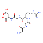
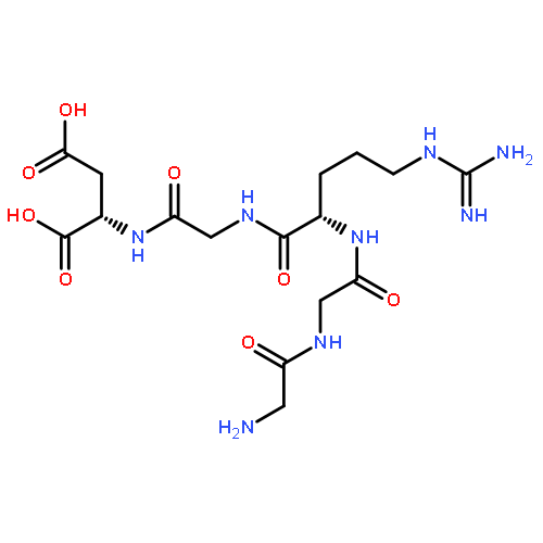
-](http://img.cochemist.com/ccimg/185700/185685-83-0.png)
-](http://img.cochemist.com/ccimg/185700/185685-83-0_b.png)


![1-Propanaminium,N,N,N-trimethyl-2,3-bis[[(9Z)-1-oxo-9-octadecen-1-yl]oxy]-](http://img.cochemist.com/ccimg/113700/113669-21-9.png)
![1-Propanaminium,N,N,N-trimethyl-2,3-bis[[(9Z)-1-oxo-9-octadecen-1-yl]oxy]-](http://img.cochemist.com/ccimg/113700/113669-21-9_b.png)


-](http://img.cochemist.com/ccimg/92500/92468-60-5.png)
-](http://img.cochemist.com/ccimg/92500/92468-60-5_b.png)

![(1R,4S,7S,10S,13R,16R)-24-hydroxy-10-(4-methoxybenzyl)-4,7,9,13,15,29-hexamethyl-22-oxa-3,6,9,12,15,29-hexaazatetracyclo[14.12.2.2~18,21~.1~23,27~]tritriaconta-18,20,23(31),24,26,32-hexaene-2,5,8,11,14,30-hexone](http://img.cochemist.com/ccimg/64800/64725-24-2.png)
![(1R,4S,7S,10S,13R,16R)-24-hydroxy-10-(4-methoxybenzyl)-4,7,9,13,15,29-hexamethyl-22-oxa-3,6,9,12,15,29-hexaazatetracyclo[14.12.2.2~18,21~.1~23,27~]tritriaconta-18,20,23(31),24,26,32-hexaene-2,5,8,11,14,30-hexone](http://img.cochemist.com/ccimg/64800/64725-24-2_b.png)
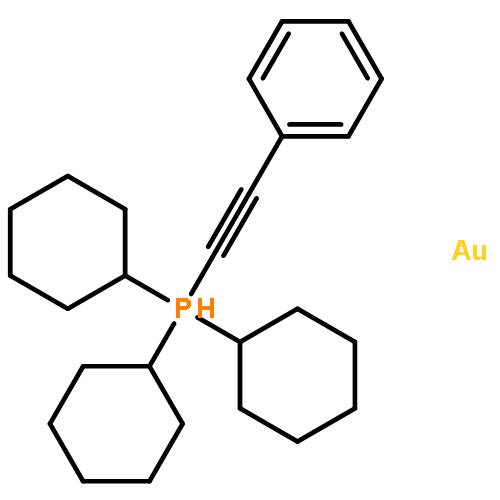


](http://img.cochemist.com/ccimg/27000/26913-06-4.png)
](http://img.cochemist.com/ccimg/27000/26913-06-4_b.png)


![Benzoxazolium,3-methyl-2-[3-(3-methyl-2(3H)-benzoxazolylidene)-1-propen-1-yl]-, iodide (1:1)](http://img.cochemist.com/ccimg/14200/14134-79-3.png)
![Benzoxazolium,3-methyl-2-[3-(3-methyl-2(3H)-benzoxazolylidene)-1-propen-1-yl]-, iodide (1:1)](http://img.cochemist.com/ccimg/14200/14134-79-3_b.png)
![Methanaminium,N-[4-[[4-(dimethylamino)phenyl]phenylmethylene]-2,5-cyclohexadien-1-ylidene]-N-methyl-](http://img.cochemist.com/ccimg/10400/10309-95-2.png)
![Methanaminium,N-[4-[[4-(dimethylamino)phenyl]phenylmethylene]-2,5-cyclohexadien-1-ylidene]-N-methyl-](http://img.cochemist.com/ccimg/10400/10309-95-2_b.png)


![5H-Benzo[a]phenoxazin-5-one,9-(diethylamino)-](http://img.cochemist.com/ccimg/7400/7385-67-3.png)
![5H-Benzo[a]phenoxazin-5-one,9-(diethylamino)-](http://img.cochemist.com/ccimg/7400/7385-67-3_b.png)